Abstract
For Salmonella, transient exposure to gastric pH prepares invading bacteria for the stresses of host-cell interactions. To resist the effects of low pH, wild-type Salmonella enterica uses the acid tolerance response and the arginine decarboxylase acid resistance system. However, arginine decarboxylase is typically repressed under routine culture conditions, and for many live attenuated Salmonella vaccine strains, the acid tolerance response is unable to provide the necessary protection. The objective of this study was to enhance survival of Salmonella enterica serovar Typhi vaccine strains at pHs 3.0 and 2.5 to compensate for the defects in the acid tolerance response imposed by mutations in rpoS, phoPQ, and fur. We placed the arginine decarboxylase system (adiA and adiC) under the control of the ParaBAD or PrhaBAD promoter to provide inducible acid resistance when cells are grown under routine culture conditions. The rhamnose-regulated promoter PrhaBAD was less sensitive to the presence of its cognate sugar than the arabinose-regulated promoter ParaBAD and provided tighter control over adiA expression. Increased survival at low pH was only observed when adiA and adiC were coregulated by rhamnose and depended on the presence of rhamnose in the culture medium and arginine in the challenge medium. Rhamnose-regulated acid resistance significantly improved the survival of ΔaroD and ΔphoPQ mutants at pHs 3 and 2.5 but only modestly improved the survival of a fur mutant. The construction of the rhamnose-regulated arginine decarboxylase system allowed us to render S. Typhi acid resistant (to pH 2.5) on demand, with survival levels approximately equivalent to that of the native arginine decarboxylase system.
INTRODUCTION
Before orally ingested enteric pathogens such as Salmonella can reach their target host cells, they must first survive their encounter with the low pH of the human stomach—∼2.0 following a fast (1). This is an extremely hostile environment, and thus Salmonella contains multiple inducible systems to aid in survival at low pH (2, 3). The best studied of these systems is the acid tolerance response (ATR). Cells exposed to moderately low pH synthesize numerous acid shock proteins. Although the specific functions of these proteins are largely unknown, jointly they mitigate the proton damage experienced by the cell during low-pH challenge (pH 3.0) (4, 5). The acid tolerance response is a complex multicomponent system coordinated by a number of global regulatory proteins. In the stationary phase, RpoS is a key regulator of the acid tolerance response. Not only does the acid tolerance response of an rpoS mutant fail to provide the same level of protection as in a wild-type strain, but rpoS mutants are unable to sustain the acid tolerance response, resulting in rapid cell death upon pH 3.0 challenge (4, 6). In log-phase cells, the Salmonella virulence proteins PhoP, PhoQ, and Fur regulate the acid tolerance response. Fur controls a subset of acid shock proteins essential for protecting the cell against organic acid challenge, while PhoP and PhoQ coordinate protection against inorganic acid challenge (7, 8).
An inability to resist low pH reduces the ability of Salmonella to reach the small intestine and increases the number of cells required to initiate a successful infection (9). To create a live attenuated Salmonella vaccine, it is necessary to introduce mutations that attenuate the virulence of the vaccine strain, and, unfortunately, these mutations often simultaneously decrease the ability to survive at low pH. The vast majority of live attenuated Salmonella vaccines for humans are constructed from Salmonella enterica serovar Typhi strain Ty2, an rpoS mutant (10). In addition to the rpoS mutation derived from its parent strain Ty2, the licensed typhoid vaccine strain Ty21a carries galE and tvi mutations as well as a number of other, less well-characterized mutations (11–13). The strain is sensitive to low pH, due at least in part to its inability to mount a functional acid tolerance response (14). Another vaccine strain, Ty800, contains a deletion of the phoPQ locus. This strain is safe and reasonably immunogenic in humans (15), but one would expect that the combination of the ΔphoPQ deletion and rpoS mutation would render this strain exquisitely sensitive to acidic pH (6, 8). A similar situation occurs for the vaccine strains χ9639(pYA4088) and χ9640(pYA4088) (16). These strains are also safe and immunogenic in humans (S. E. Frey, K. R. Lottenbach, H. Hill, T. P. Blevins, Y. Yu, Y. Zhang, K. E. Brenneman, S. M. Kelly-Aehle, C. McDonald, A. Jansen, and R. Curtiss III, submitted for publication), but the mutation in their fur locus leaves them vulnerable to low pH. For each of these vaccine strains, humans immunized with a dose of less than 109 CFU fail to develop an immune response.
Most vaccine researchers avoid the problem low gastric pH poses by coating their vaccine in a protective enteric capsule (e.g., Ty21a) or by coadministration of an antacid (usually sodium bicarbonate) at the time of immunization (17–22). Preventing vaccine exposure to low pH increases the number of viable cells that reach the intestine and improves vaccine immunogenicity (22, 23). The disadvantage of bypassing the acidic environment of the stomach is that the low-pH encounter serves as an important signal to Salmonella, allowing it to recognize entry into a host environment. Exposure to acid (along with other host signals) stimulates upregulation of the genes that confer resistance to the short-chain fatty acids (24), antimicrobial peptides (25), and osmotic stress (6) found in the intestine. Also, induction of the acid tolerance response has been linked to upregulation of SPI-1 and SPI-2 and an increase in epithelial cell invasion in the intestine (26–28). Thus, transient exposure to low pH prepares the invading bacteria for the stresses of the intestine and for host-cell interactions. Therefore, it is possible that if the survival rate of live attenuated Salmonella vaccine strains at low pH can be improved, we not only can eliminate the need for low-pH bypass strategies but also can improve the ability of the vaccine strain to interact with host tissues to enhance immunogenicity.
As a first step toward this goal, we explored methods to increase the low-pH survival of S. Typhi strains containing rpoS, ΔphoPQ, or fur mutations. Each of these mutations renders strains acid sensitive, and each has been incorporated into live attenuated vaccine strains. One robust means used by Salmonella to resist low-pH challenge is the arginine decarboxylase acid resistance system (29). This system consists of arginine decarboxylase (AdiA) and an arginine-agmatine antiporter (AdiC) (30). Acid resistance is conferred by the activity of AdiA, which consumes one proton from the intracellular environment with each reaction cycle and causes a rapid rise in intracellular pH (31, 32). AdiC then transports agmatine, the product of arginine decarboxylation, to the periplasm in exchange for another arginine substrate molecule (30, 33). The combined activities of AdiA and AdiC allow Salmonella enterica serovar Typhimurium to resist pH 2.5 for more than 2 h (2).
Because the arginine decarboxylase system functions independently of the acid tolerance response, we hypothesized that synthesis of AdiA and AdiC would confer high levels of acid resistance on strains containing mutations that affect acid tolerance, such as rpoS, phoPQ, and fur. However, the arginine decarboxylase system is tightly regulated and is not normally available to cells grown under standard vaccine culture conditions (34). Therefore, we replaced the native promoter of arginine decarboxylase with araBAD (ParaBAD) or rhaBAD (PrhaBAD) promoters. The placement of the system under the control of these promoters allows expression in vitro and during the initial stages of infection, but results in rapid downregulation once the vaccine invades host tissue and the regulatory sugar is no longer available (35). After selecting the promoter with optimal sugar-dependent expression and activity of the arginine decarboxylase system (PrhaBAD), our objectives were twofold. First, we determined whether the rhamnose-regulated arginine decarboxylase system could rescue rpoS, ΔphoPQ, and fur mutants during low-pH challenge if cells were cultured in the presence of rhamnose but without any other environmental signals that would induce either decarboxylase activity or the acid tolerance response. Second, we determined whether the rhamnose-regulated system functioned equivalently to the native arginine decarboxylase system, by comparing the level of acid resistance in strains carrying our engineered rhamnose-regulated system induced by growth with rhamnose to that in strains carrying the native arginine decarboxylase system induced by anaerobic growth in unbuffered rich medium.
MATERIALS AND METHODS
DNA manipulation and plasmid construction.
Chromosomal DNA from S. Typhi Ty2 was isolated using the Wizard genomic DNA purification kit (Promega, Madison, WI). Plasmid DNA was isolated using the QIAprep spin miniprep kit (Qiagen, Valencia, CA) or the Wizard Plus midiprep DNA purification system (Promega). DNA inserts were amplified by PCR using the Phusion DNA polymerase (New England BioLabs, Ispwich, MA) or the Easy-A high-fidelity PCR cloning enzyme (Agilent, Santa Clara, CA). Restriction and modification enzymes for cloning (New England BioLabs) were used in accordance with the manufacturer's instructions.
Construction of S. Typhi mutants.
The bacterial strains and plasmids used in this study are listed in Table 1. The primers used during the construction of plasmids are listed in Table S1 in the supplemental material. To construct the ΔaroD1299 mutant, two DNA fragments adjacent to the aroD gene were amplified from the chromosome of Ty2. Primers Aro-1 and -2 were used for the upstream fragment, while primers Aro-3 and -4 were used for the downstream fragment. These fragments were digested with BamHI, ligated using T4 DNA ligase, reamplified by PCR with primers Aro-1 and -4, and cloned into the AhdI sites of pYA4278 via TA overhangs to generate the suicide vector pYA4895. The ΔaroD1299 deletion was introduced into Ty2 by conjugation using the antibiotic resistance selection-sacB counterselection method described by Kaniga (36). The resulting strain (χ11548) exhibits aromatic amino acid auxotrophy and carries a deletion of the complete coding sequence of aroD that spans 759 bp.
Table 1.
Strains and plasmids used in this study
| Strain or plasmid | Genotypea | Source, derivation, or reference |
|---|---|---|
| Strains | ||
| E. coli | ||
| BL21(DE3) | F− ompT hsdSB (rB− mB−) gal dcm (DE3) | Novagen |
| χ7213 | thr-1 leuB6 fhuA21 lacY1 glnV44 recA1 ΔasdA4 Δ(zhf-2::Tn10) thi-1 RP4-2-Tc::Mu [λpir] | 54 |
| S. Typhi | ||
| χ3769 (Ty2) | Wild-type; cys trp rpoS | 55 |
| χ8444 | ΔphoPQ23 | 56 |
| χ11118 | ΔPfur81::TT araC ParaBAD fur | Ty2 |
| χ11500 | Δ(adiA-adiC)-4806 | Ty2 |
| χ11548 | ΔaroD1299 | Ty2 |
| χ11552 | ΔaroD1299 ΔPadiA203::TT araC ParaBAD adiA | χ11548 |
| χ11564 | ΔaroD1299 ΔPadiA276::TT rhaSR PrhaBAD adiA | χ11548 |
| χ11568 | ΔaroD1299 ΔPadiA276::TT rhaSR PrhaBAD adiA Δ(PadiY-adiY-PadiC)-119 adiC | χ11564 |
| χ11622 | ΔphoPQ23 ΔPadiA276::TT rhaSR PrhaBAD adiA Δ(PadiY-adiY-PadiC)-119 adiC | χ8444 |
| χ11623 | ΔPfur81::TT araC ParaBAD fur ΔPadiA276::TT rhaSR PrhaBAD adiA Δ(PadiY-adiY-PadiC)-119 adiC | χ11118 |
| χ11636 | ΔaroD1299 Δ(PadiY-adiY-PadiC)-119 adiC | χ11548 |
| Shigella flexneri 2457T | S. flexneri 2a, wild type; Pcr+ Mal− λr | 57 |
| Plasmids | ||
| pET28a | Protein synthesis vector, T7 promoter; Kanr | Novagen |
| pYA3700 | Vector containing tightly regulated TT araC ParaBAD cassette | 58, 59 |
| pYA4181 | Suicide vector to generate ΔPfur81::TT araC ParaBAD fur mutation | 35 |
| pYA4278 | Suicide vector; sacB mobRP4 oriR6K Cmr | 60 |
| pYA4895 | Suicide vector to generate ΔaroD1299 mutation | pYA4278 |
| pYA5066 | Suicide vector to generate Δ(adiA-adiC)-4806 mutation | pYA4278 |
| pYA5072 | Suicide vector to generate Δ(PadiY-adiY-PadiC)-119 adiC mutation | pYA4278 |
| pYA5075 | Intermediate vector for creation of ΔPadiA203::TT araC ParaBAD adiA | pYA3700 |
| pYA5081 | Suicide vector specifying tightly regulated rhaSR PrhaBAD cassette | 61 |
| pYA5085 | Protein synthesis vector with N-terminal His tag on AdiA | pET28a |
| pYA5089 | Suicide vector to generate ΔPadiA203::TT araC ParaBAD adiA mutation | pYA4278, pYA5075 |
| pYA5093 | Suicide vector to generate ΔPadiA276::TT rhaSR PrhaBAD adiA mutation | pYA5089, pYA5081 |
In genotype descriptions, the subscripted designation number refers to a composite deletion and insertion of the indicated gene. P, promoter; TT, T4 ip III transcription terminator; Cmr, chloramphenicol resistance; Kanr, kanamycin resistance.
The ΔPfur81::TT (transcription terminator) araC PBAD fur mutation was introduced into S. Typhi Ty2 via P22 HT int transduction (37) using a lysate grown on χ9269 containing a chromosomally integrated copy of pYA4181 (35) to create the S. Typhi strain χ11118. The presence of the ΔPfur81::TT araC PBAD fur mutation in S. Typhi was confirmed by PCR using the primers Fur-1 and -2. Arabinose-dependent synthesis of Fur was verified by Western blotting.
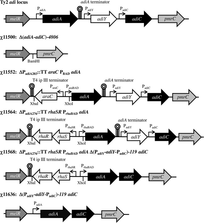
To remove the entire adi locus [Δ(adiA-adiC)-4806; hereafter Δ(adiA-adiC)], the upstream and downstream flanking regions in Ty2 were amplified using PCR primers Adi-1 and -2 and primers Adi-3 and -4, respectively. The flanking regions were digested with BamHI and ligated together with T4 DNA ligase. The resulting product was reamplified by PCR using primers Adi-1 and -4 and cloned into the AhdI sites of pYA4278 to generate the suicide vector pYA5066. The Δ(adiA-adiC) mutation carried by pYA5066 was moved into Ty2 to create χ11500. This strain carries a 4,806-bp deletion of the adi locus (complete coding sequences of adiA, adiY, and adiC and the adiY and adiC promoters) (Fig. 1). The absence of the adi locus was confirmed by PCR and by arginine decarboxylase assay.
Fig 1.
Schematic diagram of arginine decarboxylase mutations. The genes and associated regulatory sequences for the Δ(adiA-adiC)-4806, ΔPadiA203::TT araC ParaBAD adiA, ΔPadiA276::TT rhaSR PrhaBAD adiA, and Δ(PadiY-adiY-PadiC)-119 adiC mutations are shown above, along with the archetypal strain number. The wild-type arginine decarboxylase locus (adi) of S. Typhi Ty2 is depicted for comparative purposes. The diagram is approximately to scale. The promoters and transcription terminator are labeled on the figure.
To produce strains in which adiA expression could be regulated exclusively by the presence or absence of a specific sugar, we engineered the following promoter substitution mutations: ΔPadiA203::TT araC ParaBAD adiA (regulated by arabinose) and ΔPadiA276::TT rhaSR PrhaBAD adiA (regulated by rhamnose). For simplicity, these mutations will be referred to as ParaBAD adiA and PrhaBAD adiA, respectively. For the arabinose-regulated construct, the DNA regions flanking the adiA promoter were amplified by PCR from Ty2 using primers Adi-5 and -6 for the upstream region and primers Adi-7 and -8 for the downstream region. Both flanking regions were cloned into pYA3700 (using SphI and BglII for the upstream region and KpnI and SacI for the downstream region) to generate pYA5075. The DNA segment containing the flanking regions and arabinose promoter was amplified by PCR using Adi-5 and -8, and the PCR product was cloned into the AhdI sites of pYA4278 to create the suicide vector pYA5089. To generate the rhamnose-regulated construct, the araC ParaBAD promoter of pYA5089 was removed by XhoI and XbaI double digestion. The rhaSR PrhaBAD promoter from pYA5081 was amplified by PCR with the Rha-1 and -2 primers and cloned into pYA5089 using XhoI and XbaI to produce the suicide vector pYA5093. pYA5089 and pYA5093 were introduced into χ11548 by conjugation to produce χ11552 and χ11564, respectively. The juxtaposition of adiA with the appropriate promoter was verified by PCR with the Ara-1 and Adi-9 primers (χ11552) or Rha-3 and Adi-9 primers (χ11564) and by the arginine decarboxylase assay. In both strains, 203 bp of the intergenic region between melR and adiA (including the −10 and −35 sites of the adiA promoter) were deleted and replaced with either TT araC ParaBAD (χ11552) or TT rhaRS PrhaBAD (χ11564). The strong transcription terminator T4 ip III was placed between the upstream melR gene and araC or rhaSR to prevent expression of antisense RNA. A strong Shine-Dalgarno site (AGGA) was inserted 10 bp upstream of the ATG start codon of adiA (Fig. 1).
The adiC gene was fused into an operon with adiA resulting in the Δ(PadiY-adiY-PadiC)-119 adiC mutation (hereafter, adiAC). The DNA regions flanking adiY were amplified by PCR from Ty2 using primers Adi-10 and -11 for the upstream region and primers Adi-12 and -13 for the downstream region. The two DNA segments were joined by overlap PCR and reamplification with Adi-10 and -13. The final PCR product was ligated into pYA4278 at the AhdI sites to produce the suicide vector pYA5072. The suicide vector was introduced into χ11564 and χ11548 by conjugation to produce χ11568 (ΔaroD PrhaBAD adiAC) and χ11636 (ΔaroD adiAC), respectively. The presence of the adiAC operon was confirmed by PCR using Adi-14 and -15. Both strains harbor a 1,078-bp deletion that spans the transcription terminator following adiA, adiY, and the promoter of adiC. The adiA and adiC genes are separated by a 119-bp intergenic sequence expected to decrease expression of adiC from the promoter upstream of adiA (Fig. 1).
Growth conditions and culture media.
Experiments testing the regulation of arabinose- and rhamnose-controlled genes were conducted in the carbohydrate-free medium purple broth (BD Biosciences, Franklin Lakes, NJ). The use of this carbohydrate-free medium allowed us to accurately control the concentrations of rhamnose or arabinose to evaluate their effects on promoter activity.
For induction of the native arginine decarboxylase system, strains were propagated in tryptic soy broth (TSB) (BD Biosciences) with 0.4% glucose under anaerobic conditions. This causes the medium pH to fall below 5.0 during culture. In all other acid resistance experiments, strains were grown aerobically in minimal E medium (pH 7.0) with 0.4% glucose (EG medium) (38). For our experiments, 22 μg/ml l-cysteine, 20 μg/ml l-tryptophan, and 0.1% Casamino Acids were added to EG medium to supplement the growth of all strains (EGA medium). For strains with the ΔaroD1299 mutation, 20 μg/ml l-tryptophan, 2 μg/ml ρ-aminobenzoic acid, and 2.5 μg/ml 2,3-dihydroxybenzoate were added to all media. EGA medium was additionally supplemented with 50 μg/ml l-phenylalanine and 20 μg/ml l-tyrosine. Rhamnose was added to 0.1% or to 0.4% in the case of strain χ11623, as indicated. Strains containing the ΔPfur81::TT araC ParaBAD fur mutation were supplied with 0.2% arabinose unless otherwise indicated. All chemicals were purchased from Sigma-Aldrich (St. Louis, MO) or Thermo Fisher Scientific (Pittsburgh, PA) unless otherwise indicated.
Measurement of adiA expression by semiquantitative PCR.
Strains were grown in purple broth with various concentrations of rhamnose or arabinose to an optical density at 600 nm (OD600) of 0.6. Total cellular RNA was isolated using the RNeasy minikit (Qiagen) and was treated with RNase-free DNase (Qiagen). cDNA was generated via reverse transcription (RT)-PCR using 1 μg of cellular RNA with the TaqMan reverse transcriptase kit (Life Technologies, Grand Island, NY) under the following conditions: 10 min at 25°C for optimal random hexamer primer binding and then 45 min at 48°C for extension followed by 5 min at 95°C to heat inactivate the transcriptase. Semiquantitative PCR of the gapA (control) and adiA transcripts was performed using the GoTaq DNA polymerase system (Promega) using primers SQ-1 and SQ-2 for gapA and SQ-3 and SQ-4 for adiA under the following conditions: 2.5 min at 95°C for template denaturation, followed by 28 cycles of 40 s at 95°C, 30 s at 48°C for primer annealing, and 1 min at 72°C for primer extension. The semiquantitative PCR primer sequences are listed in Table S1 in the supplemental material (SQ-1 to SQ-4). PCR products were electrophoresed on a 2% agarose gel in the presence of ethidium bromide and visualized with the ChemiDoc XRS system (Bio-Rad Laboratories, Hercules, CA). Images were analyzed in Adobe Photoshop CS4 (Adobe Systems, Inc., San Jose, CA) in order to establish histogram values for the fluorescence signal intensity of the PCR products. Signal intensity values for adiA were normalized to the value obtained with the single-gene-expression control gapA for each culture.
Preparation of antiserum against arginine decarboxylase protein.
Escherichia coli BL21(DE3) harboring pYA5085 was used for the synthesis of His-tagged AdiA protein. Cells were grown in LB at 37°C to the mid-log phase (OD600 of 0.6). The growth medium was supplemented with 0.2 g/liter pyridoxine to augment protein folding and enzyme activity (39). Protein synthesis was induced with 1 mM IPTG (isopropyl β-d-1-thiogalactopyranoside) (Amresco, Solon, OH) for 4 h at 37°C. Cells were collected by centrifugation and disrupted using lysozyme (3 mg/g cells) and deoxycholic acid (120 mg/g cells) (40). His-tagged AdiA protein from the soluble fraction was purified over Talon metal affinity resin (BD Biosciences) in accordance with the manufacturer's instructions, except that 10% ethanol was added to the elution buffer. Purified protein was stored in 20 mM HEPES–50 mM NaCl (pH 8.0) (31).
One juvenile New Zealand White rabbit (Charles River Laboratories, Wilmington, MA) was immunized with 200 μg of AdiA emulsified in Freund's complete adjuvant and boosted with an additional 200 μg of AdiA emulsified in Freund's incomplete adjuvant 4 and 8 weeks after the initial injection. Serum was collected 3 weeks following the final immunization.
Western blot procedure.
Strains were grown overnight at 37°C in purple broth containing various concentrations of rhamnose or arabinose. The amount of total cellular protein in each sample was normalized by absorbance at 280 nm using the NanoDrop ND-1000 (Thermo Scientific, Wilmington, DE). Equal amounts of total cellular protein (100 μg) were mixed with 2× SDS-PAGE buffer, boiled, and electrophoresed on a 10% acrylamide gel (41). Separated proteins were transferred to a polyvinylidene difluoride (PVDF) membrane (Bio-Rad) using Towbin's wet transfer method (42), blocked in 5% skim milk, and then probed with rabbit antiserum (final dilution, 1:10,000) for the presence of AdiA (35). Bound primary antibody was detected by the addition of goat anti-rabbit IgG conjugated to alkaline phosphatase (Sigma-Aldrich). Blots were developed with NBT/BCIP (nitroblue tetrazolium–5-bromo-4-chloro-3-indolyl phosphate) (Amresco) and photographed using the ChemiDoc XRS system.
Arginine decarboxylase assays.
Arginine decarboxylase enzyme activity was measured using a modified version of the rapid glutamate decarboxylase assay previously described (43). Strains were grown overnight (18 h) to the stationary phase in purple broth, washed once in phosphate-buffered saline (PBS) (40), and normalized to an OD600 value of 0.7. Five milliliters of normalized cells was pelleted, resuspended in 2.5 ml arginine decarboxylase assay medium (1 g l-arginine, 0.05 g bromocresol green, 90 g NaCl, and 3 ml Triton X-100 per liter of distilled water [adjusted to pH 3.4]), and vortexed for 30 s. Assay tubes were incubated at 37°C for 5 to 30 min, scored, and photographed.
Acid resistance assays.
Acid resistance was determined essentially as described previously (44, 45), with the following modifications. Strains were grown overnight to stationary phase in minimal EGA medium at pH 7.0 (38) or in TSB with 0.4% glucose. Cultures were normalized to the same OD600 and then pelleted and washed once in EGA medium (pH 7.0) containing no growth supplements. Cells were pelleted a second time and resuspended at a density of 1 × 10⁹ CFU/ml in EGA medium containing 1 mM l-arginine at pH 3.0, 2.5, or 2.0. Low-pH challenge was conducted at 37°C, and samples were collected immediately after resuspension (time [t] 0) and hourly for 4 h. Samples were serially diluted and plated onto LB agar to assess bacterial viability.
Statistical analyses.
All statistical analyses were performed using GraphPad Prism version 5.04 for Windows (GraphPad Software, San Diego, CA). Survival curves for 4-h acid resistance assays were compared using two-way repeated measures (mixed model) analysis of variance (ANOVA) with Bonferroni's posttest. Data from 1-h acid resistance challenges were compared using the paired t test.
RESULTS AND DISCUSSION
Comparison of adiA regulation from arabinose- and rhamnose-regulated promoters.
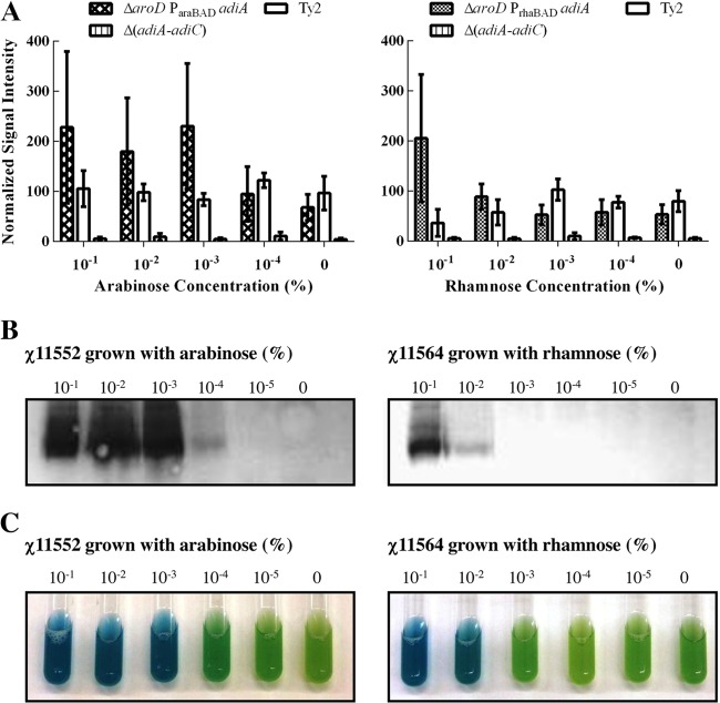
To allow expression of the arginine decarboxylase system during aerobic growth, we constructed S. Typhi strains in which adiA expression was regulated by either the ParaBAD or PrhaBAD promoter. For safety, the sugar-regulated adiA constructs were introduced into S. Typhi strain χ11548, which carries an attenuating ΔaroD mutation (18, 46). Thus, in strains χ11552 (ΔaroD ParaBAD adiA) and χ11564 (ΔaroD PrhaBAD adiA), adiA expression responds to the level of exogenous arabinose or rhamnose, respectively. In the absence of the regulating sugar, both strains expressed low levels of adiA transcript consistent with background levels observed in Ty2 cultured under noninducing conditions for adiA (Fig. 2A). Both strains increased production of the adiA mRNA transcript when 0.1% (10−1%) of the appropriate sugar was added. The two promoters drove production of essentially equivalent amounts of adiA transcript at this concentration, consistent with previous results (47). As the amount of regulatory sugar present in the culture was decreased, the activities of the two promoters decreased differentially. Strain χ11552 (ParaBAD adiA) continued to express adiA mRNA at arabinose concentrations as low as 0.001% (10−3%). Only when the arabinose concentration fell below 0.001% (10−3%) did the amount of adiA transcript return to background levels. In contrast, strain χ11564 (PrhaBAD adiA) expressed adiA transcript only in the presence of 0.1% (10−1%) rhamnose and produced background levels of adiA mRNA at lower rhamnose concentrations.
Fig 2.
Regulation of adiA by the araC ParaBAD and rhaSR PrhaBAD promoters. χ11552 (ΔaroD ParaBAD adiA) and χ11564 (ΔaroD PrhaBAD adiA) were cultured in the presence of various concentrations of arabinose or rhamnose (ranging from 10−1 to 10−5%), normalized and assayed by semiquantitative PCR for the level of adiA transcript (A), probed for the presence of AdiA by Western blotting (B), or tested for arginine decarboxylase activity via colorimetric assay (C). mRNA data are plotted as the means and standard errors of the means (SEM) from three independent experiments. Western blot and enzyme assay data are representative of three independent assays. In the colorimetric arginine decarboxylase assay, active enzyme raises the assay medium pH above 5.0, resulting in a color change from yellow-green (negative) to blue (positive).
AdiA protein synthesis and enzyme activity levels presented a pattern similar to that of the mRNA. χ11552 (ParaBAD adiA) synthesized AdiA over a wide range of arabinose concentrations (10−1 to 10−4% arabinose), while in χ11564 (PrhaBAD adiA), AdiA was detected over a narrower range of rhamnose concentrations (10−1 to 10−2% rhamnose) (Fig. 2B). Interestingly, χ11552 (ParaBAD adiA) produced greater amounts of AdiA protein in the presence of 0.1% arabinose than did χ11564 (PrhaBAD adiA) in the presence of 0.1% rhamnose. Arginine decarboxylase activity was detected in χ11552 (ParaBAD adiA) cultures grown in the presence of arabinose concentrations as low as 10−3% (Fig. 2C). An intermediate reaction suggestive of low levels of enzyme activity was observed at 10−4% arabinose. In contrast, arginine decarboxylase activity was observed in χ11564 (PrhaBAD adiA) only at rhamnose concentrations greater than 10−2%. While the largest amounts of AdiA in both strains were observed at the arabinose and rhamnose concentrations that increased levels of adiA transcript, small amounts of AdiA were also detected at sugar concentrations that did not produce a measurable increase in the amount of adiA transcript present, which could reflect differences in the sensitivities of the assays or differences in the stabilities of the adiA mRNA transcript and AdiA protein.
Comparison of the arabinose-regulated ParaBAD and rhamnose-regulated PrhaBAD promoters indicated that PrhaBAD was less sensitive to its regulatory sugar than ParaBAD. The reduced sensitivity of the PrhaBAD promoter made it an ideal choice to regulate the arginine decarboxylase system since it allowed tighter control of gene expression in media containing trace amounts of rhamnose, such as LB and TSB. In addition, the promoter will not be active if the cell encounters trace amounts of the inducing sugar in vivo, which should enhance its safety as a vaccine. An additional advantage of the PrhaBAD promoter was its decreased level of AdiA protein synthesis. Protein overproduction has been shown to impair the efficacy of Salmonella vaccines (48, 49). For these reasons, we selected the ΔPadiA276::TT rhaSR PrhaBAD adiA mutation for use in further studies.
Coregulation of adiA and adiC is necessary for survival during pH 3.0 challenge.
Our goal in introducing the PrhaBAD adiA construct into S. Typhi was to provide arginine-dependent acid resistance when cells were grown under conditions when this system is not normally induced (noninducing conditions). To test this, we performed low-pH challenges on cells grown aerobically in minimal EGA medium. However, while χ11564 (ΔaroD PrhaBAD adiA) exhibited rhamnose-inducible arginine decarboxylase activity under these conditions (data not shown), the survival profile of χ11564 (ΔaroD PrhaBAD adiA) at pH 3.0 did not differ from that of Ty2 or its parent strain, χ11548 (ΔaroD) (Fig. 3). We reasoned that the PrhaBAD promoter in strain χ11564 (ΔaroD PrhaBAD adiA) does not drive adiC expression due to the presence of a transcriptional terminator downstream of adiA and the intervening adiY gene. To coregulate expression of both adiA and adiC, the intergenic region between the two genes, including the regulatory gene adiY and the adiC promoter, was deleted, resulting in the fusion of adiA and adiC into a single operon under the control of the native adiA promoter, resulting in strain χ11636 (adiAC) (Fig. 1). The sensitivity of χ11636 to pH 3.0 challenge was not significantly different from those of Ty2 and χ11548 (ΔaroD) (P = 0.327) (Fig. 3). However, when the adiAC operon fusion was placed under transcriptional control of the PrhaBAD promoter and the resulting strain (χ11568 ΔaroD PrhaBAD adiAC) was grown in the presence of 0.1% rhamnose, it displayed a 1,000- to 10,000-fold increase over Ty2 in the number of viable cells present at all time points during pH 3.0 challenge (P < 0.0001) (Fig. 3). Thus, our ability to rescue χ11568 at low pH via rhamnose induction of the adiA system indicates that the activity of the arginine decarboxylase system alone is sufficient for low-pH survival in S. Typhi, despite the presence of the rpoS mutation in Ty2. This is not surprising, since the induction and activity of the native adiA system in Salmonella are independent of rpoS (2).
Fig 3.
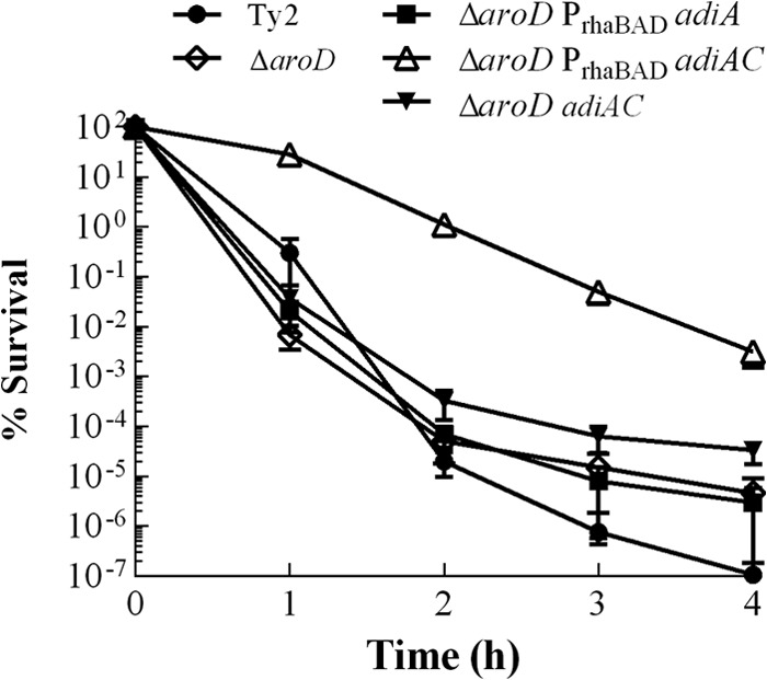
Coregulation of adiA and adiC by rhamnose is required for survival during pH 3 challenge. χ11564 (ΔaroD PrhaBAD adiA), χ11568 (ΔaroD PrhaBAD adiAC), and χ11636 (ΔaroD adiAC) were grown to the stationary phase in EGA medium in the presence of 0.1% rhamnose and then challenged with pH 3.0 EG medium containing 1 mM arginine. Survival was monitored by plating on LB agar containing all necessary supplements. The data shown are the means and SEM from three independent assays.
Survival of strain χ11568 during pH 3.0 challenge is rhamnose and arginine dependent.
A number of acid resistance and acid tolerance mechanisms have been described in stationary-phase Salmonella. To confirm that the acid resistance phenotype of χ11568 (ΔaroD PrhaBAD adiAC) was attributable to the rhamnose-regulated arginine decarboxylase system, χ11568 was tested for survival at pH 3.0 in the absence of rhamnose and arginine. When cultured in minimal EGA medium without rhamnose, χ11568 (ΔaroD PrhaBAD adiAC) displayed a survival profile during pH 3.0 challenge indistinguishable from that of the wild-type Ty2 and χ11548 (ΔaroD) strains (Fig. 4A). Addition of rhamnose to the EGA culture medium restored the acid resistance of χ11568 (ΔaroD PrhaBAD adiAC), resulting in a 1,000- to 10,000-fold higher survival rate when rhamnose was provided (P = 0.001).
Fig 4.
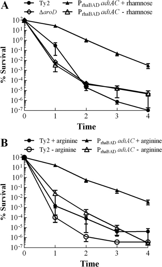
Acid resistance depends on the presence of rhamnose and arginine. Ty2, χ11548 (ΔaroD), and χ11568 (ΔaroD PrhaBAD adiAC) were grown to the stationary phase in EGA medium and then challenged with EG medium (pH 3.0). (A) Strains cultured in the presence or absence of 0.1% rhamnose. (B) Strains challenged in the presence or absence of 1 mM arginine. Survival in all assays was monitored by plating on LB agar containing all necessary supplements. The data shown are the means and SEM from three independent assays.
The acid resistance of χ11568 (ΔaroD PrhaBAD adiAC) also depended on the presence of arginine in the challenge medium (Fig. 4B). The percentage of viable χ11568 (ΔaroD PrhaBAD adiAC) cells during challenge rapidly declined over 4 h in the absence of arginine, with few survivors detected after the first 2 h. However, cells that were challenged in the presence of 1 mM arginine showed a marked increase in survival (P = 0.003). The arginine requirement for survival at low pH confirms that the acid resistance we observed was due to the Salmonella arginine decarboxylase system and not to the stationary-phase acid tolerance response or the oxidative acid resistance response (AR1), as neither of these systems requires arginine (3, 44). Interestingly, even though cells were cultured in aerobic minimal medium to prevent induction of the native arginine decarboxylase system in wild-type S. Typhi strain Ty2 (2), we observed an arginine-dependent increase (P = 0.022) in resistance to pH 3 challenge (Fig. 4B). This suggests that arginine decarboxylase is expressed at low levels in S. Typhi during stationary-phase culture—a conclusion consistent with the low, but detectable, levels of adiA transcript observed in Ty2 (Fig. 2).
The rhamnose-regulated arginine decarboxylase system also provided a substantial benefit to S. Typhi survival during pH 2.5 challenge (Fig. 5). After 1 h at pH 2.5, χ11568 (ΔaroD PrhaBAD adiAC) survived significantly better than its ΔaroD parent (χ11548) (P = 0.010), wild-type strain Ty2 (P = 0.010), and the arginine decarboxylase deletion mutant χ11500 (ΔadiA-adiC) (P = 0.035). Of the 109 CFU that were challenged, over 105 CFU of χ11568 (ΔaroD PrhaBAD adiAC) remained viable after 1 h. However, the arginine decarboxylase system did not appear to be able to protect S. Typhi for longer than 1 h at pH 2.5, as we did not detect any viable cells after the first hour of challenge (data not shown). This is quite different from what occurs in S. Typhimurium, where the arginine decarboxylase system protects cells for more than 2 h at pH 2.5 (2).
Fig 5.
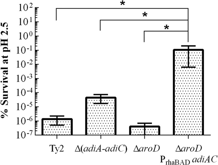
Acid resistance of a ΔaroD1299 mutant containing rhamnose-regulated arginine decarboxylase at pH 2.5. Ty2, χ11500 (ΔadiA-adiC), χ11548 (ΔaroD), and χ11568 (ΔaroD PrhaBAD adiAC) were grown to the stationary phase in EGA medium containing 0.1% rhamnose and then challenged with EG medium containing 1 mM arginine at pH 2.5 for 1 h. Survival in all assays was monitored by plating on LB agar containing all necessary supplements. The data shown are the means and SEM from three independent assays. Pairs of data marked with an asterisk are significantly different (P < 0.05).
Rhamnose-dependent acid resistance is equivalent to acid resistance in cells grown under decarboxylase-inducing conditions.
We next compared the level of acid resistance afforded by the rhamnose-regulated system to the acid resistance provided by the native system. Strains were grown anaerobically with 0.1% rhamnose in unbuffered rich medium where the pH was allowed to fall below pH 5.0 during growth (native inducing conditions). The arginine decarboxylase deletion mutant χ11500 (ΔadiA-adiC) rapidly succumbed to challenge at both pHs 3.0 and 2.5 (Fig. 6A and B). Ty2 and χ11548 (ΔaroD) displayed a high degree of acid resistance at pH 3.0 (greater than 104 CFU/ml were viable after 4 h), but succumbed to pH 2.5 after 2 h. In contrast, the highly acid-resistant Shigella flexneri strain 2457T exhibited >70% viability for 4 h at pH 3.0, and viability only decreased by 1 log after 4 h at pH 2.5. Strain χ11568 (ΔaroD PrhaBAD adiAC) was not able to match the acid resistance profile of Shigella, although it displayed a survival profile equivalent to those of S. Typhi Ty2 and χ11548 (ΔaroD) grown under these conditions (P = 0.210). No protection was afforded against pH 2.0 challenge (data not shown), consistent with previous reports for Salmonella (3, 50).
Fig 6.
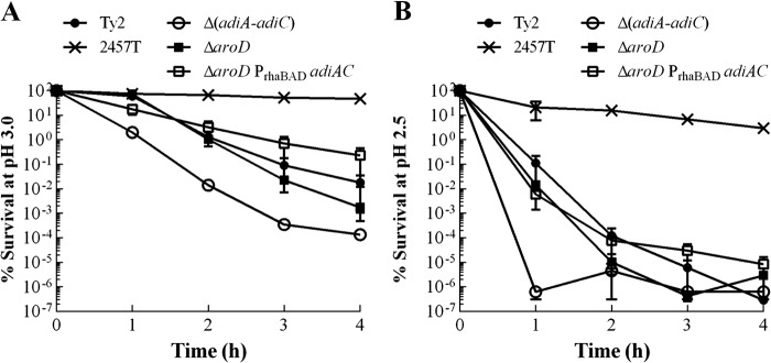
Comparison of acid resistance provided by native and rhamnose-regulated arginine decarboxylase. S. flexneri 2457T, Ty2, χ11500 (ΔadiA-adiC), χ11548 (ΔaroD), and χ11568 (ΔaroD PrhaBAD adiAC) were grown overnight in TSB medium with 0.4% glucose and 0.1% rhamnose under anaerobic conditions. Cells were challenged for 4 h with EG medium containing 1 mM arginine at pH 3.0 (A) or pH 2.5 (B). Survival in all assays was monitored by plating on LB agar containing all necessary supplements. The data shown are the means and SEM from three independent assays.
Rescue of ΔphoPQ23 and ΔPfur81::TT araC ParaBAD fur mutants at pHs 3.0 and 2.5.
Based on our successful rescue of a ΔaroD mutant with the rhamnose-regulated arginine decarboxylase system, we wanted to evaluate the system in S. Typhi strains carrying attenuating mutations known to affect low-pH survival. Therefore, we introduced the rhamnose-regulated adiA system into two S. Typhi strains containing mutations that result in well-characterized acid sensitivities (7, 8): ΔphoPQ mutant χ8444 and fur mutant χ11118 (Table 1). The resulting strains, χ11622 (ΔphoPQ PrhaBAD adiAC) and χ11623 (ParaBAD fur PrhaBAD adiAC), exhibited rhamnose-dependent arginine decarboxylase activity (data not shown).
To evaluate the ability of the rhamnose-regulated arginine decarboxylase system to rescue χ11622 (ΔphoPQ PrhaBAD adiAC), the strain was grown in aerobically in minimal EGA medium to the stationary phase at pH 7.0 in the presence of 0.1% rhamnose and then was challenged at either pH 3.0 or 2.5. Under these growth conditions, which do not induce the native adiA system, the ΔphoPQ23 mutant χ8444 displayed a survival profile similar to that of wild-type Ty2 (P = 0.996) (Fig. 7A). In contrast, the rhamnose-regulated arginine decarboxylase system in strain χ11622 (ΔphoPQ PrhaBAD adiAC) provided approximately a 1,000-fold increase in viability at pH 3.0 over both the parent ΔphoPQ mutant (χ8444) and the wild-type strain Ty2 (P = 0.034). At pH 2.5, the viability of χ11622 (ΔphoPQ PrhaBAD adiAC) after 1 h significantly exceeded that of the other S. Typhi strains: P = 0.009 for χ8444, P = 0.010 for Ty2, and P = 0.0232 for χ11500 [Δ(adiA-adiC)] (Fig. 7B). When strains were grown under native adiA-inducing conditions and challenged at pH 3.0 or 2.5, the rhamnose-inducible arginine decarboxylase system in χ11622 (grown with 0.1% rhamnose) provided protection equivalent to that of the native system (see Fig. S1 in the supplemental material).
Fig 7.
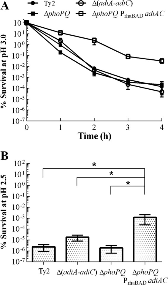
Acid resistance of a ΔphoPQ23 mutant containing rhamnose-regulated arginine decarboxylase. Ty2, χ11500 (ΔadiA-adiC), χ8444 (ΔphoPQ), and χ11622 (ΔphoPQ PrhaBAD adiAC) were grown to the stationary phase in EGA medium containing 0.1% rhamnose and then challenged with EG medium containing 1 mM arginine at pH 3.0 for 4 h (A) or pH 2.5 for 1 h (B). Survival in all assays was monitored by plating on LB agar containing all necessary supplements. The data shown are the means and SEM from three independent assays. Pairs of data marked with an asterisk are significantly different (P < 0.05).
The success of our system at rescuing the ΔphoPQ mutant may be due to two factors. First, the strains were challenged during stationary phase, when PhoP and PhoQ are less important for acid tolerance (51). Second, mutations that inactivate phoP or phoQ cause a well-characterized sensitivity to inorganic acid (8). At low pH, inorganic acids exist almost exclusively in their dissociated state (free proton with conjugate base), which makes them ideal candidates for neutralization by arginine decarboxylase (which will consume the free protons in the decarboxylase reaction, which immediately raises the intracellular pH and stops further cytoplasmic damage by the free protons). Thus, the arginine decarboxylase system is well poised to compensate for the acid sensitivity imposed by a ΔphoPQ mutation.
We next examined the impact of the arginine decarboxylase system on a fur mutant [χ11623 (ParaBAD fur PrhaBAD adiAC)]. Although fur expression in χ11118 (ParaBAD fur) is conditional, depending on the presence of arabinose in the culture medium (35), the strain displayed the phenotype of a fur knockout mutant in the acid resistance assay irrespective of the arabinose concentration (see Fig. S2 in the supplemental material). Therefore, we decided to work with it and the rhamnose-regulated arginine decarboxylase daughter strain [χ11623 (ParaBAD fur PrhaBAD adiAC)] only in the absence of arabinose. We observed no difference in survival at pH 3.0 between the wild-type strain Ty2, the arginine decarboxylase knockout χ11500 (ΔadiA-adiC), and χ11118 (ParaBAD fur) in our assay (P = 0.392) (Fig. 8A). Strain χ11623 (ParaBAD fur PrhaBAD adiAC) displayed greater survival than its parent χ11118 (ParaBAD fur) for the first hour of challenge at pH 3.0 (P = 0.010) indicating that the arginine decarboxylase system could rescue this strain to some degree. However, there was no difference between χ11623 (ParaBAD fur PrhaBAD adiAC) and χ11118 (ParaBAD fur) for the later time points (P = 0.337). A similar trend was observed at pH 2.5 (Fig. 8B). χ11623 (ParaBAD fur PrhaBAD adiAC) survived significantly better after 1 h at pH 2.5 than its acid-sensitive parent χ11118 (ParaBAD fur) (P = 0.013), but it was not significantly different from the wild-type Ty2 (P = 0.242) or the arginine decarboxylase mutant χ11500 (ΔadiA-adiC) (P = 0.122). Even when grown under native adiA-inducing conditions, χ11623 displayed very poor survival (see Fig. S1 in the supplemental material).
Fig 8.
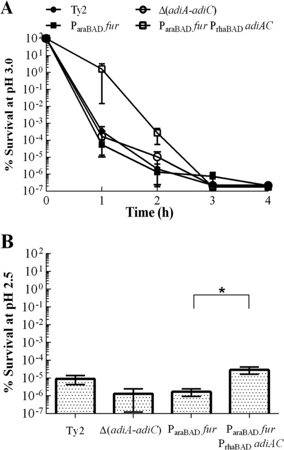
Acid resistance of a ΔPfur81::TT araC ParaBAD fur mutant containing rhamnose-regulated arginine decarboxylase. Arginine decarboxylase rescue was performed by growing strains in EGA medium to stationary phase in the absence of arabinose and presence of 0.1% rhamnose and challenging them with EG medium containing 1 mM arginine at pH 3.0 for 4 h (A) or pH 2.5 for 1 h (B). Survival in all assays was monitored by plating on LB agar containing all necessary supplements. The data shown are the means and SEM from three independent assays. Pairs of data marked with an asterisk are significantly different (P < 0.05). χ11500, Δ(adiA-adiC); χ11118, ParaBAD fur; χ11623, ParaBAD fur PrhaBAD adiAC.
There are several possible reasons for the difficulty in rescuing strain χ11623 (ParaBAD fur PrhaBAD adiAC). First, unlike phoPQ mutants, fur mutants are sensitive to organic acids. Inorganic acids such as HCl and organic acids behave quite differently inside the cell, due to differences in their dissociation constants. Our EGA challenge medium contained 10 mM citric acid (38). It is possible that the consumption of free protons by the arginine decarboxylase system is less effective at countering the effects of an organic acid, such as citric acid, than the strong inorganic acid HCl (8, 24, 38, 52). Second, because the ΔPfur81::TT araC ParaBAD fur mutation was introduced into Ty2, the strain also contains a mutation in rpoS. RpoS and Fur jointly regulate a number of key effectors responsible for protection against organic acid. Thus, the combination of fur and rpoS mutations may have rendered χ11623 more sensitive to acid than strains carrying the rpoS mutation alone or the combination of phoPQ and rpoS (4, 6). Finally, the Pfur81::TT araC ParaBAD fur mutation may have altered the ability of χ11623 to transport rhamnose, as it required four times the concentration of rhamnose to induce arginine decarboxylase activity as the ΔaroD and ΔphoPQ mutants (data not shown). Fur is known to regulate expression of a number of outer membrane proteins and other genes that may influence surface structure (53). Thus, it is possible that membrane perturbations due to the lack of Fur in the cell may have resulted in a reduction in rhamnose transport activity by RhaT.
Conclusion.
In this work, we constructed an acid resistance system whose expression and activity responded to the presence of a single sugar, either arabinose or rhamnose. Both adiA expression and adiC expression were required for acid resistance, and the rhamnose-regulated PrhaBAD promoter provided tighter control over adiA expression than the arabinose-regulated ParaBAD promoter. Rhamnose-dependent acid resistance in S. Typhi depended on three things: the presence of rhamnose in the culture medium, the presence of arginine in the challenge medium, and the fusion of adiA and adiC into an operon under the control of PrhaBAD. The absence of any of these components resulted in rapid cell death at low pH. The level of acid resistance provided by PrhaBAD adiAC grown with rhamnose under decarboxylase-inducing conditions was equivalent to the level of acid resistance observed with the native arginine decarboxylase system grown under the same conditions. However, the rhamnose-regulated adiAC system was inducible in cells otherwise unprepared for low-pH challenge, thus our rhamnose-regulated system significantly improved the survival of acid-unadapted aroD, ΔphoPQ, and fur mutants at pHs 3 and 2.5. This has far-reaching implications for vaccine development, as high levels of acid resistance can be attained without anaerobic, low-pH culture—a process which is neither efficient nor cost-effective. Instead, cells can be grown in the optimal medium for vaccine formulation as long as rhamnose is included.
The construction of the rhamnose-regulated arginine decarboxylase system allowed us to render S. Typhi acid resistant (to pH 2.5) on demand. Importantly, aerobically grown vaccine strains were protected from pHs 3 and 2.5. Since the low pH of the gastric environment poses a significant threat to the success of any live attenuated Salmonella vaccine, the rhamnose-regulated arginine decarboxylase system represents a novel means to augment survival in this in vivo compartment. Also, because low gastric pH is an important virulence signal, the ability to administer vaccines without stomach pH neutralization may also improve vaccine performance in the host. We plan to address these issues in future studies.
Supplementary Material
ACKNOWLEDGMENTS
This work was supported by R21 grants AI092307 and CA152456 and R01 grant AI093348 from the National Institutes of Health.
We are grateful to Justin Jensen for assistance with plasmid construction and AdiA purification and Jacquelyn Kilbourne for valuable advice and assistance with antibody production. We also thank Tina Hartig and Brandon Moore for technical support with the acid resistance assays and Caitlin McDonald for critical review of the manuscript.
Footnotes
Published ahead of print 3 May 2013
Supplemental material for this article may be found at http://dx.doi.org/10.1128/JB.00104-13.
REFERENCES
- 1. Verdu E, Viani F, Armstrong D, Fraser R, Siegrist HH, Pignatelli B, Idstrom JP, Cederberg C, Blum AL, Fried M. 1994. Effect of omeprazole on intragastric bacterial counts, nitrates, nitrites, and N-nitroso compounds. Gut 35:455–460 [DOI] [PMC free article] [PubMed] [Google Scholar]
- 2. Kieboom J, Abee T. 2006. Arginine-dependent acid resistance in Salmonella enterica serovar Typhimurium. J. Bacteriol. 188:5650–5653 [DOI] [PMC free article] [PubMed] [Google Scholar]
- 3. Lin J, Lee IS, Frey J, Slonczewski JL, Foster JW. 1995. Comparative analysis of extreme acid survival in Salmonella typhimurium, Shigella flexneri, and Escherichia coli. J. Bacteriol. 177:4097–4104 [DOI] [PMC free article] [PubMed] [Google Scholar]
- 4. Foster JW. 1993. The acid tolerance response of Salmonella typhimurium involves transient synthesis of key acid shock proteins. J. Bacteriol. 175:1981–1987 [DOI] [PMC free article] [PubMed] [Google Scholar]
- 5. Foster JW, Spector MP. 1995. How Salmonella survive against the odds. Annu. Rev. Microbiol. 49:145–174 [DOI] [PubMed] [Google Scholar]
- 6. Lee IS, Lin J, Hall HK, Bearson B, Foster JW. 1995. The stationary-phase sigma factor sigma S (RpoS) is required for a sustained acid tolerance response in virulent Salmonella typhimurium. Mol. Microbiol. 17:155–167 [DOI] [PubMed] [Google Scholar]
- 7. Hall HK, Foster JW. 1996. The role of fur in the acid tolerance response of Salmonella typhimurium is physiologically and genetically separable from its role in iron acquisition. J. Bacteriol. 178:5683–5691 [DOI] [PMC free article] [PubMed] [Google Scholar]
- 8. Bearson BL, Wilson L, Foster JW. 1998. A low pH-inducible, PhoPQ-dependent acid tolerance response protects Salmonella Typhimurium against inorganic acid stress. J. Bacteriol. 180:2409–2417 [DOI] [PMC free article] [PubMed] [Google Scholar]
- 9. Wilmes-Riesenberg MR, Bearson B, Foster JW, Curtiss R., III 1996. Role of the acid tolerance response in virulence of Salmonella typhimurium. Infect. Immun. 64:1085–1092 [DOI] [PMC free article] [PubMed] [Google Scholar]
- 10. Robbe-Saule V, Norel F. 1999. The rpoS mutant allele of Salmonella typhi Ty2 is identical to that of the live typhoid vaccine Ty21a. FEMS Microbiol. Lett. 170:141–143 [DOI] [PubMed] [Google Scholar]
- 11. Germanier R, Furer E. 1975. Isolation and characterization of Gal E mutant Ty 21a of Salmonella typhi: a candidate strain for a live, oral typhoid vaccine. J. Infect. Dis. 131:553–558 [DOI] [PubMed] [Google Scholar]
- 12. Germanier R, Furer E. 1983. Characteristics of the attenuated oral vaccine strain “S. typhi” Ty 21a. Dev. Biol. Stand. 53:3–7 [PubMed] [Google Scholar]
- 13. Hone D, Morona R, Attridge S, Hackett J. 1987. Construction of defined galE mutants of Salmonella for use as vaccines. J. Infect. Dis. 156:167–174 [DOI] [PubMed] [Google Scholar]
- 14. Hone DM, Harris AM, Levine MM. 1994. Adaptive acid tolerance response by Salmonella typhi and candidate live oral typhoid vaccine strains. Vaccine 12:895–898 [DOI] [PubMed] [Google Scholar]
- 15. Hohmann EL, Oletta CA, Killeen KP, Miller SI. 1996. phoP/phoQ-deleted Salmonella typhi (Ty800) is a safe and immunogenic single-dose typhoid fever vaccine in volunteers. J. Infect. Dis. 173:1408–1414 [DOI] [PubMed] [Google Scholar]
- 16. Shi H, Wang S, Roland KL, Gunn BM, Curtiss R., III 2010. Immunogenicity of a live recombinant Salmonella vaccine expressing pspA in neonates and infant mice born from naive and immunized mothers. Clin. Vaccine Immunol. 17:363–371 [DOI] [PMC free article] [PubMed] [Google Scholar]
- 17. Tacket CO, Sztein MB, Losonsky GA, Wasserman SS, Nataro JP, Edelman R, Pickard D, Dougan G, Chatfield SN, Levine MM. 1997. Safety of live oral Salmonella typhi vaccine strains with deletions in htrA and aroC aroD and immune response in humans. Infect. Immun. 65:452–456 [DOI] [PMC free article] [PubMed] [Google Scholar]
- 18. Tacket CO, Hone DM, Curtiss R, III, Kelly SM, Losonsky G, Guers L, Harris AM, Edelman R, Levine MM. 1992. Comparison of the safety and immunogenicity of ΔaroC ΔaroD and Δcya Δcrp Salmonella typhi strains in adult volunteers. Infect. Immun. 60:536–541 [DOI] [PMC free article] [PubMed] [Google Scholar]
- 19. Kirkpatrick BD, Tenney KM, Larsson CJ, O'Neill JP, Ventrone C, Bentley M, Upton A, Hindle Z, Fidler C, Kutzko D, Holdridge R, Lapointe C, Hamlet S, Chatfield SN. 2005. The novel oral typhoid vaccine M01ZH09 is well tolerated and highly immunogenic in 2 vaccine presentations. J. Infect. Dis. 192:360–366 [DOI] [PubMed] [Google Scholar]
- 20. DiPetrillo MD, Tibbetts T, Kleanthous H, Killeen KP, Hohmann EL. 1999. Safety and immunogenicity of phoP/phoQ-deleted Salmonella typhi expressing Helicobacter pylori urease in adult volunteers. Vaccine 18:449–459 [DOI] [PubMed] [Google Scholar]
- 21. Gilman RH, Hornick RB, Woodard WE, DuPont HL, Snyder MJ, Levine MM, Libonati JP. 1977. Evaluation of a UDP-glucose-4-epimeraseless mutant of Salmonella typhi as a live oral vaccine. J. Infect. Dis. 136:717–723 [DOI] [PubMed] [Google Scholar]
- 22. Black R, Levine MM, Young C, Rooney J, Levine S, Clements ML, O'Donnell S, Hugues T, Germanier R. 1983. Immunogenicity of Ty21a attenuated Salmonella typhi given with sodium bicarbonate or in enteric-coated capsules. Dev. Biol. Stand. 53:9–14 [PubMed] [Google Scholar]
- 23. Levine MM, Ferreccio C, Black RE, Germanier R. 1987. Large-scale field trial of Ty21a live oral typhoid vaccine in enteric-coated capsule formulation. Lancet i:1049–1052 [DOI] [PubMed] [Google Scholar]
- 24. Baik HS, Bearson S, Dunbar S, Foster JW. 1996. The acid tolerance response of Salmonella typhimurium provides protection against organic acids. Microbiology 142:3195–3200 [DOI] [PubMed] [Google Scholar]
- 25. Groisman EA, Kayser J, Soncini FC. 1997. Regulation of polymyxin resistance and adaptation to low-Mg2+ environments. J. Bacteriol. 179:7040–7045 [DOI] [PMC free article] [PubMed] [Google Scholar]
- 26. Rychlik I, Barrow PA. 2005. Salmonella stress management and its relevance to behaviour during intestinal colonisation and infection. FEMS Microbiol. Rev. 29:1021–1040 [DOI] [PubMed] [Google Scholar]
- 27. Durant JA, Corrier DE, Ricke SC. 2000. Short-chain volatile fatty acids modulate the expression of the hilA and invF genes of Salmonella typhimurium. J. Food Prot. 63:573–578 [DOI] [PubMed] [Google Scholar]
- 28. Lee AK, Detweiler CS, Falkow S. 2000. OmpR regulates the two-component system SsrA-SsrB in Salmonella pathogenicity island 2. J. Bacteriol. 182:771–781 [DOI] [PMC free article] [PubMed] [Google Scholar]
- 29. Richard H, Foster JW. 2004. Escherichia coli glutamate- and arginine-dependent acid resistance systems increase internal pH and reverse transmembrane potential. J. Bacteriol. 186:6032–6041 [DOI] [PMC free article] [PubMed] [Google Scholar]
- 30. Gong S, Richard H, Foster JW. 2003. YjdE (AdiC) is the arginine:agmatine antiporter essential for arginine-dependent acid resistance in Escherichia coli. J. Bacteriol. 185:4402–4409 [DOI] [PMC free article] [PubMed] [Google Scholar]
- 31. Andrell J, Hicks MG, Palmer T, Carpenter EP, Iwata S, Maher MJ. 2009. Crystal structure of the acid-induced arginine decarboxylase from Escherichia coli: reversible decamer assembly controls enzyme activity. Biochemistry 48:3915–3927 [DOI] [PubMed] [Google Scholar]
- 32. Blethen SL, Boeker EA, Snell EE. 1968. Arginine decarboxylase from Escherichia coli. I. Purification and specificity for substrates and coenzyme. J. Biol. Chem. 243:1671–1677 [PubMed] [Google Scholar]
- 33. Iyer R, Williams C, Miller C. 2003. Arginine-agmatine antiporter in extreme acid resistance in Escherichia coli. J. Bacteriol. 185:6556–6561 [DOI] [PMC free article] [PubMed] [Google Scholar]
- 34. Auger EA, Redding KE, Plumb T, Childs LC, Meng SY, Bennett GN. 1989. Construction of lac fusions to the inducible arginine- and lysine decarboxylase genes of Escherichia coli K12. Mol. Microbiol. 3:609–620 [DOI] [PubMed] [Google Scholar]
- 35. Curtiss R, III, Wanda SY, Gunn BM, Zhang X, Tinge SA, Ananthnarayan V, Mo H, Wang S, Kong W. 2009. Salmonella enterica serovar Typhimurium strains with regulated delayed attenuation in vivo. Infect. Immun. 77:1071–1082 [DOI] [PMC free article] [PubMed] [Google Scholar]
- 36. Kaniga K, Delor I, Cornelis GR. 1991. A wide-host-range suicide vector for improving reverse genetics in gram-negative bacteria: inactivation of the blaA gene of Yersinia enterocolitica. Gene 109:137–141 [DOI] [PubMed] [Google Scholar]
- 37. Kang HY, Dozois CM, Tinge SA, Lee TH, Curtiss R., III 2002. Transduction-mediated transfer of unmarked deletion and point mutations through use of counterselectable suicide vectors. J. Bacteriol. 184:307–312 [DOI] [PMC free article] [PubMed] [Google Scholar]
- 38. Vogel HJ, Bonner DM. 1956. Acetylornithinase of Escherichia coli: partial purification and some properties. J. Biol. Chem. 218:97–106 [PubMed] [Google Scholar]
- 39. De Biase D, Tramonti A, John RA, Bossa F. 1996. Isolation, overexpression, and biochemical characterization of the two isoforms of glutamic acid decarboxylase from Escherichia coli. Protein Expr. Purif. 8:430–438 [DOI] [PubMed] [Google Scholar]
- 40. Sambrook J, Russell DW. 2001. Molecular cloning: a laboratory manual, vol 3 Cold Spring Harbor Laboratory Press, Cold Spring Harbor, NY [Google Scholar]
- 41. Laemmli UK. 1970. Cleavage of structural proteins during the assembly of the head of bacteriophage T4. Nature 227:680–685 [DOI] [PubMed] [Google Scholar]
- 42. Towbin H, Staehelin T, Gordon J. 1989. Immunoblotting in the clinical laboratory. J. Clin. Chem. Clin. Biochem. 27:495–5012681521 [Google Scholar]
- 43. Rice EW, Johnson CH, Dunnigan ME, Reasoner DJ. 1993. Rapid glutamate decarboxylase assay for detection of Escherichia coli. Appl. Environ. Microbiol. 59:4347–4349 [DOI] [PMC free article] [PubMed] [Google Scholar]
- 44. Castanie-Cornet MP, Penfound TA, Smith D, Elliott JF, Foster JW. 1999. Control of acid resistance in Escherichia coli. J. Bacteriol. 181:3525–3535 [DOI] [PMC free article] [PubMed] [Google Scholar]
- 45. Berk PA, Jonge R, Zwietering MH, Abee T, Kieboom J. 2005. Acid resistance variability among isolates of Salmonella enterica serovar Typhimurium DT104. J. Appl. Microbiol. 99:859–866 [DOI] [PubMed] [Google Scholar]
- 46. Hoiseth SK, Stocker BA. 1981. Aromatic-dependent Salmonella typhimurium are non-virulent and effective as live vaccines. Nature 291:238–239 [DOI] [PubMed] [Google Scholar]
- 47. Haldimann A, Daniels LL, Wanner BL. 1998. Use of new methods for construction of tightly regulated arabinose and rhamnose promoter fusions in studies of the Escherichia coli phosphate regulon. J. Bacteriol. 180:1277–1286 [DOI] [PMC free article] [PubMed] [Google Scholar]
- 48. Galen JE, Wang JY, Chinchilla M, Vindurampulle C, Vogel JE, Levy H, Blackwelder WC, Pasetti MF, Levine MM. 2010. A new generation of stable, nonantibiotic, low-copy-number plasmids improves immune responses to foreign antigens in Salmonella enterica serovar Typhi live vectors. Infect. Immun. 78:337–347 [DOI] [PMC free article] [PubMed] [Google Scholar]
- 49. Wang S, Li Y, Shi H, Sun W, Roland KL, Curtiss R., III 2011. Comparison of a regulated delayed antigen synthesis system with in vivo-inducible promoters for antigen delivery by live attenuated Salmonella vaccines. Infect. Immun. 79:937–949 [DOI] [PMC free article] [PubMed] [Google Scholar]
- 50. Foster JW. 1999. When protons attack: microbial strategies of acid adaptation. Curr. Opin. Microbiol. 2:170–174 [DOI] [PubMed] [Google Scholar]
- 51. Foster JW, Hall HK. 1990. Adaptive acidification tolerance response of Salmonella typhimurium. J. Bacteriol. 172:771–778 [DOI] [PMC free article] [PubMed] [Google Scholar]
- 52. McHan F, Shotts EB. 1993. Effect of short-chain fatty acids on the growth of Salmonella typhimurium in an in vitro system. Avian Dis. 37:396–398 [PubMed] [Google Scholar]
- 53. Troxell B, Fink RC, Porwollik S, McClelland M, Hassan HM. 2011. The Fur regulon in anaerobically grown Salmonella enterica sv. Typhimurium: identification of new Fur targets. BMC Microbiol. 11:236. 10.1186/1471-2180-11-236 [DOI] [PMC free article] [PubMed] [Google Scholar]
- 54. Roland K, Curtiss R, III, Sizemore D. 1999. Construction and evaluation of a Δcya Δcrp Salmonella typhimurium strain expressing avian pathogenic Escherichia coli O78 LPS as a vaccine to prevent airsacculitis in chickens. Avian Dis. 43:429–441 [PubMed] [Google Scholar]
- 55. Felix A, Pitt RM. 1951. The pathogenic and immunogenic activities of Salmonella typhi in relation to its antigenic constituents. J. Hyg. (Lond.) 49:92–110 [DOI] [PMC free article] [PubMed] [Google Scholar]
- 56. Brenneman KE, McDonald C, Kelly-Aehle SM, Roland KL, Curtiss R., III 2012. Use of RapidChek SELECT Salmonella to detect shedding of live attenuated Salmonella enterica serovar Typhi vaccine strains. J. Microbiol. Methods 89:137–147 [DOI] [PubMed] [Google Scholar]
- 57. Formal SB, Dammin GJ, Labrec EH, Schneider H. 1958. Experimental Shigella infections: characteristics of a fatal infection produced in guinea pigs. J. Bacteriol. 75:604–610 [DOI] [PMC free article] [PubMed] [Google Scholar]
- 58. Wang S, Li Y, Shi H, Scarpellini G, Torres-Escobar A, Roland KL, Curtiss R., III 2010. Immune responses to recombinant pneumococcal PsaA antigen delivered by a live attenuated Salmonella vaccine. Infect. Immun. 78:3258–3271 [DOI] [PMC free article] [PubMed] [Google Scholar]
- 59. Santander J, Wanda SY, Nickerson CA, Curtiss R., III 2007. Role of RpoS in fine-tuning the synthesis of Vi capsular polysaccharide in Salmonella enterica serotype Typhi. Infect. Immun. 75:1382–1392 [DOI] [PMC free article] [PubMed] [Google Scholar]
- 60. Kong Q, Yang J, Liu Q, Alamuri P, Roland KL, Curtiss R., III 2011. Effect of deletion of genes involved in lipopolysaccharide core and O-antigen synthesis on virulence and immunogenicity of Salmonella enterica serovar Typhimurium. Infect. Immun. 79:4227–4239 [DOI] [PMC free article] [PubMed] [Google Scholar]
- 61. Kong W, Brovold M, Tully J, Benson L, Curtiss R., III 2012. Abstr. 112th Gen. Meet. Am. Soc. Microbiol American Society for Microbiology, Washington, DC: http://gm.asm.org [Google Scholar]
Associated Data
This section collects any data citations, data availability statements, or supplementary materials included in this article.


