

Abstract
Purpose
The purpose of the study was to propose a method of prone positioning for posterior cervico-dorsal spine surgeries that is easy to achieve without additional equipment and may reduce complications associated with prone positioning in patients.
Methods
41 patients underwent posterior spine surgeries using our method of prone positioning on a fluoroscopy compatible conventional operation table, and the technical difficulties and complications associated were noted. After induction under general anaesthesia in supine position, cervical tongs were applied. An assembly of two adequately padded cylindrical bolsters and two lateral brace attachments was set on a conventional operating table. The patient was then positioned prone so that the tongs as well as insertion pins of the tongs rest on the lateral brace attachments, with the face and head suspended freely in between. A neutralisation weight was then applied suspended from the tongs to stabilize the head.
Results
The time required for patient positioning was noted and was found to be nearly the same as that required for traditional prone positioning. No problems were noted during patient positioning and with anaesthesia tubing management. All surgeries went well without position related complications except for one patient who developed post-operative macroglossia. All cervical tong pin sites healed without any complications. The fluoroscope easily gained access to the operative areas.
Conclusions
Our modification appears simple, versatile and reproducible for posterior approach procedures of the cervical and upper dorsal spine in prone position. Also, the method can be easily implemented in most conventional operating room facilities with minimal surgeon effort and without the need for any additional inventory.
Keywords: Prone position, Lateral brace attachments, Facial and ocular pressure, Cervical tongs, Macroglossia
Introduction
Good patient positioning is essential for smooth surgery and should easily be achieved for most patients, allowing hassle-free access to the surgeons as well as anaesthetists. Complications resulting from patient positioning must be minimal. Prone position is universal for posterior cervical and dorsal spine surgeries. For cervical and upper dorsal spine surgeries, this requires stabilization of head and the cervical spine, usually achieved with Mayfield’s horseshoe headrest with or without skeletal traction with Gardner Well’s cervical tongs. This assembly, however, predisposes to complications resulting from localised pressure over the contact areas of the face, eyes and peri-ocular areas due to malpositioning, especially in prolonged surgeries [1–4]. The authors present a modification of the prone position using a Gardner Well’s tong for traction and two lateral brace attachments of an operating table which are readily available with most conventional operating table configurations. This modification using lateral brace attachments avoids localised pressure associated with horseshoe headrest use, and allows free access to anaesthetists for better endo-tracheal tube management.
Materials and methods
Using the modified prone position, 41 patients underwent posterior spine surgeries including cervico-dorsal kypho-scoliosis deformity correction (7 cases), post-tubercular deformity correction (17 cases), traumatic upper dorsal spine fixation (12 cases), cervical myelopathy decompression with or without instrumentation (3 cases) and reduction and stabilization with instrumentation for congenital atlanto-axial dislocation (2 cases). Age of the patients ranged from 3 to 64 years. The regions involved in these patients were cervical spine in 5, cervico-dorsal spine in 7 and dorsal spine in 29 cases.
Each patient was induced in supine position on an adjacent spine trolley and Gardner’s cervical tongs were applied. The assembly of two adequately padded cylindrical bolsters and two lateral brace attachments was set on a conventional operating table (here Mediland Amax 9000, Mediland Enterprises Co. Ltd., Taiwan) as shown in the Figs. 1 and 2. The cylindrical bolsters were adjusted so that the patient’s clavicles and the iliac crests rested upon them with adequate separation to allow free respiratory movements of thorax and abdomen. Using two radial setting clamps for side rail of the operating table, two lateral brace attachments were anchored to the table. The separation between the lateral brace attachments was so adjusted that it exceeded the maximum transverse dimension of the face by a couple of centimetres. The patient was then positioned prone so that the tongs as well as insertion pins of the tongs rested on the lateral brace attachments, with the face and head suspended freely in between (Figs. 3, 4). A neutralisation traction weight to stabilise the skull and cervical spine intra-operatively was applied. This was done through tongs using either a special pulley attachment or simply by suspending it over a wooden block fixed to the proximal end of the table (Figs. 5, 6). The weight for traction was individualised depending upon the age and weight of the patient. For adult patients weighing approximately 120 lbs, a weight of 10 lbs was used, whereas for younger patients, the weight was reduced accordingly, to approximate the weight of the skull. To flex or extend the cervical spine or to rotate the head slightly, the height of lateral brace attachments was varied. To flex the cervical spine, the height of the lateral braces was decreased equally on both sides; whereas to extend, the height was increased. Similarly to rotate the head and face to one side, for example, the left side, the lateral brace on the left was elevated and the one on the right was equally lowered down. These adjustments were done at the time of initial positioning, prior to surgery. Adjusting the position intra-operatively was avoided. The tendency of the head and tongs to translate on the lateral brace attachments is effectively neutralised by the suspended weight, at the same time maintaining in-line stabilization. This method also allowed the patient to be positioned in the reverse Trendelenburg’s position, which may be desirable in some cervical spine surgeries to reduce venous congestion and bleeding. The traction in these cases was suitably increased to neutralise the vertical vector of the body weight. Flexion or extension of the head was also adjusted pre-operatively to some extent by varying the position of insertion sites of the Gardner’s tongs (either anterior or posterior to the tragal line; thereby providing an extension or flexion moment respectively at the atlanto-occipital joint). At the end of the surgery, patient was turned supine and tongs were removed. The pin sites were dressed with tincture benzoin seals.
Figs. 1, 2.
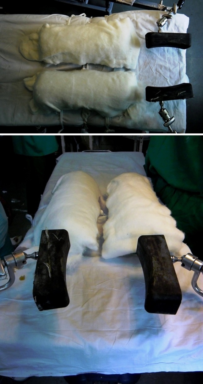
The assembly of two adequately padded cylindrical bolsters and two lateral brace attachments set on a conventional operating table as seen from different angles
Figs. 3, 4.

The patient positioned prone with cervical tongs resting on lateral brace attachments. Note that the face and head are suspended freely
Figs. 5, 6.
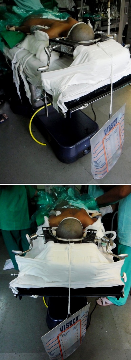
Neutralisation traction weight applied, suspended over a wooden block fixed at the head end of table
The positioning time, i.e. the time required after induction of anaesthesia from setting up the assembly, applying cervical tongs till positioning the patient prone on it was noted. The surgical time in terms of duration for which the patient was kept prone was recorded. Intra-operatively, difficulties due to patient positioning, anaesthesia tubing management and obtaining fluoroscopic images were noted. Any complications directly related to patient positioning were noted at the end of the surgery.
Results
The positioning time using this method ranged from 11 to 19 min (average 14.54 min, standard deviation 2.04). The surgical time ranged from 3 to 13 h (average 6.8 h, standard deviation 2.57). No problems were noted during patient positioning. Intra-operatively, the patient remained stable for the duration of the procedure and no re-adjustment of position was required. The method safely allowed tilting of the table to either side up to approximately 30° without slippage of the tongs. All surgeries were performed without any position related complications except for one patient who developed post-operative macroglossia and oropharyngeal oedema. All cervical tong pin sites healed without any complications.
This position facilitated easy access to anaesthesia tubings which could be taken out from either sides below the lateral brace attachments (Figs. 7, 8). Also, the Boyles apparatus was positioned towards the foot end of the patient, thereby allowing circumferential access to operating surgeons and assistants. Good unhindered fluoroscopic images could also be achieved in orthogonal planes.
Figs. 7, 8.

Note the easy access to the anaesthesia tubings facilitated by the position
Discussion
Prone position is commonly used worldwide for spine surgeries. However, numerous well-known complications have been reported from prone patient positioning, such as pressure induced skin necrosis [4–12], and ocular complications like conjunctival [13] and corneal abrasions [14, 15], post-operative visual loss (POVL) due to central retinal artery occlusion [1, 2, 5, 16–19], ophthalmoplegia [2, 20–23] and acute angle closure glaucoma [24]. Many of these complications are related to prolonged direct pressure over the contact areas.
Post-operative visual loss following non-ocular surgery is an infrequent but disastrous complication with an estimated incidence ranging from 0.001 to 1 % depending on the type of surgery [20, 25, 26]. Direct pressure on the eye, especially as a result of patient malpositioning, has been cited as a factor contributing to visual loss [2, 3, 19]. The incidence of significant visual complications after spine surgery, according to a recent review, could be of the order of one case per 100 spine surgeries per year [27].
Bekar et al. [1] reported a case of unilateral blindness due to patient positioning during cervical syringomyelia surgery in prone position. They further stated that, in spinal surgeries using horseshoe headrest with the patient in the prone position, the possibility of central retinal artery occlusion (CRAO) increases and its cause can be attributed primarily to excessive extra-ocular pressure.
Kasodekar and Chen [28] reported a case of monocular blindness as a complication of intraoperative positioning in posterior cervical spine surgery. They concluded that inappropriate pressure from padding while lying on the horseshoe headrest led to extrinsic pressure over the eyeball, causing raised intraocular pressure and ischaemic optic neuropathy. Wolfe et al. [21] reported a case of unilateral blindness as a complication of prone patient positioning for spinal surgery. They suggested that a padded Mayfield headrest may not be appropriate for all patients undergoing spine surgery, as exophthalmos or a flattened nasal bridge may allow transmission of pressure to the globe.
Direct pressure is a common cause of position related injury. Affected skin areas include the malar regions, iliac crests, chin, eyelids, nose, and tongue [4–12]. Jain et al. reported a case of pressure sores occurring in prone position under general anaesthesia on malar prominences of both cheeks. The authors speculated that pressure from the horseshoe on bony structures surrounding the eyeballs had resulted in pressure sores [4]. Members of the American Society of Anaesthesiologists (ASA) task force on peri-operative blindness agree that the use of horseshoe headrest increases the risk of ocular compression [29]. Sporadic cases of occlusion of vertebral arteries have been reported in the literature. As most of these cases involved positioning the patient prone with head rotated, it would seem prudent to maintain neutral neck alignment to minimise the risk of occluding the carotid or vertebral arteries [6].
Positioning the head of patients undergoing procedures in prone position remains a difficult task for the anaesthesiologist. Often, it is a compromise between a normal position of the head without derogating facial and neck tissues on one hand and sufficient control over airway devices on the other hand. Anaesthesia tubing management is greatly simplified using our modification. By placing the anaesthesia trolley and Boyles apparatus at the foot end of the patient, circumferential access to the operative area is facilitated. Fluoroscopic access to operative area is also easily achieved.
Prone positioning using the three-pin head holder has also been described [30]. It provides secure head positioning whilst allowing some intra-operative manoeuvrability. However, the use of three-pin head holder requires special table attachments. Application of cervical traction is not possible with it since the holder is anchored to the table. Also, the method is not without complications [31].
The positioning time in our study averaged 14.54 min, which in the authors’ experience is nearly the same as that required for conventional positioning. Although cervical tong application may appear to require additional time, it is a relatively quick procedure and the time consumed is well compensated by the reduced time required to ensure pressure-free placement of face and eyes and anaesthesia tubing re-adjustment in conventional positioning. As this was an observational study, no formal comparison with the conventional prone positioning technique was done. Also, no studies, to the best of authors’ knowledge, at present, mention or compare the positioning times for different methods of prone positioning.
As the head is freely suspended, it essentially does away with all pressure related ocular and facial complications of prone position. The similar was reflected in our study which had an average surgical time of 6.8 h. We encountered only one complication in the form of macroglossia after a prolonged surgery (13 h). Extubation had to be delayed for 24 h, but there were no long-term sequelae. There have been reports of macroglossia occurring after surgery in prone position [32, 33]. However, this complication is a dependency related phenomenon due to prolonged prone positioning, not specifically related to our method.
All techniques have downsides. The drawbacks of our technique include inability to re-adjust the position intra-operatively and inability to tilt the table beyond 30° to either side. Also at this stage, the technique described, seems to be best suited for simple posterior cervical and upper dorsal spine surgeries, which do not require significant intra-operative position changes; and as a means to stabilize the head and neck during posterior thoraco-lumbar spine surgeries. Also, complications directly related to prone position per se like dependent edema, macroglossia etc. could not be addressed by our technique.
Conclusion
Thus, our modification appears simple, versatile and reproducible for different surgical procedures of the cervical and upper dorsal spine in prone position. Also, the method can be easily implemented in most conventional operating room facilities with minimal surgeon effort and without the need for any additional inventory. However, further studies are required to conclusively establish the efficacy and safety of this method.
Conflict of interest
None.
Contributor Information
Abhijeet B. Kadam, Email: abhijeetsr71@gmail.com
Abhishek S. Jaipuria, Email: abhijai08@gmail.com
Ashok K. Rathod, Phone: +91-9820858169, Email: drakrkem@gmail.com
References
- 1.Bekar A, Türeyen K, Aksoy K. Unilateral blindness due to patient positioning during cervical syringomyelia surgery: unilateral blindness after prone position. J Neurosurg Anesthesiol. 1996;8(3):227–229. doi: 10.1097/00008506-199607000-00007. [DOI] [PubMed] [Google Scholar]
- 2.Hollenhorst RW, Svien HJ, Benoit CF. Unilateral blindness occurring during anesthesia for neurosurgical operations. Arch Ophthalmol. 1954;52:819–830. doi: 10.1001/archopht.1954.00920050825002. [DOI] [PubMed] [Google Scholar]
- 3.Claudio S, Antonio B, Giuseppe B, Vincenzo A, Aldo M. Positioning on surgical table. Eur Spine J. 2004;13:S50–S55. doi: 10.1007/s00586-004-0728-y. [DOI] [PMC free article] [PubMed] [Google Scholar]
- 4.Jain V, Bithal PK, Rath GP. Pressure sore on malar prominences by horseshoe headrest in prone position. Anaesth Intensive Care. 2007;35:304–305. [PubMed] [Google Scholar]
- 5.Roth S, Tung A, Ksiazek S. Visual loss in a prone-positioned spine surgery patient with the head on a foam headrest and goggles covering the eyes: an old complication with a new mechanism. Anesth Analg. 2007;104(5):1185–1187. doi: 10.1213/01.ane.0000264319.57758.55. [DOI] [PubMed] [Google Scholar]
- 6.Edgcombe H, Carter K, Yarrow S. Anaesthesia in the prone position. Br J Anaesth. 2008;100:165–183. doi: 10.1093/bja/aem380. [DOI] [PubMed] [Google Scholar]
- 7.Anderton JM. The prone position for the surgical patient: a historical review of the principles and hazards. Br J Anaesth. 1991;67:452–463. doi: 10.1093/bja/67.4.452. [DOI] [PubMed] [Google Scholar]
- 8.Drummond JC. Macroglossia [comment] Anesth Analg. 1999;89:534–535. doi: 10.1097/00000539-199908000-00064. [DOI] [PubMed] [Google Scholar]
- 9.Moore DC, Edmunds LH. Prone position frame. Surgery. 1950;27:276–279. [PubMed] [Google Scholar]
- 10.Smith RH. One solution to the problem of the prone position for surgical procedures. Anesth Analg. 1974;53:221–224. [PubMed] [Google Scholar]
- 11.Weis K. Threatening necrosis of the tip of the tongue during long-term anaesthesia in the prone position. Der Anaesthetist. 1964;13:241. [PubMed] [Google Scholar]
- 12.Grisell Margaret, Place H. Face tissue pressure in prone positioning: a comparison of three face pillows while in the prone position for spinal surgery. Spine. 2007;7(5):84S–85S. doi: 10.1097/BRS.0b013e31818b9029. [DOI] [PubMed] [Google Scholar]
- 13.Biswas BK, Bithal PK, Dash M, et al. Keratoconjunctival injury in the prone position: a prospective study in neurosurgical patients. Eur J Anaesthesiol. 2004;21:663–665. doi: 10.1017/s0265021504218130. [DOI] [PubMed] [Google Scholar]
- 14.Stambough JL, Dolan D, Werner R, Godfrey E. Ophthalmologic complications associated with prone positioning in spine surgery. J Am Acad Orthop Surg. 2007;15:156–165. doi: 10.5435/00124635-200703000-00005. [DOI] [PubMed] [Google Scholar]
- 15.Cucchiara R, Black S. Corneal abrasion during anesthesia and surgery. Anesthesiology. 1988;69:978–979. doi: 10.1097/00000542-198812000-00034. [DOI] [PubMed] [Google Scholar]
- 16.Kamming D, Clarke S. Postoperative visual loss following prone spinal surgery. Br J Anaesth. 2005;95(2):257–260. doi: 10.1093/bja/aei173. [DOI] [PubMed] [Google Scholar]
- 17.Nakra D, Bala I, Pratap M. Unilateral postoperative visual loss due to central retinal artery occlusion following cervical spine surgery in prone position. Br J Anaesth. 2005;95(5):719–720. doi: 10.1111/j.1460-9592.2007.02222.x. [DOI] [PubMed] [Google Scholar]
- 18.Hélaine L, Cadic A, Magro E, et al. Vision loss after spine surgery: a case report. Ann Fr Anesth Reanim. 2009;28(2):165–167. doi: 10.1016/j.annfar.2008.12.003. [DOI] [PubMed] [Google Scholar]
- 19.Slocum HC, O’Neal KC, Allen CR. Neurovascular complications from malposition on the operating table. Surg Gynecol Obstet. 1948;86:729–734. [PubMed] [Google Scholar]
- 20.Chung M-S, Son J-H. Visual loss in one eye after spinal surgery. Korean J Ophthalmol. 2006;20(2):139–142. doi: 10.3341/kjo.2006.20.2.139. [DOI] [PMC free article] [PubMed] [Google Scholar]
- 21.Wolfe SW, Lospinuso MF, Burke SW. Unilateral blindness as a complication of patient positioning for spinal surgery. A case report. Spine. 1992;17:600–605. doi: 10.1097/00007632-199205000-00023. [DOI] [PubMed] [Google Scholar]
- 22.West J, Askin G, Clarke M, Vernon SA. Loss of vision in one eye following scoliosis surgery. Br J Ophthalmol. 1990;74:243–244. doi: 10.1136/bjo.74.4.243. [DOI] [PMC free article] [PubMed] [Google Scholar]
- 23.Halfon MJ, Bonardo P, Valiensi S, et al. Central retinal artery occlusion and ophthalmoplegia following spinal surgery. Br J Ophthalmol. 2004;88:1350–1352. doi: 10.1136/bjo.2003.039651. [DOI] [PMC free article] [PubMed] [Google Scholar]
- 24.Michael MS, Salim S. Bilateral acute angle-closure glaucoma as a complication of facedown spine surgery. Spine. 2010;10(9):e7–e9. doi: 10.1016/j.spinee.2010.07.006. [DOI] [PubMed] [Google Scholar]
- 25.Warner ME, Warner MA, Garrity JA, et al. The frequency of perioperative vision loss. Anesth Analg. 2001;93:1417–1421. doi: 10.1097/00000539-200112000-00013. [DOI] [PubMed] [Google Scholar]
- 26.Williams EL, Hart WM, Jr, Tempelhoff R. Postoperative ischemic optic neuropathy. Anesth Analg. 1995;80:1018–1029. doi: 10.1097/00000539-199505000-00029. [DOI] [PubMed] [Google Scholar]
- 27.Myers MA, Hamilton SR, Bogosian AJ, et al. Visual loss as a complication of spine surgery. Spine. 1997;22:1325–1329. doi: 10.1097/00007632-199706150-00009. [DOI] [PubMed] [Google Scholar]
- 28.Kasodekar VB, Chen JLT. Monocular blindness: a complication of intraoperative positioning in posterior cervical spine surgery. Singapore Med J. 2006;47(7):631. [PubMed] [Google Scholar]
- 29.American Society of Anesthesiologists Task Force on Perioperative Blindness Practice Advisory for perioperative visual loss associated with spine surgery: a report by the American Society of Anesthesiologists Task Force on Perioperative Blindness. Anesthesiology. 2006;104(6):1319–1328. doi: 10.1097/00000542-200606000-00027. [DOI] [PubMed] [Google Scholar]
- 30.Konkorde P, Basligin Y. Placement of three-pin head holders in the concorde position. Turk Neurosurg. 2010;20(2):136–141. doi: 10.5137/1019-5149.JTN.2982-10. [DOI] [PubMed] [Google Scholar]
- 31.Martínez-Lage JF, Almagro MJ, Serrano C, Mena L. Depressed skull fracture by a three-pin head holder: a case illustration. Childs Nerv Syst. 2011;27(1):163–165. doi: 10.1007/s00381-010-1213-z. [DOI] [PubMed] [Google Scholar]
- 32.Pivalizza EG, Katz J, Singh S, et al. Massive macroglossia after posterior fossa surgery in the prone position. J Neurosurg Anesthesiol. 1998;10:34–36. doi: 10.1097/00008506-199801000-00008. [DOI] [PubMed] [Google Scholar]
- 33.Szabo M, Denman W, Marota J, Roberts J. Evaluation of airway edema in patients operated on in the prone position. J Neurosurg Anaesthesiol. 1997;9:380. doi: 10.1097/00008506-199710000-00021. [DOI] [Google Scholar]