Abstract
MicroRNAs (miRNAs) are a type of endogenous noncoding small RNAs involved in the regulation of multiple biological processes. Recently, miR-29 was found to participate in myogenesis. However, the underlying mechanisms by which miR-29 promotes myogenesis have not been identified. We found here that miR-29 was significantly upregulated with age in postnatal mouse skeletal muscle and during muscle differentiation. Overexpression of miR-29 inhibited mouse C2C12 myoblast proliferation and promoted myotube formation. miR-29 specifically targeted Akt3, a member of the serine/threonine protein kinase family responsive to growth factor cell signaling, to result in its post-transcriptional downregulation. Furthermore, knockdown of Akt3 by siRNA significantly inhibited the proliferation of C2C12 cells, and conversely, overexpression of Akt3 suppressed their differentiation. Collectively and given the inverse endogenous expression pattern of rising miR-29 levels and decreasing Akt3 protein levels with age in mouse skeletal muscle, we propose a novel mechanism in which miR-29 modulates growth and promotes differentiation of skeletal muscle through the post-transcriptional downregulation of Akt3.
Keywords: miR-29, myoblast, proliferation, differentiation, Akt3
MicroRNAs (miRNAs) are endogenous noncoding small RNAs comprising 21–23 base pairs. The miRNA sequence is complementary to the 3′ untranslated region (UTR) of its target mRNA, and the resulting complementary binding inhibits gene expression at the post-transcriptional level by mediating mRNA breakdown or translation inhibition.1, 2 miRNA has been confirmed to be involved in multiple physiological and biochemical processes, including tumorigenesis, hematopoiesis, neurogenesis, immunity, cell proliferation, and apoptosis.3, 4, 5, 6 miRNA has also been shown to participate in muscle development or repair in mice, rats, pigs, and humans.7, 8, 9, 10, 11 Previous studies have identified numerous muscle-associated miRNAs, including miR-1, miR-29, miR-133, miR-206, and miR-214, which have important roles in regulating myogenesis and differentiation of skeletal or cardiac muscle.12, 13, 14, 15
The miR-29 family comprises three members, miR-29a, mir-29b, and miR-29c. Their seed sequences are identical, and they have similar expression patterns and functions. Functional studies have shown that miR-29 participates in different physiological processes. It represses Tcl1 expression, thereby inhibiting B cell chronic lymphocytic leukemia in mice.16, 17 Overexpression of miR-29a/b/c causes insulin resistance, indicating its possible role in type 2 diabetes mellitus.18 miR-29a/b has also been shown to promote osteogenesis by participating in the Wnt signaling circuit or targeting inhibitors of osteoblast differentiation.19, 20 However, most studies have focused on the role of miR-29 as a tumorigenic inhibitor. For example, miR-29 restores normal patterns of DNA methylation by targeting DNA methyltransferase 3A and 3B, thus inhibiting tumorigenesis in lung cancer.21 miR-29 directly targets p85 alpha and CDC42 in HeLa cells, suppresses Mcl-1 and Bcl-2 in hepatocellular carcinoma, and induces apoptosis in a p53-dependent manner or through a mitochondrial pathway.22, 23 A recent study shows that miR-29 acts as a tumor suppressor in rhabdomyosarcoma by regulating the expression of CCND2 and E2F7.24
Importantly, the miR-29 family has been found to be involved in muscle development. Knockdown of miR-29b in vivo induced cardiac fibrosis in mice.15 Repression of miR-29a/b upregulated the expression of ADAMTS-7, and subsequently induced vascular smooth muscle cell (VSMC) calcification.25 These results indicate that miR-29 is important in the function of cardiac muscle and VSMCs. We previously found that miR-29a, miR-29b, and miR-29c were significantly upregulated in longissimus muscle of adult pigs, compared with 33- and 65-day old fetuses.26 In myotonic dystrophy type 1, miR-29b and miR-29c were found to be downregulated.27 Furthermore, miR-29 is downregulated in Duchenne muscular dystrophy, and restoration of its expression could improve dystrophy by promoting myogenic differentiation and inhibiting fibrogenic differentiation.28, 29 These results provide evidence for the role of miR-29 as a critical regulator in skeletal muscle, but its effector mechanism is not clear. Current evidence indicates that in undifferentiated myoblasts, TGF-beta-Smad3 signaling enhances the recruitment of a complex, which contains Yin Yang1 (YY1), Rybp (Ring1 and YY1-binding protein), and Polycomb protein (recruited by YY1), thereby negatively regulating miR-29 expression.30, 31 On the other hand, under normal myogenic differentiation conditions, miRNA-29 promotes myogenic differentiation of C2C12 cells by inhibiting Smad3, which in turn inhibits TGF-beta-mediated regulation of HDAC4, or YY1, Rybp, Polycomb-repressive complex, Ezh2, collagens, Lims1, and Mfap5 (microfibrillar-associated protein 5), thereby preventing the transdifferentiation of C2C12 cells into myofibroblasts.28, 30, 32, 33 These studies show that miR-29 indirectly promotes myogenesis through inhibiting the conversion of myoblasts to myofibroblasts; however, it is unclear if miR-29 could perform a more direct myogenic role by targeting effector genes that mediate skeletal muscle cell proliferation or differentiation.
Akt kinase family comprises three members: Akt1, Akt2, and Akt3. Akt3 is mainly known for its role in tumorigenesis. Raised Akt3 is found to associate with breast and prostate cancers,34 and ovarian cancer cells.35 The probable underlying mechanism is that AKT3 promotes cell proliferation by accelerating G2–M phase transition.36 High expression of Akt3 is also associated with malignant melanomas. Knockdown or inhibition of Akt3, but not Akt1 or Akt2, induced apoptosis in melanoma cells; therefore, the Akt3 signaling cascade is considered a therapeutic target in melanoma treatment.37, 38, 39 Downregulation of AKT3 by miRNAs, such as miR-15a and miR-16-1 or miR-93, resulted in the inhibition of proliferation in multiple myeloma and breast cancer stem cells.40, 41 Recent studies found that Akt3 has a potential role in promoting the proliferation of human and rat aortic VSMCs.42, 43 Moreover, marked cardiac hypertrophy was found in cardiac-specific Akt3 transgenic mice, suggesting a role for Akt3 in cardiac muscle development.44 However, there is no clear evidence of a functional link between the Akt3 isoform and skeletal muscle or myoblasts.
The aim of this study is to elucidate the underlying mechanism by which miR-29 modulates the proliferation and differentiation of myoblasts, and its relationship with Akt3. We demonstrated that in aging mouse skeletal muscles, miR-29 was upregulated, but Akt3 expression was downregulated. Akt3 promoted myoblast proliferation and delayed muscle differentiation, and was downregulated by miR-29 at the post-transcriptional level.
Results
miR-29 was significantly upregulated during postnatal skeletal muscle growth in mice
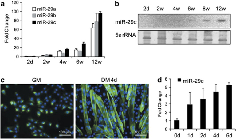
Our previous study showed that expression of miR-29 family members was significantly higher in the longissimus muscle of adult pigs than those from 33- and 65-day fetuses,26 indicating its potential role in skeletal muscle development. In order to confirm the expression profile of miR-29, we collected hindleg muscles from postnatal Balb/c mice at 2 days, 2 weeks, 4 weeks, 6 weeks, and 12 weeks, and analyzed the expression of miR-29 in these samples by Q-PCR. As anticipated, we found that the expression of miR-29a, miR-29b, and miR-29c was upregulated in an age-dependent manner. Expression of miR-29 in the skeletal muscle of 12-week-old mice was over 60-fold higher than in 2-day-old mice (Figure 1a). The three miR-29 members showed similar rising expression patterns with age. Furthermore, northern blotting confirmed the upregulation of miR-29c during postnatal muscle growth (Figure 1b). Next, we investigated the expression profile of miR-29c in vitro in murine C2C12 myoblasts and myotubes (Figure 1c). miR-29c was upregulated during C2C12 differentiation (Figure 1d). These results indicated that miR-29 expression was upregulated during postnatal skeletal muscle growth and muscle differentiation.
Figure 1.
Upregulation of miR-29 in mouse skeletal muscle with age. (a) Expression of miR-29 in the hindleg muscles of postnatal Balb/c mice was determined by Q-PCR. The ages of the mice were 2 days, 2 weeks, 4 weeks, 6 weeks, and 12 weeks. Fold change was in relation to expression in 2-day-old mice. (b) Northern blotting analysis of miR-29 in thigh muscles from postnatal Balb/c mice. 5s rRNA was used as the reference gene. (c) Immunofluorescence detection of fast skeletal myosin (Alexa Fluor 488 dye, green) in C2C12 myoblasts (GM) and myotubes at 4 days of differentiation (DM 4d). Cell nuclei were positive for DAPI (blue). Scale bar=100 μm. (d) miR-29c expression in C2C12 cells differentiated for 0, 1, 2, 4, and 6 days. Fold change in relation to expression on 0 day. Expression of miR-29 was normalized to U6 and is presented as mean±S.E.M. (from three independent individuals or replicates per time point)
miR-29 repressed proliferation of C2C12 cells
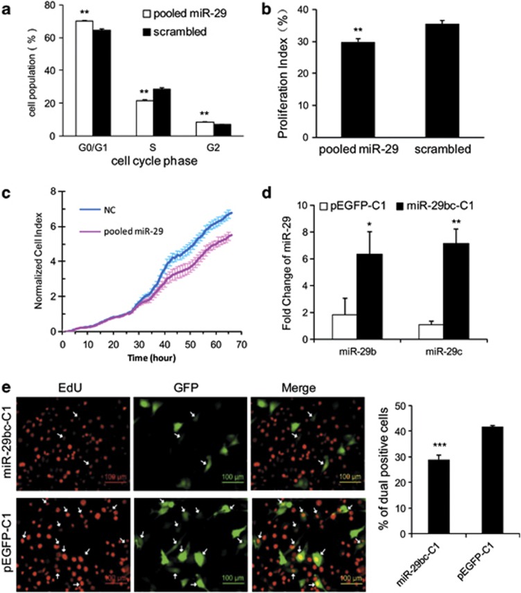
In order to detect the effects of miR-29 on muscle proliferation, C2C12 myoblasts transfected with synthetic pooled miR-29 mimics (miR-29a/b/c mimics mixed equally) or scrambled negative control (NC) miRNA were grown to near confluence and subjected to propidium iodide-cell cycle flow cytometry (Supplementary Figure 1). There was a significantly higher number of cells in the G0/G1 and G2 stages in the miR-29-transfected group than in the NC group (Figure 2a). This suggests that miR-29 could arrest C2C12 cells in the G0/G1 stage. The proliferation index was lower in miR-29-transfected cells than in NC-transfected cells (Figure 2b). xCELLigence system-based real-time monitoring of C2C12 cell proliferation found that miR-29-transfected cells displayed a reduced growth rate compared with corresponding NC miRNA-transfected cells from 24 h post transfection (Figure 2c). To perform overexpression of miR-29b and miR-29c, a 1007-bp DNA fragment, housing the primary transcript of mmu-miR-29b-2 and the mmu-miR-29c cluster, was cloned into the pEGFP-C1 vector (miR-29bc-C1). Twenty-four hours post transfection with the recombinant GFP-miR vector, Q-PCR showed that miR-29b and miR-29c were successfully overexpressed in C2C12 cells (Figure 2d). miR-29bc-C1 and pEGFP-C1 (control) transfected cells were nuclear stained with 5-Ethynyl-2′-deoxyuridine (EdU) (a dye only inserts in the replicating DNA molecules) and subjected to flow cytometry to identify cell populations that were dual positive for EdU and GFP. miR-29bc-C1-transfected cells showed significantly less mitotic activity, as evident by a significant reduction in dual EdU- and GFP-labeled cells (Figure 2e). Collectively, these results indicated that miR-29 overexpression inhibited C2C12 cell proliferation.
Figure 2.
miR-29 repressed proliferation of C2C12 cells. C2C12 cells were transiently transfected with pooled miR-29a/b/c mimics or scrambled NC, and the cell cycle phase (a) and proliferation index (b) were analyzed by propidium iodine flow cytometry. (c) C2C12 cells were transfected with pooled miR-29 mimics or NC upon the cell index reaching 1.0, and cell growth dynamics were continuously monitored using the xCELLigence system. (d) miR-29bc-C1 or pEGFP-C1 plasmids were transfected into C2C12 myoblasts and 24 h later, the cells were processed for Q-PCR detection of miR-29 b/c. Expression of miR-29c was normalized by U6. (e) After transfection with miR-29bc-C1 or pEGFP-C1, C2C12 cells were stained with EdU. Cells dual positive for EdU and GFP are indicated by white arrows (left graph), and the percentage of dual positive cells was determined by flow cytometry (right graph). The scale bar stands for 100 μm. The results are presented as mean±S.E.M. (three independent replicates per group). *P<0.05; **P<0.01; ***P<0.001
miR-29 promoted C2C12 myoblast differentiation
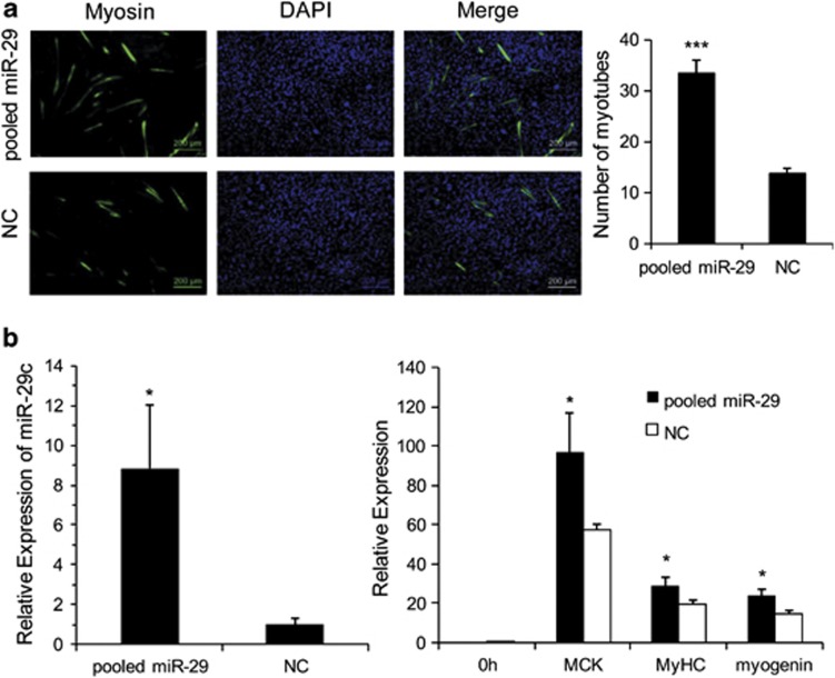
To establish the involvement of miR-29 in muscle cell differentiation, C2C12 myoblasts were transfected with synthetic pooled miR-29 mimics or NC miRNA. Twenty-four hours later, growth medium was replaced with differentiation medium to induce cell differentiation. Immunofluorescence detection of fast skeletal myosin showed that more cells underwent differentiation and myotube formation in the pooled miR-29 group than in the NC group at 72 h (Figure 3a). Several myoblast differentiation marker genes were also detected at 48 h of differentiation by Q-PCR. Along with the successful overexpression of miR-29, expression of the myogenin, MyHC IId, and MCK genes was significantly upregulated in cells transfected with the pooled miR-29 mimics than in the cells transfected with NC miRNA (Figure 3b). Together, these results showed that miR-29 promoted myoblast differentiation.
Figure 3.
Promotion of C2C12 cell differentiation by miR-29. (a) C2C12 myoblasts transfected with pooled miR-29 mimics or NC were differentiated for 3 days. Immunofluorescence detection with fast myosin antibody was used to identify myotubes (green). The nuclei were stained blue with DAPI. The scale bar stands for 200 μm. The number of myotubes is presented as mean±S.E.M. (14 random fields were captured for each treatment group). (b) C2C12 myoblasts transfected with pooled miR-29 mimics or NC were induced to undergo differentiation for 48 h. Subsequent detection of miR-29c, myogenin, MyHC IId, and MCK was determined by Q-PCR. The results are presented as mean±S.E.M. (three independent replicates per group). *P<0.05; ***P<0.001
miR-29 directly targeted 3′ UTR of Akt3
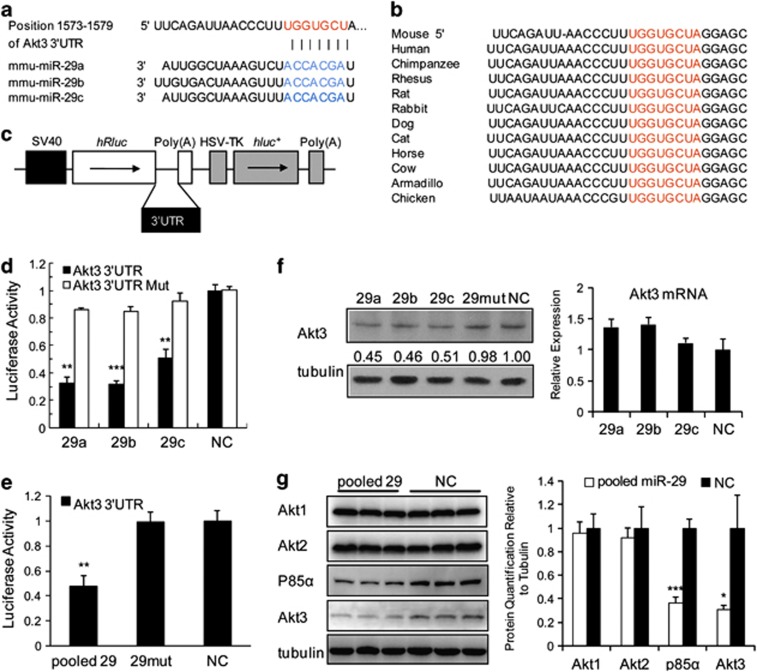
In order to uncover the mechanisms by which miR-29 suppresses proliferation in myoblasts, we examined potential targets of miR-29 involved in the cell cycle. Akt3 was identified by TargetScan analysis as a prime target, with a highly conserved complementary miR-29-binding site in its 3′ UTR across vertebrates from chickens to humans (Figures 4a and b).
Figure 4.
Akt3 3′ UTR as a target of miR-29. The predicted binding site of miR-29 in the 3′ UTR of Akt3 (a) is highly conserved among vertebrates (b). The Akt3 3′ UTR was inserted into the dual-luciferase reporter vector psi-CHECK2 at the 3′ end of the Renilla luciferase gene (hRluc). The constitutive firefly luciferase (hluc+) expression was used as an internal normalization control (c). The Akt3 3′ UTR or Akt3 3′ UTR Mut (containing a 2-base substitution in the binding site for miR-29) construct was co-transfected with miR-29a, miR-29b, miR-29c, or NC, as indicated, into BHK-21 cells, and normalized Renilla luciferase activity was determined (d). Pooled miR-29 mimics, miR-29 mut (housing a 2-base mutation), or NC was co-transfected with the Akt3 3′ UTR dual-luciferase vector, as indicated, into BHK-21 cells, and normalized Renilla luciferase activity was assayed (e). Akt3 protein expression in C2C12 myoblasts was detected at 48 h post transfection with miR-29a/b/c, miR-29 mut, or NC. Comparable tubulin levels served as loading controls in all western blotting assays (f). (g) Protein detection of members of the Akt3 family and p85a in C2C12 myoblasts transfected with pooled miR-29a/b/c and NC. Results are presented as mean±S.E.M. (three independent replicates per group). *P<0.05; **P<0.01; ***P<0.001
To establish miR-29 function at the Akt3 3′ UTR, a fragment of Akt3 3′ UTR containing the binding site of miR-29 was spliced to the 3′-end of the Renilla luciferase reporter gene in the dual-luciferase assay cassette (Figure 4c). Renilla luciferase expression was significantly reduced in BHK-21 cells when the construct was co-transfected with miR-29a, miR-29b, or miR-29c (Figure 4d). However, miR-29 had no appreciable inhibitory effect on a mutated Akt3 3′ UTR dual-luciferase construct (containing 2-base substitutions in the binding site of miR-29) (Figure 4d). Likewise, a 2-base mutation introduced into the seed sequence of miR-29 (29mut) abrogated any inhibition of the Akt3 3′ UTR construct, although the significant suppression effect was still observed in the cells transfected with the pooled miR-29 mimics (Figure 4e). These results demonstrated the specific inhibition of Akt3 expression by miR-29.
The inhibitory effect of miR-29a/b/c on endogenous Akt3 protein expression was evident in C2C12 myoblasts, even though no significant inhibition was detected at the mRNA level (Figure 4f). The impact of miR-29 on the protein expression of other members of the Akt family, and p85a, a regulatory subunit of phosphatidylinositol 3-kinase (PI3K), was evaluated by western blotting. Akt1 and Akt2 were not inhibited in C2C12 myoblasts transfected with pooled miR-29 mimics (Figure 4h). Protein p85α, previously shown to be a target of miR-29 in HeLa cells,22 like Akt3 protein, was downregulated by miR-29 (Figure 4h). Therefore Akt3 protein expression was directly inhibited by miR-29.
Inverse expression relationship between miR-29 and Akt3 protein in vivo
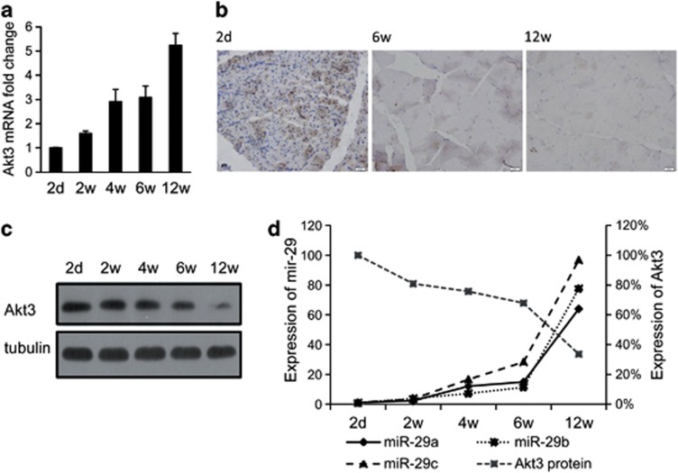
The functional relationship between the miR-29 and Akt3 expression in Balb/c mouse skeletal muscle was examined. Akt3 mRNA expression in murine muscle showed a rising trend with age, from 2-day to 12-week-old animals (Figure 5a). However, Akt3 protein expression was significantly downregulated with age, as determined by western blotting (Figure 5b). Immunohistochemical detection of Akt3 in murine muscle sections corroborated the finding of reduced Akt3 protein expression with age across the same age range (Figure 5c). The post-transcriptional (protein) regulation of Akt3 by miR-29 therefore showed an inverse relationship of expression, such that the increase of miR-29 in skeletal muscle with age (Figures 1a and b) coincided with the corresponding decrease in Akt3 protein expression (Figure 5d).
Figure 5.
Inverse in vivo expression relationship between miR-29 and Akt3. Rising mRNA expression of Akt3 in thigh muscles of postnatal Balb/c mice of ages 2 days, 2 weeks, 4 weeks, 6 weeks, and 12 weeks (a). Akt3 expression was normalized to tubulin expression. Reduction in Akt3 protein expression in thigh muscles of Balb/c mice with increasing age. Immunohistochemical detection of Akt3 protein in Balb/c thigh muscles of different ages (b). Scale bar=20 μm. Inverse relationship with age between endogenous miR-29a/b/c and Akt3 protein expression in postnatal mouse muscles (c). Relative expression of Akt3 was normalized to tubulin levels. Expression of miR-29 was normalized to U6 levels (d). The results are presented as mean±S.E.M. (three independent individuals per group)
Akt3 inhibited myogenic differentiation and promoted proliferation of C2C12 cells
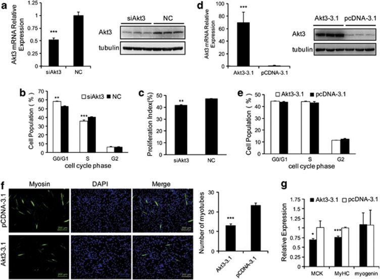
Transfection of siAkt3 into C2C12 myoblasts substantially knocked down the expression of Akt3 mRNA and protein (Figure 6a), which resulted in increased cell arrest in the G0/G1 stage and decreased proliferation rate (Figures 6b and c, and Supplementary Figure 2), an outcome similar to miR-29 overexpression (Figures 2a and b). Overexpression of Akt3 by transfection of Akt3-3.1, as evidenced by raised expression of Akt3 mRNA and protein (Figure 6d), resulted in no apparent change in cell cycle (Figure 6e). However, there was a significant reduction in the number of myotubes inferred by the immunofluorescence detection of fast skeletal myosin expression when Akt3 was overexpressed (Figure 6f). Myoblast differentiation marker genes were also detected at 48 h of differentiation by Q-PCR. Expressions of the MyHC IId and MCK genes were significantly downregulated in cells transfected with Akt3-3.1, relative to cells transfected with pcDNA-3.1 empty plasmid (Figure 6g). Together, these results show that Akt3 promoted myoblast proliferation at the expense of muscle differentiation.
Figure 6.
Akt3 inhibited differentiation and promoted proliferation of C2C12 cells. Transfection of siAkt3 into C2C12 myoblasts knocked down the expression of Akt3 mRNA (left) and protein (right), detected by Q-PCR and western blotting (a), which resulted in raised cell population in the G0/G1 phase and in reduced cell population in the S phase (b), and overall reduction in cell proliferation (c) as determined by flow cytometry. Overexpression of Akt3 via transfection of Akt3-3.1 expression vector into C2C12 myoblasts was detected as raised Akt3 mRNA (left) and protein (right) levels (d). Overexpression of Akt3 in C2C12 myoblasts had no appreciable effect on cell interphase cycle (G0/G1, S, and G2), as determined by flow cytometry (e). Immunofluorescence detection of fast skeletal myosin (green myotubes) in Akt3 expression plasmid-transfected C2C12 cells after 3 days of differentiation (f). Nuclei were stained blue with DAPI. Scale bar=200 μm. Number of myotubes graphically presented as mean±S.E.M. (right). A minimum of nine random fields were captured per group and numerically quantified. C2C12 myoblasts transfected with Akt3-3.1 or pcDNA-3.1 were induced to undergo differentiation for 48 h. Subsequent RNA detection of myogenin, MyHC IId, and MCK was determined by Q-PCR, normalized to tubulin expression (g). All the Q-PCR and flow cytometry results are presented as mean±S.E.M. (three independent replicates per group). *P<0.05; **P<0.01; ***P<0.001
Akt3 could restore cell cycle and block myoblasts' differentiation in miR-29 overexpression
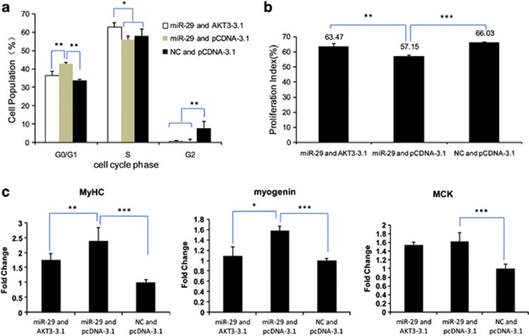
Since Akt3 was shown to be a target of miR-29 during cell cycle and differentiation, its principal role was further evaluated by its overexpression in C2C12 myoblasts in the presence of transfected pooled miR-29, which resulted in increased number of cells that progressed from G0/G1 stage to S and M stages (Figures7a and b and Supplementary Figure 3). As expected, the presence of pooled miR-29 with control empty plasmid (pCDNA-3.1) in C2C12 myoblasts led to increased cell arrest at the G0/G1 stage. Furthermore, the increase in muscle gene expression (MCK, MyHC IId, and myogenin) as a consequence of miR-29 transfection was attenuated by the coexpression of Akt3 (Figure 7c). Therefore, the overexpression of Akt3 could restore cell cycle or proliferation index, over-riding the differentiation effect of miR-29.
Figure 7.
Akt3 overexpression could overcome the cell cycle and differentiation effects of miR-29 in C2C12 cells. C2C12 myoblasts were transiently co-transfected with pooled miR-29 and Akt3 expression vector without the 3′ UTR miR-29 recognition site (Akt3-3.1), miR-29 and pCDNA-3.1, or NC and pCDNA-3.1 as control, followed by 3 days of differentiation. The cell cycle phase (a) and proliferation index (b) were analyzed by propidium iodine flow cytometry. RNA expression of myogenin, MyHC IId, and MCK from corresponding transfections was quantified by Q-PCR (c). Fold change calculation was with reference to control transfection (NC and pCDNA-3.1). The results are presented as mean±S.E.M. (three independent replicates per group). *P<0.05; **P<0.01; ***P<0.001
Discussion
miRNAs were recently discovered as a class of regulators in skeletal muscle development in mice, rats, pigs, and human.7, 8, 9, 10, 11 The most notable muscle-specific miRNAs are miR-1, -133, and -206. miR-1 is highly expressed in adult pig muscle. miR-133 is moderately expressed in adult muscle, but barely detectable in fetal and neonate muscles. miR-206 is abundant in porcine skeletal muscle of different ages, but is lowly expressed in proliferating satellite cells.8 The principal effects of miRNAs are on the modulation of proliferation and differentiation of muscle cells. For instance, miR-206 promotes differentiation by inhibiting Id1-3 and MyoR, both of which are inhibitors of myogenic transcription factors.13 miR-1 promotes myogenesis by repressing HDAC4, whereas miR-133 enhances myoblast proliferation by inhibiting serum response factor.14 Besides, miR-100, -192, -335, -214, and -221/222 were also proved to be participating in muscle proliferation and differentiation.11, 12, 45 Based on these, miRNAs could have therapeutic application.3
We previously showed that miR-29 expression is developmentally upregulated in porcine skeletal muscle from fetal to adult growth.26 Similar developmental expression of miR-29 was observed in mice and humans.27, 32, 33 These results suggest that miR-29 is transcriptionally regulated by similar mechanisms in the skeletal muscle among mammals. Activated NF-κB has been shown to stimulate the expression of YY1 transcription factor that in turn binds to a consensus site upstream of the miR-29b-2/c cluster, thereby inhibiting its transcription.33, 46 Additionally, others have reported direct transcriptional suppression of the miR-29b-1/miR-29a cluster promoter by NF-κB.47 Activation of NF-κB was shown to decrease during mouse skeletal muscle differentiation.48 Indeed, NF-κB activation signaled by a range of inflammatory cytokines (including tumor necrosis factor-like weak inducer of apoptosis) could inhibit myogenic differentiation.49, 50 Conversely, inhibition of NF-κB stimulates skeletal muscle differentiation and regeneration, making NF-κB a potential therapeutic target for Duchenne muscular dystrophy and other muscle-wasting disorders.51, 52, 53 Additionally, putative binding sites of MyoD, myogenin, and Mef2 are present upstream of the miR-29b-2/c cluster,36 indicating their possible roles in myogenic differentiation via miR-29 regulation.
miR-29 is widely recognized as an activator of tumor suppression and apoptosis. In cancer biology, DNMT3A, DNMT3B, p85 alpha, CDC42, Bcl-2, Mcl-1, CCND2, and E2F7 are established targets of miR-29.21, 22, 23, 24, 54 So far, the effector targets of miR-29 that mediate skeletal muscle differentiation are still needed to be identified. In the present study, miR-29 was shown to inhibit myoblast proliferation by targeting Akt3. All members of the Akt family are known to be intimately involved in numerous biological programs, whereas Akt3 isoform is specifically involved in some cancers such as melanomas.34, 35, 36, 37, 38, 40 There is evidence for Akt3 in the regulation of cell growth in VSMCs and heart.42, 43, 44 Presently, we demonstrated that Akt3 can promote myoblast proliferation and is a direct target of miR-29. Knocking down Akt3 expression mimicked the overexpression of miR-29 in prohibiting the proliferation of C2C12 cells (Figures 6b and c). In addition, p85α, another target of miR-29 in tumor suppression, was inhibited in the miR-29-overexpressed myoblasts22 (Figure 4g). Collectively, miR-29 inhibits myoblast proliferation by targeting the proliferation-associated genes Akt3 and p85α.
Previous work has shown that miR-29 targets collagen, Mfap5, Lims1, polycomb group members, and other key factors such as YY1, HDAC4, and Rybp, to inhibit myoblast conversion to myofibroblasts, thereby promoting myogenesis in an indirect manner.28, 30, 31, 32, 33 Conversely, decrease in miR-29 could suppress myogenesis in chronic kidney-disease mice.10 We showed here that miR-29-mediated silencing of Akt3 in myoblasts could delay cell proliferation, and that Akt3 overexpression suppressed myoblast differentiation. In addition, we found that the cell cycle and differentiation effects of miR-29 could be significantly reduced in the presence of Akt3 overexpression (Figure 7). This is a more direct effect of miR-29-induced differentiation, compared with the indirect effect of inhibition of the above-mentioned fibrogenic route.
In conclusion, our study has provided new insights into the fundamental role of miR-29 in the promotion of muscle differentiation, at least in part, by inhibiting Akt3 at the post-transcriptional level to mediate its myogenic effects. miR-29 and its target genes may therefore be potential candidates for intervention aimed at improving muscle growth in farm animals, as well as in skeletal muscle-wasting conditions.
Materials and Methods
Animals and cells
Male Balb/c mice were obtained from Hubei Centre for Disease Control and Prevention. Five groups of mice, three per group, at different ages were used: 2 days, 2 weeks, 4 weeks, 6 weeks, and 12 weeks of age. Three muscle samples from the hindleg of each mouse were collected. Two muscle samples from each mouse were frozen in liquid nitrogen, stored at −80 °C for use in real-time Q-PCR and western blotting. The third sample was stored in 4% paraformaldehyde for immunohistochemistry. The experiments were performed in accordance with the Guide for the Care and Use of Laboratory Animals (1996), and protocols approved by The Hubei Province for Biological Studies Animal Care and Use Committee.
Mouse C2C12 myoblasts were cultured in high-glucose Dulbecco's modified Eagle's medium (DMEM) (Hyclone, Logan, UT, USA) with 10% (v/v) fetal bovine serum. To induce differentiation, culture medium was switched to DMEM with 2% horse serum.
Quantitative (Q)-PCR
Total RNA (including miRNA) was extracted from tissues or cells with TRIzol reagent (Invitrogen, Carlsbad, CA, USA) and treated with DNase I (Fermentas, Ottawa, ON, Canada). The concentration and quality of RNA were assessed by the NanoDrop 2000 (Thermo, Waltham, MA, USA) and denatured gel electrophoresis. Reverse transcription was performed using M-MLV reverse transcriptase (Invitrogen). Random primers, oligo (dT)12–18 or miRNA-specific primers were added to initiate cDNA synthesis. The Q-PCR reaction was carried out in the LightCycler 480 II (Roche, Basel, Switzerland) system, and the reaction mixture used LightCycler 480 SYBR Green I Master (Roche). The sequence of miRNA-specific primers and Q-PCR detection primers can be found in Supplementary Table 1.
Immunofluorescence
After transfection and differentiation, C2C12 cells cultured in six-well plate were washed with phosphate-buffered saline (PBS) twice, and fixed in 4% paraformaldehyde for 15 min. They were then washed twice with PBS, incubated in ice-cold 0.25% Triton X-100 at room temperature for 10 min, and further washed thrice. The cells were incubated in blocking solution (3% bovine serum albumin, 0.3% TritonX-100, 10% fetal bovine serum in PBS) at 4 °C overnight. The next day, after three washes with PBS, the cells were incubated in primary monoclonal anti-myosin (skeletal, fast) antibody (Sigma Life Science, St Louis, MO, USA) at room temperature for 1 h. The cells were washed three times, and incubated with secondary Alexa Fluor 488 IgG, IgM (H+L) antibody (Invitrogen). After 1 h, the secondary antibody was removed, and the cell nuclei were stained with 4′,6-diamidino-2-phenylindole (DAPI) in the dark. After washing three times, images were captured with a Nikon ECLIPSE TE2000-S (Nikon, Tokyo, Japan).
Northern blotting
Total RNA was separated on 15% urea-PAGE gel and transferred to positively charged nylon membrane (PerkinElmer, Waltham, MA, USA) using Trans-Blot SD Semi-Dry Transfer Cell (Bio-Rad, Hercules, CA, USA). Oligonucleotide probes used for hybridization with miRNA were labeled with [32P] at their 5′-end. 5s rRNA was used as the loading control. The sequences of the probes were: miR-29c: 5′-TAACCGATTTCAAATGGTGCTA-3′ 5s rRNA: 5′-CGGTATTCCCAGGCGGTCT-3′.
Cloning for dual-luciferase assay
The psiCHECK-2 dual-luciferase reporter vector (Promega, Madison, WI, USA) housing the 3′ UTR of Akt3, which was XhoI and NotI cloned to the 3′-end of the Renilla gene was used to examine the effect of miR-29 on Renilla production. Akt3 3′ UTR was amplified with the use of forward primer 5′-CGCCTGAGACTGATTCCTG-3′ and reverse primer 5′-CTTTACACCCCAGCAGTC-3′. Site-directed mutation was used to introduce a 2-base substitution into the miR-29-binding site of Akt3 3′ UTR (Akt3 3′ UTR Mut) by mutagen primers 5′-AGCTCCTAGCCACAAAGGGTTAATC-3′ and 5′-ACCCTTTGTGGCTAGGAGCTGACAC-3′. A 2-base substitution in the seed sequence of miR-29 was introduced to create mutant forms of miR-29 (miR-29 Mut) when synthesized. The miRNA mimics and 3′ UTR dual-luciferase vector were co-transfected into cells using Lipofectamine 2000 (Invitrogen). Medium was replaced with fresh growth medium 6 h after transfection. Cells were incubated for 24 h, and assayed with the Dual-Luciferase Reporter Assay System (Promega).
Transfections
One day before transfection, cells were plated in growth medium without antibiotics. Cells were transfected with miRNA mimics or NC-nonspecific miRNA (GenePharma, Shanghai, China) using the Lipofectamine 2000 transfection reagent (Invitrogen). Opti-MEM I Reduced Serum Medium (Gibco, Grand Island, NY, USA) was used to dilute Lipofectamine 2000 and nucleic acids. Transfection procedure was performed according to the manufacturer's instructions. The miRNA mimics were synthesized as duplexes. The miRNA sequences were: mmu-miR-29a: 5′-UAGCACCAUCUGAAAUCGGUUA-3′, 3′-ACCGAUUUCAGAUGGUGCUAUU-5′ mmu-miR-29b: 5′-UAGCACCAUUUGAAAUCAGUGUU-3′ 3′-CACUGAUUUCAAAUGGUGCUAUU-5′ mmu-miR-29c: 5′-UAGCACCAUUUGAAAUCGGUUA-3′, 3′-ACCGAUUUCAAAUGGUGCUAUU-5′ mut-miR-29: 5′-UAGACCCAUUUGAAAUCGGUUA-3′, 3′-UAACCGAUUUCAAAUGGGUCUA-5′. The NC was similar to miRNA in composition, but not in specificity: 5′-UUCUCCGAACGUGUCACGUTT-3′ 3′-TTAAGAGGCUUGCACAGUGCA-5′. The sequence of siAkt3 was 5′-AACCAGGATCATGAGAAACTT-3′.
Cell proliferation analysis
Cell cycle flow cytometry
Thirty six hours after transfection, trypsinised cells were fixed in 70% (v/v) ethanol overnight at −20 °C overnight. Following incubation in 50 μg/ml propidium iodide solution (which contains 100 μg/ml RNase A and 0.2% (v/v) Triton X-100) for 30 min at 4 °C, the cells were analyzed using a FACSCalibur (Becton Dickinson, Franklin Lakes, NJ, USA) and the ModFit software (Verity Software House, Topsham, ME, USA). The proliferative index (PI) stands for the proportion of mitotic cells from a total of 20 000 cells examined.
EdU assay
Six hours after transfection with pEGFP-C1 or miR-29bc-C, C2C12 cells were cultured with fresh growth medium containing EdU (final concentration, 10 μM) for 24 h. Cells were trypsinized, fixed with 4% paraformaldehyde, permeabilized, and analyzed by flow cytometry to detect the population of GFP-positive cells and EdU-stained cells.
xCELLigence real-time cell analyzer
C2C12 cells were seeded on a 16-well E-Plate and allowed to grow for 24 h. When the cell index reached 1.0∼1.5, they were transfected with miRNA mimics using the FuGENE HD transfection reagent (Roche). Cell growth and proliferation were monitored by an xCELLigence RTCA DP instrument (Roche).
Western blotting
Protein lysates were generated with the use of M-PER Mammalian Protein Extraction Reagent (Pierce, Rockford, IL, USA). Sodium dodecyl sulphate-polyacrylamide gel electrophoresis (SDS-PAGE) was performed, followed by protein transfer to polyvinylidene fluoride membranes (Millipore, Billerica, MA, USA) using Mini Trans-Blot Cell (Bio-Rad). Primary antibodies specific for Akt1, Akt2, β-tubulin (Cell Signaling Technology, Boston, MA, USA; 1 : 1000 dilution), Akt3 (Santa Cruz Biotechnology, Santa Cruz, CA, USA; 1 : 1000 dilution), p85α (Abcam, Hong Kong, China; 1 : 1000 dilution), and HRP-labeled anti-rabbit/mouse/goat IgG secondary antibody (Beyotime, Jiangsu, China; 1 : 2000 dilution) were used to detect protein expression.
Immunohistochemistry
After fixation with 4% paraformaldehyde, the skeletal muscle samples were embedded in paraffin, and 6-μm thick serial sections were made. The streptavidin–biotin–peroxidase complex method was used to determine the expression of Akt3. The primary antibody was rabbit anti-Akt3 (Cell Signaling Technology, 1 : 200 dilution), and the secondary antibody was biotinylated sheep anti-rabbit antibody. Diaminobenzidine and Harris' hematoxylin were used to counterstain the sections. Images were captured using Olympus BH-2 System (Olympus, Shinjuku, Tokyo, Japan).
pcDNA-3.1-Akt3 expression vector without the 3′ UTR miR-29 recognition site (Akt3-3.1)
Akt3-coding sequence was amplified by the forward primer 5′-GGATCACAGATGCAGCTACC-3′ and reverse primer 5′-GTAGAAAGGCAACCTTCCACAC-3′, and inserted into pcDNA-3.1. The primary transcript of the mmu-miR-29b-2 and mmu-miR-29c cluster was amplified by the forward primer 5′-CTACCGACACTATGCATCTTG-3′ and reverse primer 5′-AGTGATCTCTAACCTTGGCAG-3′, and cloned into the pEGFP-C1 vector. A stop codon was added between the EGFP gene and multiple cloning sites to stop the translation of EGFP.
Statistical analysis
All results are presented as mean±S.E.M. based on at least three replicates for each treatment. Unpaired Student's t-test was used for P-value calculations.
Ethics standards
The experiments were performed in accordance with the Guide for the Care and Use of Laboratory Animals (1996), and protocols approved by the Hubei Province for Biological Studies Animal Care and Use Committee.
Acknowledgments
This work was supported by grants from the National Key Basic Research Program of China (2012CB124702), National Outstanding Youth Foundation of the National Natural Science Foundation of China (31025026) and Youth Foundation of the National Natural Science Foundation of China (30901020). This work is partly supported by the Biotechnology and Biological Sciences Research Council (UK) China Partnering Award.
Glossary
- miRNAs
microRNAs
- UTR
untranslated region
- VSMC
vascular smooth muscle cell
- YY1
Yin Yang1
- Rybp
Ring1 and YY1-binding protein
- Mfap5
microfibrillar-associated protein 5
- Q-PCR
quantitative polymerase chain reaction
- EdU
5-Ethynyl-2′-deoxyuridine
The authors declare no conflict of interest.
Footnotes
Supplementary Information accompanies this paper on Cell Death and Disease website (http://www.nature.com/cddis)
Edited by M Agostini
Supplementary Material
References
- Hutvagner G, Zamore PD. A microRNA in a multiple-turnover RNAi enzyme complex. Science. 2002;297:2056–2060. doi: 10.1126/science.1073827. [DOI] [PubMed] [Google Scholar]
- Bartel DP. MicroRNAs: genomics, biogenesis, mechanism, and function. Cell. 2004;116:281–297. doi: 10.1016/s0092-8674(04)00045-5. [DOI] [PubMed] [Google Scholar]
- Kloosterman WP, Plasterk RH. The diverse functions of microRNAs in animal development and disease. Dev Cell. 2006;11:441–450. doi: 10.1016/j.devcel.2006.09.009. [DOI] [PubMed] [Google Scholar]
- Ambros V. The functions of animal microRNAs. Nature. 2004;431:350–355. doi: 10.1038/nature02871. [DOI] [PubMed] [Google Scholar]
- Yoon AR, Gao R, Kaul Z, Choi IK, Ryu J, Noble JR, et al. MicroRNA-296 is enriched in cancer cells and downregulates p21WAF1 mRNA expression via interaction with its 3' untranslated region. Nucleic Acids Res. 2011;39:8078–8091. doi: 10.1093/nar/gkr492. [DOI] [PMC free article] [PubMed] [Google Scholar]
- Liu K, Liu Y, Mo W, Qiu R, Wang X, Wu JY, et al. MiR-124 regulates early neurogenesis in the optic vesicle and forebrain, targeting NeuroD1. Nucleic Acids Res. 2010;39:2869–2879. doi: 10.1093/nar/gkq904. [DOI] [PMC free article] [PubMed] [Google Scholar]
- Xie SS, Huang TH, Shen Y, Li XY, Zhang XX, Zhu MJ, et al. Identification and characterization of microRNAs from porcine skeletal muscle. Anim Genet. 2009;41:179–190. doi: 10.1111/j.1365-2052.2009.01991.x. [DOI] [PubMed] [Google Scholar]
- McDaneld TG, Smith TP, Doumit ME, Miles JR, Coutinho LL, Sonstegard TS, et al. MicroRNA transcriptome profiles during swine skeletal muscle development. BMC Genomics. 2009;10:77. doi: 10.1186/1471-2164-10-77. [DOI] [PMC free article] [PubMed] [Google Scholar]
- Nakasa T, Ishikawa M, Shi M, Shibuya H, Adachi N, Ochi M. Acceleration of muscle regeneration by local injection of muscle-specific microRNAs in rat skeletal muscle injury model. J Cell Mol Med. 2009;14:2495–2505. doi: 10.1111/j.1582-4934.2009.00898.x. [DOI] [PMC free article] [PubMed] [Google Scholar]
- Wang XH, Hu Z, Klein JD, Zhang L, Fang F, Mitch WE. Decreased miR-29 suppresses myogenesis in CKD. J Am Soc Nephrol. 2011;22:2068–2076. doi: 10.1681/ASN.2010121278. [DOI] [PMC free article] [PubMed] [Google Scholar]
- Sylvius N, Bonne G, Straatman K, Reddy T, Gant TW, Shackleton S. MicroRNA expression profiling in patients with lamin A/C-associated muscular dystrophy. FASEB J. 2011;25:3966–3978. doi: 10.1096/fj.11-182915. [DOI] [PubMed] [Google Scholar]
- Feng Y, Cao JH, Li XY, Zhao SH. Inhibition of miR-214 expression represses proliferation and differentiation of C2C12 myoblasts. Cell Biochem Funct. 2011;29:378–383. doi: 10.1002/cbf.1760. [DOI] [PubMed] [Google Scholar]
- Kim HK, Lee YS, Sivaprasad U, Malhotra A, Dutta A. Muscle-specific microRNA miR-206 promotes muscle differentiation. J Cell Biol. 2006;174:677–687. doi: 10.1083/jcb.200603008. [DOI] [PMC free article] [PubMed] [Google Scholar]
- Chen JF, Mandel EM, Thomson JM, Wu Q, Callis TE, Hammond SM, et al. The role of microRNA-1 and microRNA-133 in skeletal muscle proliferation and differentiation. Nat Genet. 2006;38:228–233. doi: 10.1038/ng1725. [DOI] [PMC free article] [PubMed] [Google Scholar]
- van Rooij E, Sutherland LB, Thatcher JE, DiMaio JM, Naseem RH, Marshall WS, et al. Dysregulation of microRNAs after myocardial infarction reveals a role of miR-29 in cardiac fibrosis. Proc Natl Acad Sci USA. 2008;105:13027–13032. doi: 10.1073/pnas.0805038105. [DOI] [PMC free article] [PubMed] [Google Scholar]
- Efanov A, Zanesi N, Nazaryan N, Santanam U, Palamarchuk A, Croce CM, et al. CD5+CD23+ leukemic cell populations in TCL1 transgenic mice show significantly increased proliferation and Akt phosphorylation. Leukemia. 2010;24:970–975. doi: 10.1038/leu.2010.46. [DOI] [PMC free article] [PubMed] [Google Scholar]
- Pekarsky Y, Santanam U, Cimmino A, Palamarchuk A, Efanov A, Maximov V, et al. Tcl1 expression in chronic lymphocytic leukemia is regulated by miR-29 and miR-181. Cancer Res. 2006;66:11590–11593. doi: 10.1158/0008-5472.CAN-06-3613. [DOI] [PubMed] [Google Scholar]
- He A, Zhu L, Gupta N, Chang Y, Fang F. Overexpression of micro ribonucleic acid 29, highly up-regulated in diabetic rats, leads to insulin resistance in 3T3-L1 adipocytes. Mol Endocrinol. 2007;21:2785–2794. doi: 10.1210/me.2007-0167. [DOI] [PubMed] [Google Scholar]
- Kapinas K, Kessler C, Ricks T, Gronowicz G, Delany AM. miR-29 modulates Wnt signaling in human osteoblasts through a positive feedback loop. J Biol Chem. 2010;285:25221–25231. doi: 10.1074/jbc.M110.116137. [DOI] [PMC free article] [PubMed] [Google Scholar]
- Li Z, Hassan MQ, Jafferji M, Aqeilan RI, Garzon R, Croce CM, et al. Biological functions of miR-29b contribute to positive regulation of osteoblast differentiation. J Biol Chem. 2009;284:15676–15684. doi: 10.1074/jbc.M809787200. [DOI] [PMC free article] [PubMed] [Google Scholar]
- Fabbri M, Garzon R, Cimmino A, Liu Z, Zanesi N, Callegari E, et al. MicroRNA-29 family reverts aberrant methylation in lung cancer by targeting DNA methyltransferases 3A and 3B. Proc Natl Acad Sci USA. 2007;104:15805–15810. doi: 10.1073/pnas.0707628104. [DOI] [PMC free article] [PubMed] [Google Scholar]
- Park SY, Lee JH, Ha M, Nam JW, Kim VN. miR-29 miRNAs activate p53 by targeting p85 alpha and CDC42. Nat Struct Mol Biol. 2009;16:23–29. doi: 10.1038/nsmb.1533. [DOI] [PubMed] [Google Scholar]
- Xiong Y, Fang JH, Yun JP, Yang J, Zhang Y, Jia WH, et al. Effects of microRNA-29 on apoptosis, tumorigenicity, and prognosis of hepatocellular carcinoma. Hepatology. 2010;51:836–845. doi: 10.1002/hep.23380. [DOI] [PubMed] [Google Scholar]
- Li L, Sarver AL, Alamgir S, Subramanian S. Downregulation of microRNAs miR-1, -206 and -29 stabilizes PAX3 and CCND2 expression in rhabdomyosarcoma. Lab Invest. 2012;92:571–583. doi: 10.1038/labinvest.2012.10. [DOI] [PubMed] [Google Scholar]
- Du Y, Gao C, Liu Z, Wang L, Liu B, He F, et al. Upregulation of a disintegrin and metalloproteinase with thrombospondin motifs-7 by mir-29 repression mediates vascular smooth muscle calcification. Arterioscler Thromb Vasc Biol. 2012;32:2580–2588. doi: 10.1161/ATVBAHA.112.300206. [DOI] [PubMed] [Google Scholar]
- Huang TH, Zhu MJ, Li XY, Zhao SH. Discovery of porcine microRNAs and profiling from skeletal muscle tissues during development. PLoS One. 2008;3:e3225. doi: 10.1371/journal.pone.0003225. [DOI] [PMC free article] [PubMed] [Google Scholar]
- Perbellini R, Greco S, Sarra-Ferraris G, Cardani R, Capogrossi MC, Meola G, et al. Dysregulation and cellular mislocalization of specific miRNAs in myotonic dystrophy type 1. Neuromuscul Disord. 2010;21:81–88. doi: 10.1016/j.nmd.2010.11.012. [DOI] [PubMed] [Google Scholar]
- Wang L, Zhou L, Jiang P, Lu L, Chen X, Lan H, et al. Loss of miR-29 in myoblasts contributes to dystrophic muscle pathogenesis. Mol Ther. 2012;20:1222–1233. doi: 10.1038/mt.2012.35. [DOI] [PMC free article] [PubMed] [Google Scholar]
- Cacchiarelli D, Martone J, Girardi E, Cesana M, Incitti T, Morlando M, et al. MicroRNAs involved in molecular circuitries relevant for the Duchenne muscular dystrophy pathogenesis are controlled by the dystrophin/nNOS pathway. Cell Metab. 2010;12:341–351. doi: 10.1016/j.cmet.2010.07.008. [DOI] [PubMed] [Google Scholar]
- Zhou L, Wang L, Lu L, Jiang P, Sun H, Wang H. A novel target of microRNA-29, Ring1 and YY1-binding protein (Rybp), negatively regulates skeletal myogenesis. J Biol Chem. 2012;287:25255–25265. doi: 10.1074/jbc.M112.357053. [DOI] [PMC free article] [PubMed] [Google Scholar]
- Zhou L, Wang L, Lu L, Jiang P, Sun H, Wang H. Inhibition of miR-29 by TGF-beta-Smad3 signaling through dual mechanisms promotes transdifferentiation of mouse myoblasts into myofibroblasts. PLoS One. 2012;7:e33766. doi: 10.1371/journal.pone.0033766. [DOI] [PMC free article] [PubMed] [Google Scholar]
- Winbanks CE, Wang B, Beyer C, Koh P, White L, Kantharidis P, et al. TGF-beta regulates miR-206 and miR-29 to control myogenic differentiation through regulation of HDAC4. J Biol Chem. 2011;286:13805–13814. doi: 10.1074/jbc.M110.192625. [DOI] [PMC free article] [PubMed] [Google Scholar]
- Wang H, Garzon R, Sun H, Ladner KJ, Singh R, Dahlman J, et al. NF-kappaB-YY1-miR-29 regulatory circuitry in skeletal myogenesis and rhabdomyosarcoma. Cancer Cell. 2008;14:369–381. doi: 10.1016/j.ccr.2008.10.006. [DOI] [PMC free article] [PubMed] [Google Scholar]
- Nakatani K, Thompson DA, Barthel A, Sakaue H, Liu W, Weigel RJ, et al. Up-regulation of Akt3 in estrogen receptor-deficient breast cancers and androgen-independent prostate cancer lines. J Biol Chem. 1999;274:21528–21532. doi: 10.1074/jbc.274.31.21528. [DOI] [PubMed] [Google Scholar]
- Liby TA, Spyropoulos P, Buff Lindner H, Eldridge J, Beeson C, Hsu T, et al. Akt3 controls vascular endothelial growth factor secretion and angiogenesis in ovarian cancer cells. Int J Cancer. 2011;130:532–543. doi: 10.1002/ijc.26010. [DOI] [PMC free article] [PubMed] [Google Scholar]
- Cristiano BE, Chan JC, Hannan KM, Lundie NA, Marmy-Conus NJ, Campbell IG, et al. A specific role for AKT3 in the genesis of ovarian cancer through modulation of G(2)-M phase transition. Cancer Res. 2006;66:11718–11725. doi: 10.1158/0008-5472.CAN-06-1968. [DOI] [PubMed] [Google Scholar]
- Madhunapantula SV, Robertson GP. The PTEN-AKT3 signaling cascade as a therapeutic target in melanoma. Pigment Cell Melanoma Res. 2009;22:400–419. doi: 10.1111/j.1755-148X.2009.00585.x. [DOI] [PMC free article] [PubMed] [Google Scholar]
- Stahl JM, Sharma A, Cheung M, Zimmerman M, Cheng JQ, Bosenberg MW, et al. Deregulated Akt3 activity promotes development of malignant melanoma. Cancer Res. 2004;64:7002–7010. doi: 10.1158/0008-5472.CAN-04-1399. [DOI] [PubMed] [Google Scholar]
- Sharma A, Sharma AK, Madhunapantula SV, Desai D, Huh SJ, Mosca P, et al. Targeting Akt3 signaling in malignant melanoma using isoselenocyanates. Clin Cancer Res. 2009;15:1674–1685. doi: 10.1158/1078-0432.CCR-08-2214. [DOI] [PMC free article] [PubMed] [Google Scholar]
- Liu S, Patel SH, Ginestier C, Ibarra I, Martin-Trevino R, Bai S, et al. MicroRNA93 regulates proliferation and differentiation of normal and malignant breast stem cells. PLoS Genet. 2012;8:e1002751. doi: 10.1371/journal.pgen.1002751. [DOI] [PMC free article] [PubMed] [Google Scholar]
- Roccaro AM, Sacco A, Thompson B, Leleu X, Azab AK, Azab F, et al. MicroRNAs 15a and 16 regulate tumor proliferation in multiple myeloma. Blood. 2009;113:6669–6680. doi: 10.1182/blood-2009-01-198408. [DOI] [PMC free article] [PubMed] [Google Scholar]
- Fox TE, Houck KL, O'Neill SM, Nagarajan M, Stover TC, Pomianowski PT, et al. Ceramide recruits and activates protein kinase C zeta (PKC zeta) within structured membrane microdomains. J Biol Chem. 2007;282:12450–12457. doi: 10.1074/jbc.M700082200. [DOI] [PubMed] [Google Scholar]
- Sandirasegarane L, Kester M. Enhanced stimulation of Akt-3/protein kinase B-gamma in human aortic smooth muscle cells. Biochem Biophys Res Commun. 2001;283:158–163. doi: 10.1006/bbrc.2001.4739. [DOI] [PubMed] [Google Scholar]
- Taniyama Y, Ito M, Sato K, Kuester C, Veit K, Tremp G, et al. Akt3 overexpression in the heart results in progression from adaptive to maladaptive hypertrophy. J Mol Cell Cardiol. 2005;38:375–385. doi: 10.1016/j.yjmcc.2004.12.002. [DOI] [PubMed] [Google Scholar]
- Cardinali B, Castellani L, Fasanaro P, Basso A, Alema S, Martelli F, et al. Microrna-221 and microrna-222 modulate differentiation and maturation of skeletal muscle cells. PLoS One. 2009;4:e7607. doi: 10.1371/journal.pone.0007607. [DOI] [PMC free article] [PubMed] [Google Scholar]
- Wang H, Hertlein E, Bakkar N, Sun H, Acharyya S, Wang J, et al. NF-kappaB regulation of YY1 inhibits skeletal myogenesis through transcriptional silencing of myofibrillar genes. Mol Cell Biol. 2007;27:4374–4387. doi: 10.1128/MCB.02020-06. [DOI] [PMC free article] [PubMed] [Google Scholar]
- Mott JL, Kurita S, Cazanave SC, Bronk SF, Werneburg NW, Fernandez-Zapico ME. Transcriptional suppression of mir-29b-1/mir-29a promoter by c-Myc, hedgehog, and NF-kappaB. J Cell Biochem. 2010;110:1155–1164. doi: 10.1002/jcb.22630. [DOI] [PMC free article] [PubMed] [Google Scholar]
- Bakkar N, Wang J, Ladner KJ, Wang H, Dahlman JM, Carathers M, et al. IKK/NF-kappaB regulates skeletal myogenesis via a signaling switch to inhibit differentiation and promote mitochondrial biogenesis. J Cell Biol. 2008;180:787–802. doi: 10.1083/jcb.200707179. [DOI] [PMC free article] [PubMed] [Google Scholar]
- Dogra C, Changotra H, Mohan S, Kumar A. Tumor necrosis factor-like weak inducer of apoptosis inhibits skeletal myogenesis through sustained activation of nuclear factor-kappaB and degradation of MyoD protein. J Biol Chem. 2006;281:10327–10336. doi: 10.1074/jbc.M511131200. [DOI] [PubMed] [Google Scholar]
- Langen RC, Schols AM, Kelders MC, Wouters EF, Janssen-Heininger YM. Inflammatory cytokines inhibit myogenic differentiation through activation of nuclear factor-kappaB. FASEB J. 2001;15:1169–1180. doi: 10.1096/fj.00-0463. [DOI] [PubMed] [Google Scholar]
- Micheli L, Leonardi L, Conti F, Maresca G, Colazingari S, Mattei E, et al. PC4/Tis7/IFRD1 stimulates skeletal muscle regeneration and is involved in myoblast differentiation as a regulator of MyoD and NF-kappaB. J Biol Chem. 2010;286:5691–5707. doi: 10.1074/jbc.M110.162842. [DOI] [PMC free article] [PubMed] [Google Scholar]
- Thaloor D, Miller KJ, Gephart J, Mitchell PO, Pavlath GK. Systemic administration of the NF-kappaB inhibitor curcumin stimulates muscle regeneration after traumatic injury. Am J Physiol. 1999;277 (2 Pt 1:C320–C329. doi: 10.1152/ajpcell.1999.277.2.C320. [DOI] [PubMed] [Google Scholar]
- Malik V, Rodino-Klapac LR, Mendell JR. Emerging drugs for Duchenne muscular dystrophy. Expert Opin Emerg Drugs. 2012;17:261–277. doi: 10.1517/14728214.2012.691965. [DOI] [PMC free article] [PubMed] [Google Scholar]
- Mott JL, Kobayashi S, Bronk SF, Gores GJ. mir-29 regulates Mcl-1 protein expression and apoptosis. Oncogene. 2007;26:6133–6140. doi: 10.1038/sj.onc.1210436. [DOI] [PMC free article] [PubMed] [Google Scholar]
Associated Data
This section collects any data citations, data availability statements, or supplementary materials included in this article.


