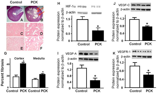
Figure 3.

Histological and protein expression analysis of kidneys from controls and PCK animals. A and B showed Hematoxolin & Eosin staining of kidneys from control and PCK rats showing cystic structures in PCK (3B) and not in control (3A). Furthermore, Picrosirius red staining for fibrosis demonstrates increased fibrosis in both the cortex and medulla in PCK rats (3D and 3F, respectively) compared to control (3C and 3E, respectively), quantified in 3G. H–K shows cortical protein expression, densitometry and quantitation normalized to β-actin, of angiogenic factors, showing decreased expression of hypoxia inducible factor (HIF) 1-α and vascular endothelial growth factor (VEGF) pathways. * p<0.05 compared to control. VEGFR-1: VEGF receptor-1.
