Abstract
Background The dorsal capsuloligamentous scapholunate septum (DCSS) is a confluence of the dorsal capsule, the dorsal intercarpal ligament (DIC), and the scapholunate interosseous ligament (SLIOL). It appears to play a role in the stability of the scapholunate articulation. The purpose of this study was to describe the anatomical basis for this structure and to investigate its role in scapholunate instability through sectioning of this structure followed by an arthroscopic and fluoroscopic analysis.
Material and Methods In the anatomical part of the study we dissected 3 fresh cadaver wrists to examine the anatomy of the DCSS. In the arthroscopic part of the study we assessed the EWAS grade of SL instability before and after sectioning the DCSS and measured the scapholunate and radiolunate angles fluoroscopically.
Results Sectioning the DCSS increased the EWAS grade of SL instability but did not affect the scapholunate gap, the scapholunate angle or radiolunate angle.
Conclusion We have demonstrated that there is a distinct structure that is separate from the dorsal capsule, which we have labeled the Dorsal Capsuloligamentous Scapholunate Septum. We believe that the DCSS is a previously unreported secondary stabilizer of the SL joint which may have therapeutic and prognostic implications.
Keywords: scapholunate complex, scaphoLunate Instability, capsuloScapholunate Septum, DCSS
Our understanding of carpal biomechanics is evolving.1,2 The stability of the scapholunate articulation is dependent upon the scapholunate interosseous ligament which is the primary stabilizer,3,4,5,6,7,8 and by several extrinsic ligaments which act as secondary stabilizers.6,7,9,10,11,12,13,14,15,16,17
In this paper we describe an attachment between the dorsal wrist capsule, the dorsal part of the scapholunate interosseous ligament (SLIOL) and the dorsal intercarpal ligament (DIC) which we have termed the Dorsal Capsuloligamentous Scapholunate Septum (DCSS). It inserts on to the scaphoid, the lunate and the SLIOL. It is our belief that the DCSS may play an important role in the stabilization of the scapholunate articulation.
The purpose of this study was to describe the anatomical basis for this structure and to investigate its role in scapholunate instability through serial sectioning of this structure followed by an arthroscopic and fluoroscopic analysis.
Material and Methods
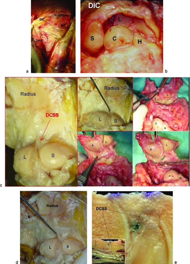
In the anatomical part of the study we dissected 3 fresh cadaver wrists to examine the anatomy of the DCSS. In the first specimen we performed a dorsal approach and excised the extensor tendons to expose, the dorsal radiocarpal (DRC) and the dorsal intercarpal (DIC) ligaments (Fig. 1a). We then opened the midcarpal joint and examined the relations between the DIC and the proximal row (Fig. 1b). In the other 2 wrists, a volar approach was performed and the volar radial carpal ligaments were sectioned then the wrists were then hyperextended to study the dorsal capsular structures (Fig. 1c).
Fig. 1.
(a) DRC and DIC anatomic relations. (b) Relation between DIC and proximal row. (c) Volar radiocarpal intrarticular of Dorsal Capsuloligamentous Scapholunate Septum (DCSS). The volar capsule has been cut while the dorsal capsule has been left intact. The wrist is extended in a gunshot fashion to expose the distal radius and the proximal articular surfaces of the scaphoid and lunate. The arrow is pointing to the DCSS. Volar radiocarpal intraarticular approach of DCSS. (d) Probe checking the DCSS. (e) DCSS on sagittal slice of the wrist.
In the arthroscopic part of the study we studied Ten wrists from 5 fresh frozen cadavers with no previous history of any wrist disorder. There were 3 female and 2 male specimens. The mean age was 52.8 years (range, 50–57 years).
The status of the bone, cartilage and ligaments for each wrist was assessed arthroscopically by a single surgeon and radiographically by another single surgeon. The specimens were mounted and wrist arthroscopy was performed through the 3–4, 4–5, and midcarpal radial and ulnar portals using a 2.7 mm scope, a 1 mm diameter hook, and 5 kg of counter-traction.
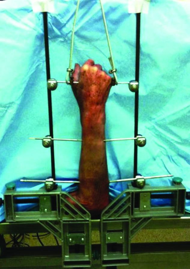
The Stability of the scapholunate and lunotriquetral intervals was assessed arthroscopically and then graded using the European Wrist Arthroscopy Society (EWAS) classification [Table 1] (from J Messina. abstract Congress of FESSH Poznan 2009). Midcarpal arthroscopy was then performed to test the scapholunate and lunotriquetral intervals. The scapholunate gap was measured in each wrist using fluoroscopy (Xi SCAN 440® - FM control - Madrid - Spain). Thereafter, each specimen was fixed in a bridging external fixator frame with 2 transfixing Steinman pins immobilizing the proximal forearm and one transverse metacarpal pin which was free to slide along the fixator frame. A 10 lbkg axial load was applied to the carpus by hanging a 10 lb kg weight from the metacarpal pin. (Fig. 2).
Table 1. EWAS stages of scapholunate interosseous instability24,25.
| Stage | Arthroscopical findings |
|---|---|
| Scope through MCR and probe through MCU | |
| Stage I | No passage of the probe in the interosseous Scapholunate (SL) space, but synovitis. |
| Stage IIA | Volar passage of the probe in the SL space without the ability to turn the probe (widening less than one mm). |
| Stage IIB | Dorsal passage of the probe in the SL space without the ability to turn the probe (widening less than one mm). |
| Stage IIC | Complete passage of the probe in the SL space without the ability to turn the probe (widening less than one mm). |
| Stage IIIA | Volar widening of the SL space with the ability to turn the probe (widening more than one mm, anterior laxity). |
| Stage IIIB | Dorsal widening of the SL space with the ability to turn the probe (widening more than one mm, posterior laxity). |
| Stage IIIC | Complete widening of the SL space with the ability to turn the probe (widening more than one mm, global laxity). |
| Stage IV | SL gap more than 2.7 mm with passage of the scope from MC to RC joint. |
Fig. 2.

View of the wrist under axial load.
A 1.8 mm K-wire was placed in the 3rd metacarpal as a reference to control the rotation of the wrist in the coronal plane. A dorsal subcutaneous 45 mm needle was used to determine the radiographic magnification. Posteroanterior and lateral views were taken, without and with an axial load (Fig. 2). The scapholunate gap was measured according to Lee and Coll.18 The scapholunate and the radiolunate angles were measured by Watson's method.19 Each space and angle were measured independently by two reviewers and then compared. They used the software “Image J 1.45s” (National Institute of Health. USA. 2012). Measurements showing a difference more than 0.5 mm or 5° between the two observers were recalculated. The final value was the average of the two measurements. After each ligament section, the scapholunate stability was assessed arthroscopically and fluorsocopically.
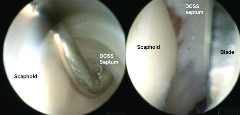
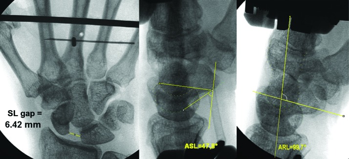
Arthroscopic sectioning of the DCSS was performed under direct vision. This was achieved by introducing the arthroscope through the 4–5 radiocarpal portal and a 15 scalpel blade through the 3–4 radiocarpal portal directed radially to avoid cutting the dorsal radiocarpal ligament (Fig. 3). After sectioning, the scapholunate gap on the posteroanterior view was measured under fluoroscopy, along with a measurement of the scapholunate and the radiolunate angles on the lateral view (Fig. 4) The scapholunate gap was considered normal under 3 mm20. The scapholunate angle was considered normal between 30° and 60°. The radiolunate angle was considered normal between +15° and −15°. By convention, we attributed a positive sign if the lunate was extended, and a negative sign if the lunate was flexed in relation to the radius.
Fig. 3.
Arthroscopic Section of the DCSS.
Fig. 4.
Fluoroscopic measures (SL gap, SL angle and RL angle).
Results
Anatomy of the DCSS
The DCSS is a fibrous structure, that arises from the dorsal capsule inserts in a bifid manner at the bone-ligament junction of the dorsal superior aspect of the scaphoid and lunate. At this level, the DCSS seems mechanically the most resistant when it's palpated with a hook probe (Fig. 1d). The capsule's insertion on the dorsal scaphoid and lunate is thin and clearly distinguishable from the DCSS (Fig. 1e). The DCSS in the sagittal plane is oriented obliquely in a distal and volar direction.
Arthroscopic Section of DCSS
Before sectioning, arthroscopically there were five EWAS stage 1, two stage 2A, one stage 2B, one stage 2C and one stage 3A. After DCSS sectioning, there were arthroscopically one EWAS stage 1, three stage 2B, five stage 3B and one stage 3C. Nine wrists demonstrated increased laxity. After DCSS sectioning, the majority of wrists had some dorsal widening of the SL joint (Table 2). The mean scapholunate gap prior to ligament sectioning and without axial load was 1.2 mm (range, 0.25 to 1.5 mm), under load 1.5 mm (range, 0.9 mm to 2.4 mm). The mean scapholunate angle without axial loading was 46.0° (range, 31.5° to 57.2°) and 46.1° (range, 25.1° to 63.2°) under load. Without load, no specimen had a SL angle greater than 60° and under load, two specimens had a scapholunate angle greater than 60°. The mean radiolunate angle without axial load was 6.5° (range, −5.6° to16.9°) and under load 0.3° (range, −18.7° to 22.9°) The DCSS sectioning didn't affect the SL gap, SL angle or RL angle. None of the wrists had a SL angle greater than 60° on the lateral view with or without loading.
Table 2. Arthroscopic SL instability before and after DCSS sectioning.
| Specimens | Arthroscopic SL laxity before DCSS sectioning | Arthroscopic SL laxity after DCSS sectioning |
|---|---|---|
| 1R | 2C | 3B |
| 2R | 1 | 2B |
| 3R | 1 | 2B |
| 4R | 2B | 3B |
| 5R | 3A | 3C |
| 1L | 2A | 3B |
| 2L | 1 | 1 |
| 3L | 2A | 3B |
| 4L | 1 | 3B |
| 5L | 1 | 2B |
Discussion
The role of the dorsal capsular insertion and its effect on stabilizing the scapholunate interval has not been well defined. In an anatomical study of 90 cadaver wrists, Viegas et al15 noted that the DIC ligament was comprised of 2 sections: a more distal section of generally thinner fibers extending from the dorsal tubercle of the triquetrum to the dorsal aspect of the trapezoid or capitate and a more proximal, thicker section extending from the dorsal tubercle of the triquetrum to the dorsal distal aspect of the lunate, on to the dorsal groove of the scaphoid, and then to the proximal rim of the trapezium. Nagao et al21 in a 3-D study of 8 cadaver wrists showed the DIC branched to distal attachments on the distal part of the dorsal aspect of the lunate (6 cases), on the proximal part of the dorsal aspect of the scaphoid (all cases), on the waist of the dorsal aspect of the scaphoid (6 cases), on the dorsal aspect of the trapezium (1 case), and on the dorsal aspect of the trapezoid (all cases). Buijze et al22 however reported that the DIC attachment to the lunate was found in only 2 of 8 specimens. We have demonstrated that there is a distinct structure that is separate from the dorsal capsule, which we have labeled the Dorsal Capsuloligamentous Scapholunate Septum which attaches between the dorsal wrist capsule and the dorsal part of the SLIOL and the DIC ligament. Sectioning the DCSS didn't affect the static SL gap, SL angle or RL angle under a 10 kg axial load, but there was noted to be an increase in the EWAS stage of SL instability, which suggests this structure has a stabilizing effect on the scapholunate joint. We believe that the DCSS is a previously unreported secondary stabilizer of the SL joint which may have therapeutic and prognostic implications. Ruch and Poeling23 alluded to this when they wrote that the existence of a tear of the dorsal capsular reflection when combined with a scapholunate interosseous ligament (SLIL) tear connotes a greater degree of carpal instability and they advocated an open SLIL repair versus arthroscopic pinning when there was an associated tear of the dorsal capsular reflection.
Limitations of the current study include the fact that this was a cadaver study which may not correlate with in vivo findings and that the biomechanical model of a 10 kg axial load is not physiologic and does not replicate the in vivo forces. The small numbers of cadaver specimens as well as the lack of biomechanical data correlating the EWAS stage with ligament tear or attenuation as well as the lack of data on intraobserver agreement of the EWAS stage introduces a source of bias.
Conflict of Interest None
Note
Work performed at IRCAD Strasbourg, France.
References
- 1.Camus E J, Millot F, Lariviere J, Raoult S, Rtaimate M. Kinematics of the wrist using 2D and 3D analysis: biomechanical and clinical deductions. Surg Radiol Anat. 2004;26(5):399–410. doi: 10.1007/s00276-004-0260-0. [DOI] [PubMed] [Google Scholar]
- 2.Camus E J, Millot F, Larivière J, Rtaimate M, Raoult S. [The double-cup carpus: a demonstration of the variable geometry of the carpus] (in French) Chir Main. 2008;27(1):12–19. doi: 10.1016/j.main.2007.10.005. [DOI] [PubMed] [Google Scholar]
- 3.Mayfield J K. Patterns of injury to carpal ligaments. A spectrum. Clin Orthop Relat Res. 1984;(187):36–42. [PubMed] [Google Scholar]
- 4.Berger R A, Imeada T, Berglund L, An K N. Constraint and material properties of the subregions of the scapholunate interosseous ligament. J Hand Surg Am. 1999;24(5):953–962. doi: 10.1053/jhsu.1999.0953. [DOI] [PubMed] [Google Scholar]
- 5.Short W H, Werner F W, Fortino M D, Palmer A K, Mann K A. A dynamic biomechanical study of scapholunate ligament sectioning. J Hand Surg Am. 1995;20(6):986–999. doi: 10.1016/S0363-5023(05)80147-9. [DOI] [PubMed] [Google Scholar]
- 6.Short W H, Werner F W, Green J K, Weiner M M, Masaoka S. The effect of sectioning the dorsal radiocarpal ligament and insertion of a pressure sensor into the radiocarpal joint on scaphoid and lunate kinematics. J Hand Surg Am. 2002;27(1):68–76. doi: 10.1053/jhsu.2002.30074. [DOI] [PubMed] [Google Scholar]
- 7.Short W H, Werner F W, Green J K, Masaoka S. Biomechanical evaluation of the ligamentous stabilizers of the scaphoid and lunate: Part II. J Hand Surg Am. 2005;30(1):24–34. doi: 10.1016/j.jhsa.2004.09.015. [DOI] [PubMed] [Google Scholar]
- 8.Ritt M J, Linscheid R L, Cooney W P III, Berger R A, An K N. The lunotriquetral joint: kinematic effects of sequential ligament sectioning, ligament repair, and arthrodesis. J Hand Surg Am. 1998;23(3):432–445. doi: 10.1016/S0363-5023(05)80461-7. [DOI] [PubMed] [Google Scholar]
- 9.Short W H, Werner F W, Green J K, Masaoka S. Biomechanical evaluation of ligamentous stabilizers of the scaphoid and lunate. J Hand Surg Am. 2002;27(6):991–1002. doi: 10.1053/jhsu.2002.35878. [DOI] [PMC free article] [PubMed] [Google Scholar]
- 10.Short W H, Werner F W, Green J K, Sutton L G, Brutus J P. Biomechanical evaluation of the ligamentous stabilizers of the scaphoid and lunate: part III. J Hand Surg Am. 2007;32(3):297–309. doi: 10.1016/j.jhsa.2006.10.024. [DOI] [PMC free article] [PubMed] [Google Scholar]
- 11.Sennwald G R, Zdravkovic V, Oberlin C. The anatomy of the palmar scaphotriquetral ligament. J Bone Joint Surg Br. 1994;76(1):147–149. [PubMed] [Google Scholar]
- 12.Viegas S F, Yamaguchi S, Boyd N L, Patterson R M. The dorsal ligaments of the wrist: anatomy, mechanical properties, and function. J Hand Surg Am. 1999;24(3):456–468. doi: 10.1053/jhsu.1999.0456. [DOI] [PubMed] [Google Scholar]
- 13.Moritomo H, Viegas S F, Nakamura K, Dasilva M F, Patterson R M. The scaphotrapezio-trapezoidal joint. Part 1: An anatomic and radiographic study. J Hand Surg Am. 2000;25(5):899–910. doi: 10.1053/jhsu.2000.4582. [DOI] [PubMed] [Google Scholar]
- 14.Moritomo H, Viegas S F, Elder K, Nakamura K, Dasilva M F, Patterson R M. The scaphotrapezio-trapezoidal joint. Part 2: A kinematic study. J Hand Surg Am. 2000;25(5):911–920. doi: 10.1053/jhsu.2000.8637. [DOI] [PubMed] [Google Scholar]
- 15.Mitsuyasu H, Patterson R M, Shah M A, Buford W L, Iwamoto Y, Viegas S F. The role of the dorsal intercarpal ligament in dynamic and static scapholunate instability. J Hand Surg Am. 2004;29(2):279–288. doi: 10.1016/j.jhsa.2003.11.004. [DOI] [PubMed] [Google Scholar]
- 16.Berger R A. The anatomy of the ligaments of the wrist and distal radioulnar joints. Clin Orthop Relat Res. 2001;383(383):32–40. doi: 10.1097/00003086-200102000-00006. [DOI] [PubMed] [Google Scholar]
- 17.Elsaidi G A, Ruch D S, Kuzma G R, Smith B P. Dorsal wrist ligament insertions stabilize the scapholunate interval: cadaver study. Clin Orthop Relat Res. 2004;425(425):152–157. doi: 10.1097/01.blo.0000136836.78049.45. [DOI] [PubMed] [Google Scholar]
- 18.Lee S K, Desai H, Silver B, Dhaliwal G, Paksima N. Comparison of radiographic stress views for scapholunate dynamic instability in a cadaver model. J Hand Surg Am. 2011;36(7):1149–1157. doi: 10.1016/j.jhsa.2011.05.009. [DOI] [PubMed] [Google Scholar]
- 19.Watson H, Ottoni L, Pitts E C, Handal A G. Rotary subluxation of the scaphoid: a spectrum of instability. J Hand Surg [Br] 1993;18(1):62–64. doi: 10.1016/0266-7681(93)90199-p. [DOI] [PubMed] [Google Scholar]
- 20.Kuo C E, Wolfe S W. Scapholunate instability: current concepts in diagnosis and management. J Hand Surg Am. 2008;33(6):998–1013. doi: 10.1016/j.jhsa.2008.04.027. [DOI] [PubMed] [Google Scholar]
- 21.Nagao S, Patterson R M, Buford W L Jr, Andersen C R, Shah M A, Viegas S F. Three dimensional description of ligamentous attachments around the lunate. J Hand Surg. 2005;30A:685–692. doi: 10.1016/j.jhsa.2005.03.002. [DOI] [PubMed] [Google Scholar]
- 22.Buijze G A, Lozano-Calderon S A, Strackee S D, Blankevoort L, Jupiter J B. Osseous and ligamentous scaphoid anatomy: Part I. A systematic literature review highlighting controversies. J Hand Surg Am. 2011;36(12):1926–1935. doi: 10.1016/j.jhsa.2011.09.012. [DOI] [PubMed] [Google Scholar]
- 23.Ruch D S, Poehling G G. Philadelphia: Churchill Livingstone; 1999. Wrist Arthroscopy: Ligamentous Instability; pp. 200–206. [Google Scholar]
- 24.Messina J C, Dreant N, Luchetti R, Lindau T, Mathoulin C. Scapholunate tears: a new arthroscopic classification. Chirurgie de la Main. 2009;28(6):339–340. doi: 10.1016/j.main.2008.12.005. [DOI] [PubMed] [Google Scholar]
- 25.Messina J C, Dreant N, Luchetti R, Lindau T, Mathoulin C. Nuova classificazione delle lesioni del legamento scafolunato. Rivista di Chirurgia della Mano. 2009;(46):156–157. [Google Scholar]


