Abstract
Background The palmar aspect of the capsuloligamentous complex of the wrist is relatively inaccessible to surgery through an open approach. An all-inside arthroscopic suturing technique is presented that allows suturing of the palmar scapholunate or lunotriquetral ligaments or plication of the space of Poirier.
Methods Eight palmar scapholunate ligaments and four major tears of the volar capsuloligamentous complex of the wrist due to a perilunate dislocation have been repaired.
Results No complications occurred during or after the procedure. It was not possible to separate the effects of the palmar repair from the disparate treatment of the associated pathology.
Conclusions The technique described allows one to suture the palmar aspects of the SL and LT ligaments safely and to repair the space of Poirier structures without need for any special equipment.
Keywords: wrist arthroscopy, scapholunate ligament repair, space of Poirier, all-inside technique
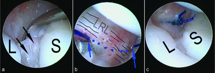
The palmar aspect of the capsuloligamentous complex of the wrist is relatively inaccessible to open surgery. When the palmar aspect of the scapholunate (SL) or the palmar aspect of the lunotriquetral (LT) ligament is torn, it is either repaired through an open surgical approach or left unrepaired. I have previously published an all-arthroscopic technique for suture repair of the palmar aspect of the SL ligament (Fig. 1a–c)1 that can also be used to close rents in the space of Poirier and to repair the palmar aspect of the lunotriquetral ligament.
Fig. 1a–c.

Intraoperative view of the operation in a clinical case (left wrist, scope in MCU portal). (a) The remnants of the SL ligament (arrows) can be seen through the anterior SL gap. (b) The thread is seen passing palmarly (marked in dots) from one side of the SL space to the other. (c) After tightening of the knot, the SL space is closed (© The American Society for Surgery of the Hand 2011).
Indications
The procedure can be used to repair any of the palmar structures of the capsuloligamentous complex. In perilunate dislocation and perilunate fracture dislocation, I have used a similar technique to repair the palmar portion of the scapholunate ligament, to close the space of Poirier, and to repair the palmar portion of the lunotriquetral ligament.
Contraindications
This technique cannot be used if there is no remaining viable ligament that is amenable to suturing.
Surgical Technique
In my experience, palpation of bony prominences is crucial in determining the proper entry sites of the Tuohy needle and Kirschner wires (K-wires). For this reason, I prefer using a dry technique for wrist arthroscopy so that fluid distension will not distort the anatomy.2,3 An understanding of the all-inside technique and its possible variations is required.4,5
The ligament stumps are first debrided with a full-radius resector and the adjacent bony surfaces refreshed with a burr inserted through the midcarpal ulnar (MCU) and midcarpal radial (MCR) portals. The scope is then positioned in the MCU portal to have an unimpeded view of the palmar SL space, while the MCR is left free for instrumentation. A 22 gauge needle inserted immediately ulnar to the flexor carpi radialis (FCR), ∼1 cm proximal to the distal wrist crease, will penetrate the midcarpal joint in the vicinity of the SL space. As this may require several attempts before the position is satisfactory, an intramuscular needle, which has a smaller bore than the Tuohy, may first be used to minimize the local insult. Next, the Tuohy needle is inserted following the same path.
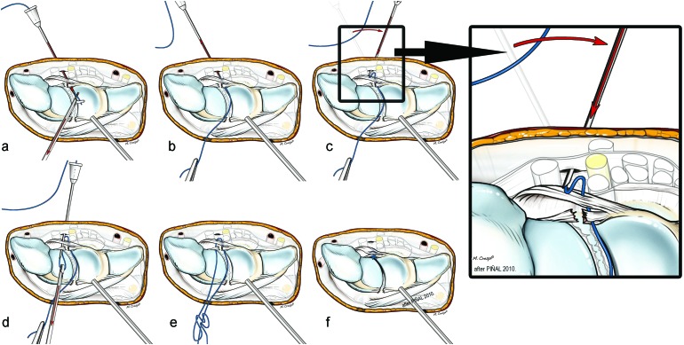
A (colored) 2–0 polydioxanone (PDS) suture is then threaded through the Tuohy needle, retrieved from the midcarpal joint with a grasper via the MCR portal, and pulled out of the joint (Fig. 2).
Fig. 2.
Steps of the operative procedure. On the right close-up of the critical step of the needle being slid over the palmar surface of the capsule. (see text for further details) (© The American Society for Surgery of the Hand, 2011).
The Tuohy needle is then withdrawn slightly outside of the capsule (but still subcutaneous), then slid just palmar to the capsule radially, and then reinserted into the joint distal to the palmar edge of the scaphoid.
The suture is again advanced through the Tuohy needle, creating a suture loop inside the joint, which is then retrieved with the grasper and withdrawn from the joint via the MCR portal so that both ends of the suture are outside the joint. In this way a suture loop is created palmar to the capsule on both sides of the SL interval, including the long radiolunate (LRL) ligament, although this has not been verified through cadaver dissection. A gliding knot is then tied over the palmar capsule and tightened with a knot pusher, apposing the palmar aspects of the scaphoid and lunate and closing any palmar gap (Fig. 2).
It is crucial that both ends of the suture are pulled out of the joint through the same path in the MCR portal. If any tissue is interposed, the gliding knot will be blocked and cannot be advanced. A suture hook can be used to make sure that both suture ends follow the same path.4
Complications
At present we have not had any complications with this technique. The palmar cutaneous branch of the median nerve is at risk, which can be minimized by making a 1-cm incision and using wound spread technique prior to introducing the Tuohy needle.
Personal Outcomes
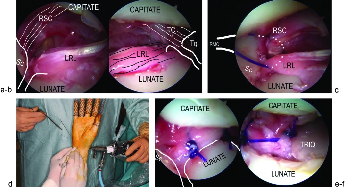
To date we have performed eight palmar SL repairs using this technique. We combined this repair with a dorsal capsuloligamentous plication, as described by Mathoulin et al,6 in six cases. We also have used a similar technique to repair major tears at the space of Poirier after perilunate dislocations in four cases (Fig. 3a–f).
Fig. 3a–f.
Closure of the space of Poirier in a perilunate dislocation. (a) Radial and (b) ulnar view after reduction of a perilunate dislocation. A major tear in the palmar capsuloligamentous system can be seen. K-wires already maintain the proximal row reduced. (c) Intraoperative view while suturing the palmar capsuloligamentous complex in this patient. The assistant has one of the suture ends exiting from the dorsal capsule while the surgeon is suturing palmarly. (d) Corresponding intraarticular view. The hitch has already been placed while both thread ends are now out of the joint through the MCR portal. (e), (f) The palmar capsuloligamentous rent is now closed by means of three stitches.Abbreviations: Sc, scaphoid; Tq, triquetrum; RSC, radioscaphocapitate ligament; LRL, long radiolunate ligament; TQ: Triquetrumcapitate ligament. (© Dr. Piñal 2012.)
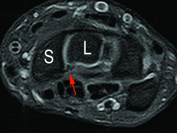
Because these repairs were performed in conjunction with other arthroscopic procedures for combined wrist pathology, it is not possible to separate out the effects of the palmar capsuloligamentous repair. It is my belief, however, that it is beneficial to repair these torn palmar ligaments, which is the rationale for this technique (Fig. 4).
Fig. 4.

Magnetic resonance image (MRI) of a repaired palmar SL at one year. (© Dr. Piñal 2012.)
Footnotes
Conflict of interest None
References
- 1.del Piñal F, Studer A, Thams C, Glasberg A. An all-inside technique for arthroscopic suturing of the volar scapholunate ligament. J Hand Surg Am. 2011;36(12):2044–2046. doi: 10.1016/j.jhsa.2011.09.021. [DOI] [PubMed] [Google Scholar]
- 2.del Piñal F, García-Bernal F J, Pisani D, Regalado J, Ayala H, Studer A. Dry arthroscopy of the wrist: surgical technique. J Hand Surg Am. 2007;32(1):119–123. doi: 10.1016/j.jhsa.2006.10.012. [DOI] [PubMed] [Google Scholar]
- 3.del Piñal F. Dry arthroscopy and its applications. Hand Clin. 2011;27(3):335–345. doi: 10.1016/j.hcl.2011.05.011. [DOI] [PubMed] [Google Scholar]
- 4.del Piñal F, García-Bernal F J, Cagigal L, Studer A, Regalado J, Thams C. A technique for arthroscopic all-inside suturing in the wrist. J Hand Surg Eur Vol. 2010;35(6):475–479. doi: 10.1177/1753193409361014. [DOI] [PubMed] [Google Scholar]
- 5.del Piñal F. Berlin: Springer Verlag; 2012. Appendix: All-inside technique; pp. 349–364. [Google Scholar]
- 6.Mathoulin C L Dauphin N Wahegaonkar A L Arthroscopic dorsal capsuloligamentous repair in chronic scapholunate ligament tears Hand Clin 2011274563–572., xi [DOI] [PubMed] [Google Scholar]


