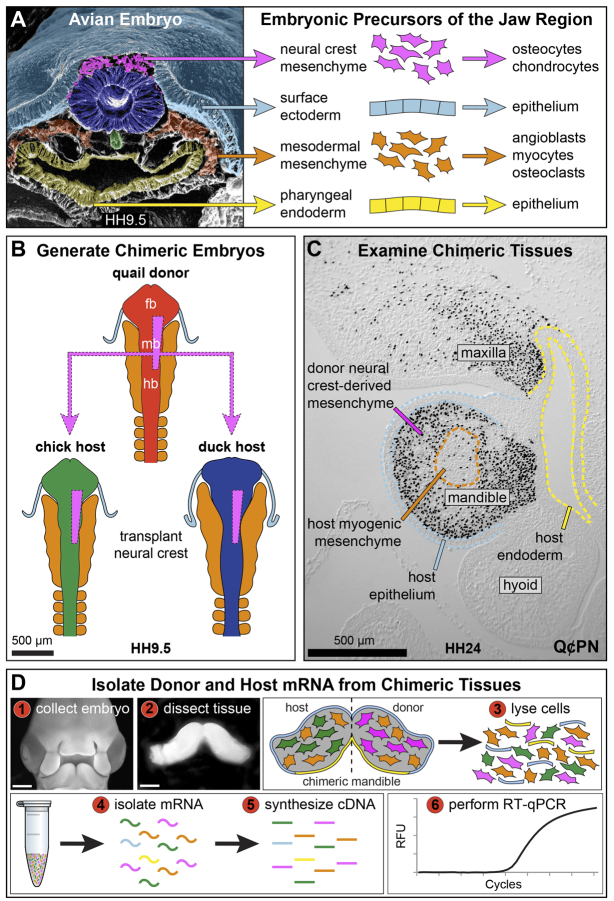
Fig. 1.
Generation and examination of chimeric tissue. (A) Pseudocolored scanning electron micrograph showing precursors of the jaw complex. Image courtesy of K. Tosney. (B) NCM transplants from quail into either chick or duck. (C) Sagittal section through the jaw region showing quail cells stained with Qc/PN (black nuclei). Duck-host ectoderm (light blue), endoderm (yellow), and myogenic mesoderm (orange) remain unstained. (D) Work flow for quantifying gene expression.

