Abstract
Phytic acid (InsP6) is considered to be the major source of phosphorus and inositol phosphates in most cereal grains. However, InsP6 is not utilized efficiently by monogastric animals due to lack of phytase enzyme. Furthermore, due to its ability to chelate mineral cations, phytic acid is considered to be an antinutrient that renders these minerals unavailable for absorption. In view of these facts, reducing the phytic acid content in cereal grains is a desired goal for the genetic improvement of several crops. In the present study, we report the RNAi-mediated seed-specific silencing (using the Oleosin18 promoter) of the IPK1 gene, which catalyzes the last step of phytic acid biosynthesis in rice. The presence of the transgene cassette in the resulting transgenic plants was confirmed by molecular analysis, indicating the stable integration of the transgene. The subsequent T4 transgenic seeds revealed 3.85-fold down-regulation in IPK1 transcripts, which correlated to a significant reduction in phytate levels and a concomitant increase in the amount of inorganic phosphate (Pi). The low-phytate rice seeds also accumulated 1.8-fold more iron in the endosperm due to the decreased phytic acid levels. No negative effects were observed on seed germination or in any of the agronomic traits examined. The results provide evidence that silencing of IPK1 gene can mediate a substantial reduction in seed phytate levels without hampering the growth and development of transgenic rice plants.
Introduction
Phytic acid (myo-inositol-1,2,3,4,5,6-hexakisphosphate or IP6) is known as the major source of phosphorus in cereal grains, comprising approximately 1–2% of the dry weight and accounting for approximately 65–80% of the total seed phosphorus [1]. In most cereals, with the exception of maize (Zea mays), approximately 80% of the total phytic acid (IP6) accumulates in the aleurone layer of the grains. In general, IP6 accumulates in the protein storage bodies as mixed salts called phytate that chelate a number of mineral cations. During the process of germination, endogenous grain phytase is activated, which degrades phytate, releasing stored phosphorus, myo-inositol and bound mineral cations [1] that are further utilized by the developing seedlings. However, due to the lack of microbial phytase enzymes [2], monogastric animals are unable to remove the phosphates from the myo-inositol ring and are, therefore, incapable of utilizing the phosphorus present in cereals [3]. Phytate has six negatively charged ions, making it a potent chelator of such divalent cations as Fe2+, Zn2+, Ca2+, and Mg2+ and rendering these ions unavailable for absorption by monogastric animals [4]. In view of these adverse effects, many attempts have been made to reduce the phytic acid content in cereals.
Among the different approaches for reducing the phytate levels in cereals, the exogenous expression of recombinant microbial phytase is common [5], [6], [7], [8], and another promising strategy is the generation of cereal mutants exhibiting a low phytic acid (lpa) phenotype [4]. Several lpa mutant lines have been generated in rice [9], [10], wheat [11], and maize [1], [12], [13]; although effective, these strategies are sometimes associated with downstream impacts on crop yield and other parameters of agronomic performance [4]. Therefore, a different strategy was pursued, whereby transgenic crops were developed by manipulating the phytic acid biosynthetic pathway [3], [14]. Recently, twelve genes from rice (Oryza sativa L.) have been identified that catalyze intermediate steps of inositol phosphate metabolism in seeds [15].
The first step of phytic acid biosynthesis in the developing rice seed is catalyzed by myo-inositol-3-phosphate synthase (MIPS, EC 5.5.1.4) [16], and various attempts have been made to silence the expression of the myo-inositol-3-phosphate synthase (MIPS) gene under the control of constitutive [17] or different rice seed-specific promoters [3], [14]. In the case of constitutive promoters (CaMV35S), expression of the MIPS gene was also suppressed in vegetative tissues in addition to the seeds, causing detrimental effects to the plant. Hence, seed-specific promoters, e.g., GlutelinB-1 (GluB-1) and Oleosin18 (Ole18), have been used to mediate suppression only in the seeds. The resulting transgenic rice plants exhibited more pronounced silencing with the Ole18 promoter, as it drives expression specifically in the aleurone layer and embryo of seeds [18], the site of maximum phytate accumulation. However, the inadvertent change in seed myo-inositol content was not considered and might have a negative impact on plant inositol metabolism, as myo-inositol-3-phosphate, the product of MIPS, is known to be the only precursor for the de novo synthesis of myo-inositol [19], [20], [21]. Therefore, to reduce the phytate content in seeds without disturbing related pathways, enzymes involved at a later stage in phytic acid biosynthesis (i.e., IPK; Inositol phosphate kinases) in rice should be targeted and the effects analyzed.
IPK1 (Inositol 1,3,4,5,6-pentakisphosphate 2-kinase) is believed to catalyze the final step in phytic acid biosynthesis, whereby the InsP5 molecule is phosphorylated at the 2nd position [22], [23], [24]. The InsP6 biosynthetic pathway was previously described in Saccharomyces cerevisiae [24], and the pathway was found to share a common final step with that in Dictyostelium discoideum: the phosphorylation of Ins (1,3,4,5,6)P5 to InsP6 by a 2-kinase enzyme designated as IPK1 (EC 2.7.1.158). The S. cerevisiae IPK1Δ mutant showed an almost complete inability to synthesize InsP6 and showed a reduction in the ability to export mRNA from the nucleus. Several myo-inositol kinase enzymes have since been identified in plants, including myo-inositol kinase [13], Ins(1,3,4)P35/6-kinase [25], Ins(1,4,5)P36/3/5-kinase, and Ins (1,3,4,5,6) P5 2-kinase [26], [27]. Recent reports examined the Ins(1,4,5)P36/3/5-kinase (AtIpk2β-1) and Ins(1,3,4,5,6)P5 2-kinase (AtIpk1-1) genes using T-DNA insertion mutants in Arabidopsis [28], and the phytate content was reduced in the AtIpk2β-1 mutant by 35% in the AtIpk1-1 by 83% and by more than 95% in the double mutant.
In the present study, we generated transgenic rice plants by silencing the last step of phytic acid biosynthesis in the Pusa Sugandhi II rice cultivar by manipulating the expression of the IPK1 gene using a seed-specific promoter, Ole18, in an RNAi-mediated approach. The resulting T4 transgenic plants were analyzed at the molecular and biochemical levels, revealing substantial reductions in phytate levels and an increase in the amount of inorganic phosphate (Pi). In addition, we also estimated the change in the concentration of different metals in the rice grains after milling, as metal ions may be affected by reduced seed phytate levels. Different agronomic traits of the transgenic plants were also analyzed and compared with non-transgenic rice plants.
Materials and Methods
Plant Material and Growth Conditions
Oryza sativa L. subspecies indica cv. Swarna and IR-36, procured from Chinsurah Rice Research Station, Hooghly, West-Bengal, were used for cloning purposes. For purposes of genetic transformation, Oryza sativa L. subspecies indica cv. Pusa Sugandhi II was obtained from IARI, ICAR, India. Following surface sterilization, the seeds were germinated on distilled water-soaked filter paper in a plant growth chamber (FLI-2000, Eyela, Japan) maintained at 30°C and 75% relative humidity.
Low-phytate transgenic (T0–T4) rice generated and respective non-transgenic control (non-transformed Pusa Sugandhi II rice) plants were grown in pots containing fertilizer-enriched paddy field soil (N: P: K = 80∶40: 40 kg/ha) under greenhouse conditions. The day/night temperature regime of 30/25°C under condition of natural illumination, and a relative humidity of 70–80% was maintained throughout the experiment.
Construction of Plasmids
Total RNA was extracted from indica rice cultivar using the RNeasy Plant mini kit following the manufacturer’s protocol (Qiagen). After RNA quantification, cDNA was synthesized from the purified RNA using the Superscript III reverse transcriptase two-step RT-PCR kit (Invitrogen, USA). The RT-PCR product of the IPK1 (GenBank accession no. AK102842; LOC_Os04g56580) gene amplified using gene-specific primers (IPK1F, 5′-CTGCTCTTCTAA TTTCTGACC-3′, and IPK1R, 5′-CTTCTTAATGTTTGTCTACTG-3′) was purified and cloned into the pENTR-D TOPO entry vector (Invitrogen) and sequenced. The 1.1-kb fragment of the IPK1 gene from the entry clone (pENTR-IPK1) was then introduced into the binary destination vector pIPKb006 using an LR clonase (Invitrogen, USA) based recombination reaction [29]. Lastly, the Ole18 promoter (GenBank accession no. AF019212) cloned from the IR36 rice cultivar using a specific primer pair (Ole18F, 5′-TCAGCCAATACATTGATCCG-3′, and Ole18R, 5′-GCAAGATGAATGCAACGAAG-3′) was ligated to the MCS of the recombined vector at the SpeI and HindIII sites. The complete RNAi vector (pOle18-IPK1-006) containing the IPK1 gene under the control of the Ole18 promoter was used for the rice transformation experiments.
Genetic Transformation and Selection of Transgenic Plants
Biolistic transformation was performed following the protocol described in previous reports [30]. Immature embryos of indica rice cultivar Pusa Sugandhi II were used for genetic transformation of the prepared plant transformation vector construct (pOle18-IPK1-006) with a particle delivery system (PDS-1000/He system, BIORAD, Hercules, CA, USA) following the manufacturer’s instructions. Following bombardment, the immature embryos were transferred to callus induction medium (MS with 30 g L−1 sucrose, 2 mg L−1 2, 4-D, and 8 g L−1 agar) supplemented with 50 mg L−1 hygromycin B for selection and maintained in the dark at 27°C for 45 days. The tissue was passed through three successive selection cycles of two weeks each. Hygromycin-resistant embryogenic calli were selected and transferred to regeneration medium (MS with 30 g L−1 sucrose, 2 mg L−1 kinetin, 0.5 mg L−1 NAA, and 8 g L−1 agar) and maintained under a 16/8-hour photoperiod at 28°C for 20 days. The regenerated plants were then transferred to rooting medium (MS without hormone) for 15 days. After the development of a proper root system, individual plants were transferred to the greenhouse and grown to maturity. All the plants were fertile and exhibited a normal phenotype.
Southern Hybridization
Genomic DNA was isolated from positive T4 transgenic plants and non-transgenic control plants using the DNeasy Plant mini kit following the manufacturer’s protocol (Qiagen). The DNA was quantified using a Nanodrop spectrophotometer (Thermo Fisher, USA), and Southern hybridization was performed according to a standard protocol [31]. Genomic DNA (10 µg) was digested separately with EcoRI and HindIII (Fermentas), separated on a 1% agarose gel, and transferred to a nylon membrane (Hybond N+, Amersham, GE Healthcare). The RGA2 intron (PCR product) labeled with [α-32P]-dCTP radioisotope (BARC, India) using the Decalabel DNA labeling kit (Fermentas) was used as the probe for hybridization. X-ray film was exposed in the dark at −80°C.
Quantitative RT-PCR Analysis
Total RNA was isolated from mature, dehusked T4 seeds using a TRIZOL (Invitrogen, USA)-based modified RNA isolation protocol [32]. The purified RNA was treated with DNase (Roche, USA) to eliminate genomic DNA contamination. First-strand cDNA was synthesized using 4 µg of total RNA and the transcriptor high-fidelity cDNA synthesis kit (Roche, USA) following the manufacturer’s instructions. The qRT-PCR reaction was performed in triplicate in 96-well optical plates using gene-specific primer pairs (InIPK1F, 5′-TGAGAAGATTGTCAGGGACTT TC-3′, and InIPK1R, 5′-CGTACTCAGAATCTGTTGTTCCA-3′; InIPK2F, 5′-GATTAAACG GTCCAACAT-3′, and InIPK2R, 5′-GGTATCAGTTGCCGTAAG-3′; and InITP5/6KF, 5′-GAT TTGCATACAGGCGACAA-3′, and InITP5/6KR, 5′-ATCGCAAGCAGTTCCACAA-3′) and SYBR Green (Fermentas). The optimized cycle (40 cycles) was as follows: 95°C for 30 s, Tm°C for 30 s, and 72°C for 30 s. The procedure was according to the manufacturer’s instructions (CFX 96 Real time system, Bio-Rad). The quantitative variation was evaluated in different samples using the ΔΔCt method, and the amplification of the β-tubulin gene (tubulinF, 5′-ATG CGTGAGATTCTTCACATCC-3′, and tubulinR, 5′-TGGGTACTCTTCACGGATCTTAG-3′) was used as the internal control to normalize all the data.
Determination of Seed Phosphorus Levels
The total phosphorus in the seeds was extracted using the alkaline peroxodisulfate digestion method [33]. An equal number of individual seed samples (transgenic and non-transgenic) was crushed, and 2 mL of digestion reagent (0.27 M potassium peroxodisulfate/0.24 M sodium hydroxide) and 10 mL of deionized water were added. The sample mixture was autoclaved at 120°C for 60 min. A 1-mL aliquot of the extract of each sample was centrifuged at 20,000 × g for 10 min, followed by spectrophotometric assay at 800 nm [34].
For the analysis of the inorganic phosphate (Pi) levels, individual seed sample of T4 transgenic and non-transgenic control were ground to powder. The crushed powder was further extracted with 12.5% (w/v) trichloroacetic acid containing 25 mM MgCl2 and centrifuged at 20,000×g for 10 min. The supernatant was collected, and the Pi level was determined using 4 mL of freshly prepared Chen’s reagent (6 N H2SO4, 2.5% ammonium molybdate, and 10% ascorbic acid). The absorbance of the resulting colored complex was measured at 800 nm [34].
Phytic Acid Content Analysis by HPLC
HPLC analysis of phytic acid was based on metal replacement reaction of the phytic acid from colored complex of iron (III)–thiocyanate and the decrease in concentration of the colored complex was monitored [35]. Prior to extraction, each sample (transgenic and non-transgenic control seeds) was homogenized well in a mortar using a pestle. A 200 mg of each sample was weighed and extracted with 0.5 M HCl by continuous stirring for 1 hr at RT followed by centrifugation at 4000 rpm for 15 min. The supernatant was collected and stored at 4°C for further use. For HPLC analysis, 0.1 mL of sample extract was placed in a 3-mL glass tube and mixed with 0.9 mL ultra-pure water and 2 mL of iron (III)–thiocyanate complex solution. The mixture was stirred in a 40°C water bath for 2.5 hr and cooled at room temperature. After centrifuging the mixture for 5 min, 20 µL of the supernatant was injected onto the column of a reverse-phase HPLC system (Waters, USA). The mobile phase was a mixture of 30% acetonitrile in water including 0.1 M HNO3, and the flow was adjusted to 1 mL min−1. The peak of iron (III)–thiocyanate was detected at 460 nm. The phytic acid concentration was calculated using the calibration curve prepared with a phytic acid standard (Sigma Aldrich; P0109).
Analysis of Seed myo-inositol Content
Non-transgenic control and T4 transgenic seeds were ground to powder and extracted with 10 volumes of 50% aqueous ethanol. The myo-inositol derivative was prepared by dissolving the residue in 50 µL of pyridine and 50 µL of trimethylsilylimidazole: trimethylchlorosilane (100∶1). Following incubation at 60°C for 15 min, 1 ml of 2,2,4-trimethylpentane and 0.5 mL of distilled water were added. The sample was then vortexed and centrifuged for 5 min, and the upper organic layer was transferred to a 2-mL glass vial [21]. The myo-inositol content was quantified as a hexa-trimethylsilyl ether derivative by GC-MS (Trace GC Ultra, Thermo Scientific). The samples were injected in the split mode (split ratio 10) with the injector temperature at 250°C and the oven at 70°C. The oven temperature was ramped at 25°C min−1 to 170°C after 2 min, continuing at 5°C min−1 to 215°C, increased at 25°C min−1 to 250°C, and reverted to the initial temperature. The electron impact mass spectra from m/z 50–500 were attained at −70 eV after a 5-min solvent delay [21]. Myo-inositol hexa-trimethylsilyl ether was identified using the database library NIST07 (MS Library Software) by comparing the mass fragmentation pattern. At the same time myo-inositol standards (Sigma Aldrich; 57569) in aqueous solution were dried, derivatized, and analyzed.
Quantification of Metals
An equal number of mature T4 transgenic and non-transgenic seeds were dehusked and then milled in a rice miller (Satake, Japan) for 30 s. The milled seeds were weighed (2 g) and then digested using a modified protocol of dry-ashing digestion [36]. The acidic ash solution was filtered through Whatman no. 42, and the final volume was brought up to 25 mL. The metal content (i.e., Ca, Fe, Zn, and Mg) of the sample extract (clear filtrate) was analyzed using an Atomic Absorption Spectrometer (AAS, AAnalyst 200, Perkin Elmer, USA) with respective hollow cathode lamps (HCLs, Perkin Elmer).
Amino Acid Analysis
The amino acid analysis of the rice samples (transgenic and non-transgenic control seeds) was according to the AccQ-tag method following the manufacturer’s instructions (Waters, USA). Approximately 20 mg of ground rice powder from each sample was digested with 2 mL of 6 N HCl containing 0.1% phenol at 110°C for 16 hours. The digested rice samples were filtered through 0.22-µM filters (Millipore), and the filtrate was neutralized with freshly prepared 6 M NaOH solution. The neutralized samples and diluted amino acid standard (10 µL) were derivatized using the AccQ-Fluor reagent kit (WAT052880-Waters Corporation, Milton, MA, USA) according to the manufacturer’s protocol. The AccQ-Fluor amino acid derivatives were separated on a Waters 2695 Separations Module HPLC System attached to a Waters 2996 fluorescence detector. A 10-µL aliquot of the sample was injected onto a Waters AccQ-Tag Column (150 mm×3.9 mm). The mobile phase was a mixture of Waters AccQ Tag Eluent A diluted (1∶10, Eluent A, WAT052890) and 60% acetonitrile (Eluent B) in a separation gradient according to the manufacturer’s protocol.
Activity of Enzymes during Seed Germination
The activity of α-amylase [37], β-amylase [37], [38], and α-glucosidase [39] were analyzed at different time periods after germination in non-transgenic and transgenic seeds.
a. α-Amylase assay
Germinating seeds were collected at 0, 12, 24, 36, 48, 60, 72, 84, and 96 hours and stored frozen at −80°C. The seed samples of both transgenic and non-transgenic control plants were crushed in 50 mM phosphate buffer (pH 7.0) and centrifuged at 4°C for 15 min. The supernatant was collected, and the enzyme assay was performed by incubating 100 µL of the enzyme extract with 1 mL of soluble starch (1%) at 50°C for 15 min. The reducing sugar released was estimated by the addition of the dinitrosalicylic acid (DNS) reagent [40].
b. β-Amylase assay
β-amylase was measured following a reported protocol [37]. The seeds were homogenized with 4 mL ice-cold 16 mM sodium acetate buffer, pH 4.8. The homogenate was centrifuged at 12,000×g for 15 min, and the supernatant was used for determining the β-amylase activity. A 0.5-mL aliquot of the enzyme extract was added to 0.5 mL of 1% potato starch in 16 mM sodium acetate buffer equilibrated at 37°C for 2 min, vortexed, and incubated with shaking for 5 min at 37°C. A 0.5-mL aliquot of 3,5-dinitrosalicylic acid (DNSA) reagent was added to the reaction mixture and boiled for 5 min. The absorbance at 540 nm was measured after adding 4.5 mL distilled water. The DNSA reagent consisted of 1% 3,5-dinitrosalicylic acid, 0.4 M NaOH, and 1 M potassium sodium tartrate. A standard curve using maltose solution was prepared in a similar manner.
c. α-Glucosidase assay
The determination of the α-glucosidase activity was performed as per a modified protocol [39], [41]. One gram of finely ground seeds collected at 0, 12, 24, 36, 48, 60, 72, 84, and 96 hours after germination was extracted for crude α-glucosidase by adding 10 mL of 10 mM acetate buffer (pH 5.0, containing 5 mM DTT and 90 mM NaCl). The mixture was maintained at room temperature for 30 min and then centrifuged at 3,000×g at 4°C for 15 min. After filtration, the supernatant was assayed for enzyme activity: 100 µL of crude enzyme was mixed with 1 mL of 6 mM p-nitrophenyl-α-D-glucopyranoside (PNPG) in 100 mM acetate buffer, pH 4.5. The reaction was performed at 40°C for 10 min and terminated by adding 0.5 mL of 200 mM Na2CO3. The amount of p-nitrophenol liberated from PNPG was measured using a spectrophotometer at 400 nm. Blanks for the reaction were prepared in the same manner, but 0.5 mL of 200 mM Na2CO3 was added before mixing with the crude enzyme.
Morphological Analysis of Transgenic Plants
Seed germination assay
A controlled germination test (CGT) and accelerated ageing test (AAT) were performed to assess the germination capability of the T4 transgenic seeds compared to the non-transgenic control [42]. For CGT, the seeds were soaked in water for 8 hr at 30°C and then transferred to fresh water (CGT) at 30°C for an additional 12 hr. The seeds were then rinsed two to three times in distilled water and germinated on filter paper soaked with distilled water at 30°C in the dark. For AAT, seeds were incubated in a growth chamber with 80% relative humidity at 45°C for 48 or 96 hr and then allowed to germinate at 30°C in the dark, as in case of CGT. The germination percentage was recorded at regular intervals and analyzed. The experiment was repeated three times to confirm the observations.
Agronomic performance of transgenic plants
The agronomic performance of the transgenic rice plants growing under greenhouse conditions was evaluated with respect to the non-transgenic control. Different agronomic parameters, such as plant height (cm), number of effective tillers, number of panicles per plant, panicle length, 1000 grain weight (DW), seed length, and breadth, were considered during the study. The height of individual plants was measured as the distance from the soil surface to the tip of the panicle of the longest tiller. The panicle length was averaged from 5 randomly selected panicles of each plant. After harvest, up to 100 dried mature grains from each plant were weighed, and the 1000-grain weight was calculated accordingly. Five randomly chosen plants from each transgenic line were evaluated for each parameter studied.
Statistical Analysis
All of the statistical analyses were performed using the Graph Pad Prism 5 software. The experimental data values are presented as the means ± standard error (SE) based on three to five replications. The means were compared by ANOVA, and the significant differences between group means were calculated following Bonferroni Post-tests.
Results
Generation of Transgenic Rice Plants
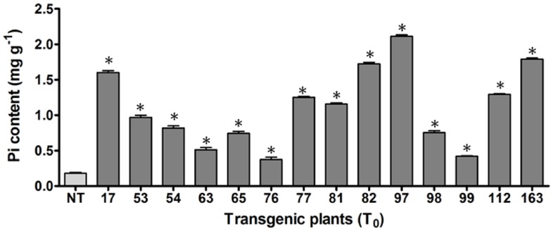
The transgenic plants generated were screened for the presence of the transgene cassette (Fig. 1) by PCR analysis using wheat RGA2 intron-specific primer pairs (RGA2F, 5′-CCTGAAATTGGT AAAAGTAGA-3′, and RGA2R, 5′-TGTATCTTCATACTGCATTTG-3′). The genomic DNA from 21-day-old plants showed amplification of the wheat RGA2 intron only in the transgenic-positive plants, whereas no amplification was observed in the non-transgenic control (non-transformed Pusa Sugandhi II rice cultivar) plants. In the T0 generation, forty-five individual putative transgenic rice plants were generated of which approximately thirty were positive for the corresponding transgene, as confirmed by genomic PCR analysis. Among the positive transgenic plants screened, fourteen plants (T0) showing higher Pi levels were selected, and the T1 generation was produced (Fig. 2). The transgenic plants (T1) exhibiting higher Pi levels (IO6–17, 82, 97, 112, and 163) were further selected, and subsequent generations (T2–T3) were grown under greenhouse conditions until maturity. After successive screening of the consequent generations (T1–T3), the progeny of IO6-97 (IO6-97-9-4) and IO6-163 (IO6-163-10-5) were selected. In the T3 generation, IO6-97-9-4 and IO6-163-10-5 showed a maximum Pi content (data not shown), thus all of the analysis were performed with the progeny of these two transgenic lines in the T4 generation.
Figure 1. Schematic diagram showing partial map of RNAi vector construct.

pOle18-IPK1-006 vector construct showing the IPK1 gene cloned in sense and antisense orientation separated by wheat RGA2 intron. HPT gene was used as the plant selection marker. (T = CaMV 35S terminator).
Figure 2. Screening of transgenic plants based on inorganic phosphate (Pi) content.
Pi fractions in non-transgenic (NT) and T0 transgenic rice plants were analyzed from the seeds. The symbol * indicates significant differences at P = 0.05 (n = 3).
Expression Analysis of the Transgenic Plants
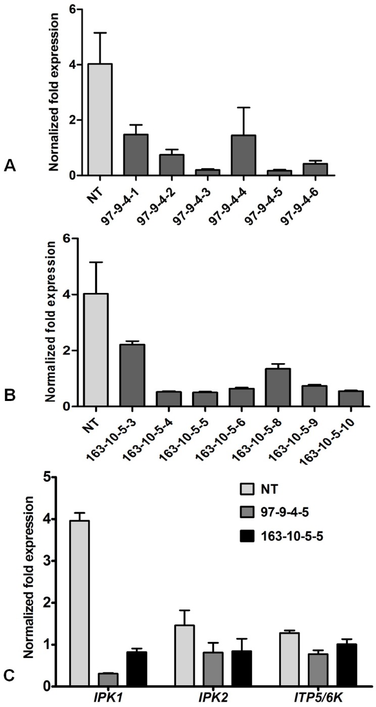
To quantify the level of down-regulation in the transgenic seeds (T4), a quantitative real-time PCR analysis was performed using SYBR Green (Fermentas). The β-tubulin gene was used as the reference gene to normalize all the data. The normalized fold reduction in the levels of IPK1 transcripts varied widely among the different progeny of IO6-97-9-4 (Fig. 3A) and IO6-163-10-5 (Fig. 3B). However, a maximum reduction of 3.85-fold was observed in the transgenic line IO6-97-9-4-5 (T4), revealing a distinct down-regulation of IPK1 and suggesting successful silencing mediated by the RNAi vector construct (pOle18-IPK1-006). Furthermore, the gene expression of other inositol phosphate kinase genes, i.e., IPK2 (inositol 1,4,5-tris-phosphate kinase/inositol polyphosphate kinase) and ITP5/6K (inositol 1,3,4-triskisphosphate 5/6-kinase) involved in phytate biosynthesis along with IPK1, was analyzed in the selected RNAi transgenic line IO6-97-9-4-5 and IO6-163-10-5-5 (Fig. 3C). The results clearly showed that, although the IPK1 gene displayed a maximum down-regulation, the expression of IPK2 and ITP5/6K was not significantly affected (P≥0.05).
Figure 3. Expression analysis of transgenic rice plants.
qRT-PCR analysis of T4 transgenic seeds of (A) IO6-97-9-4 and (B) IO6-163-10-5, as compared to the internal control β tubulin reveals down-regulation in the transcript level of IPK1. The normalized fold expression clearly indicates varied level of silencing, the maximum reduction being 3.85-fold as observed in 97-9-4-5. (C) Expression levels of IPK1, IPK2 and ITP5/6K genes in selected RNAi transgenic lines IO6-97-9-4-5 and IO6-163-10-5-5. (NT = Non-transgenic control).
Southern Blot Analysis of the Transgenic Plants
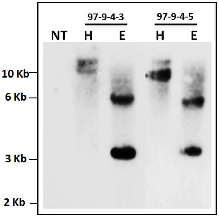
The integration of the transgene cassette in the genome of line IO6-97 was confirmed by Southern blot analysis using two different restriction enzymes: EcoRI and HindIII. The results revealed the stable integration of the transgene cassette into the progeny (T4). The Southern hybridization pattern showed two bands (in the case of both EcoRI and HindIII) corresponding to the transgene (RGA2 intron) in transgenic plants of the examined line. However, no hybridization signal was detected for the non-transgenic control plants (Fig. 4).
Figure 4. Southern blot analysis of T4 progenies of line IO6-97.
Stable integration of RGA2 intron was detected in transgenic rice plants, no hybridization signal was observed in the respective non-transgenic control. Each lane consists of 10 µg genomic DNA, digested with EcoRI or HindIII. The position and sizes of markers are indicated (NT = Non-transgenic control, E = EcoRI and H = HindIII).
Analysis of Seed Phosphorus and Phytic Acid Levels
The total phosphorus and inorganic Pi levels in the seeds of the non-transgenic rice and T4 seeds of transgenic lines IO6-97-9-4-5 and IO6-163-10-5-5 were estimated, and the average total phosphorus content of the T4 transgenic and non-transgenic seeds was found to be 3.971 mg g−1 (IO6-97-9-4-5), 3.907 mg g−1 (IO6-163-10-5-5) and 4.162 mg g−1, respectively. No significant difference was observed between the different transgenic sublines and the non-transgenic control seeds (P≥0.05). The Pi levels in the non-transgenic and T4 transgenic seeds were analyzed to further determine the storage form of phosphorus in the seeds. The average Pi concentration in the non-transgenic seeds constituted 4.33% of the seed total phosphorus. However, the average Pi content in the IO6-97-9-4-5 and IO6-163-10-5-5 seeds constituted 55.78% and 45.46% of the seed total phosphorus, which was significantly higher than that of the non-transgenic seeds (Fig. 5A). Although the seeds exhibited higher Pi levels, they displayed a normal phenotype, and no aberrations were observed.
Figure 5. Analysis of Phosphorus and phytic acid content in the transgenic rice seeds.
(A) Total phosphorus and Pi content in non-transgenic (NT) and T4 low phytate transgenic seeds and (B) amount of phytic acid in non-transgenic (NT) as compared to T4 transgenic seeds. The symbols * and *** indicates significant differences at P = 0.05 and 0.001 respectively (n = 3).
HPLC analysis was performed to quantify the phytic acid levels in the seed extracts of the transgenic and non-transgenic control, with the determination of phytic acid based on the replacement of phytic acid with the thiocyanate ligand from the iron (IIII)–thiocyanate complex. The chromatogram obtained from the HPLC/UV-vis method indicated that the transgenic seeds (showing larger peaks of iron (IIII)–thiocyanate complex) had lower phytate levels compared to the respective non-transgenic control, which exhibited a smaller peak, signifying a higher concentration of phytic acid in the seeds. The mean phytic acid values, as calculated from the corresponding peak area, were 10.28 mg g−1 for the non-transgenic seeds, 3.16 mg g−1 for the transgenic line IO6-97-9-4-5, and 5.23 mg g−1 for IO6-163-10-5-5 (Fig. 5B). These results represented an average reduction in the seed phytic acid content of 69% for line IO6-97-9-4-5 and approximately 50% for line IO6-163-10-5-5.
Seed myo-inositol Content
It has previously been established that phytic acid biosynthesis is closely related to myo-inositol synthesis [43], [21]. Hence, we also examined the effect of silencing the last step of phytate biosynthesis on the seed myo-inositol levels. The GC/MS analyses clearly showed that there was no significant difference between the myo-inositol content of the transgenic (T4) and non-transgenic control seeds (Fig. 6). Therefore, it can be considered that silencing the expression of IPK1 does not have any effect on the seed myo-inositol level, which is desirable because of the important role of myo-inositol in plant metabolism and other developmental processes.
Figure 6. Effect of IPK1 silencing on seed myo-inositol content.
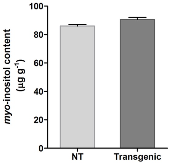
Myo-inositol content of T4 transgenic seeds (IO6-97-9-4-5) as compared to non-transgenic (NT) seeds showed no significant difference (P≥0.05).
Quantification of Metal Content in Seeds
Phytic acid is known to be a potent chelator of divalent cations and, therefore, renders these metal cations unavailable. Therefore, we examined the level of different metals (Fe, Zn, Ca, and Mg) in the milled seeds of low-phytate T4 transgenic seeds compared to that of the non-transgenic control using atomic absorption spectroscopy (AAS, Perkin Elmer). The amount of different metal cations was found to be higher in the low-phytate rice seeds when compared to their respective non-transgenic control (Table 1). Among the different metals analyzed, iron increased to a maximum of 12.61 µg g−1 in the transgenic seeds, whereas the level was 7.03 µg g−1 in the non-transgenic control seeds (Table 1). These results indicate a 1.8-fold increase in the levels of iron in milled low-phytate transgenic seeds.
Table 1. Metal content as analyzed by Atomic Absorption Spectroscopy from T4 milled seeds of greenhouse grown plants.
| Metals | Non-transgenic | Transgenic |
| Calcium (µg g−1) | 5.32±0.06 | 7.52±0.08 |
| Iron (µg g−1) | 7.03±0.07 | 12.61±0.22 |
| Zinc (µg g−1) | 22.30±0.37 | 26.62±0.29 |
| Magnesium (mg g−1) | 0.57±0.01 | 0.73±0.01 |
Values are mean ± SE, n = 3.
Amino Acid Analysis
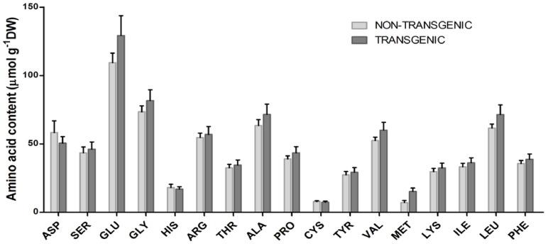
To assess the effect of silencing IPK1 on the different seed storage proteins of rice, we quantified the individual amino acids by HPLC analysis using the AccQ-tag method. The results showed no significant difference (P≥0.05) between the amount of individual amino acids analyzed from the seeds of the T4 transgenic (IO6-97-9-4-5 and IO6-163-10-5-5) and non-transgenic control plants (Fig. 7). Therefore, it can be suggested that the seed-specific suppression of IPK1 did not lead to any deleterious effects on other seed storage proteins.
Figure 7. Amino acid analysis in mature grains of non-transgenic and T4 transgenic plants.
Diagram representing the individual amino acid content of non-transgenic and the transgenic rice grains calculated with respect to the amino acid standard. The error bars indicate SE of three biological replicates for each sample. The data represented here for the transgenics is averaged from the observations of both IO6-97-9-4-5 and IO6-163-10-5-5.
Seed Germination Analysis
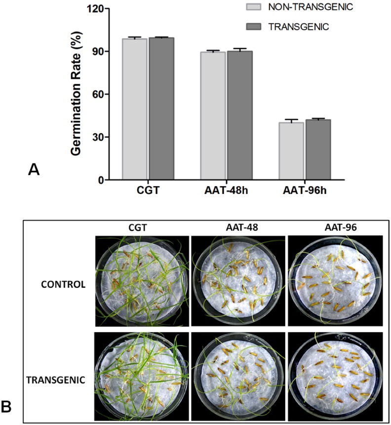
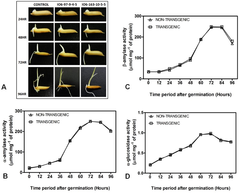
A reduction of phytate levels is often correlated to pleiotropic effects that frequently affect seed development and germination. However, no abnormality was observed at any stage of development or germination in the transgenic seeds with low phytate levels. We also examined the seed germination potential by performing both a control germination test and accelerated ageing test. The germination rate of both the transgenic (T4) and non-transgenic control seeds were recorded at regular intervals (Fig. 8A), and the morphological analysis of seed germination revealed similar phenotypes in CGT and AAT of both the transgenic and control seeds (Fig. 8B). In addition to this, the activities of the important starch-degrading enzymes involved in seed germination under optimum conditions were also analyzed (Fig. 9A). Both the transgenic and non-transgenic seeds exhibited similar activities of α-amylase (Fig. 9B), β-amylase (Fig. 9C), and α-glucosidase (Fig. 9D), giving a clear indication that the down-regulation of phytic acid did not interfere with seed germination in the transgenic plants.
Figure 8. Analysis of seed germination potential in non-transgenic and T4 transgenic low phytate seeds.
(A) Rate of germination as observed during control germination test (CGT) and accelerated ageing test (AAT) in both non-transgenic and the transgenic rice seeds. (B) Picture showing the morphology of transgenic seeds with respect to the non-transgenic control as recorded at 8th day of germination during the CGT and AAT.
Figure 9. Enzyme activity analysis during germination in T4 transgenic and non-transgenic control seeds.
(A) Picture showing the phenotype of the seeds during the course of germination at different time intervals. (B) α-amylase, (C) β-amylase and (D) α-glucosidase enzyme activity analyzed at different time intervals after germination in non-transgenic and the transgenic seeds showing no significant differences (P≥0.05). The open triangles represent response of non-transgenic (NT) and opened squares represent response of transgenics. (The data represented here for the transgenics is averaged from the observations of both IO6-97-9-4-5 and IO6-163-10-5-5).
Morphological Traits of Transgenic Plants
The agronomic performance of the T3 transgenic plants was compared with that of the non-transgenic plants (Table 2), and different morphological traits were considered and evaluated to analyze the phenotypic alterations in the transgenic plants. All the T3 and non-transgenic plants showed similar morphologies at the juvenile stage, and no significant difference was observed in the plant height, number of tillers, and panicles between mature T3 and non-transgenic plants (P≥0.05). In addition, the weight of 1000 dry seeds of the transgenic plants was not significantly different from that of the non-transgenic seeds (P≥0.05). All other morphological parameters considered were similar in both the T3 transgenic and non-transgenic plants.
Table 2. Different parameters considered for agronomic evaluation of T3 transgenic plants grown in greenhouse.
| Parameters | Non-transgenic control | IO6-97-9-4 | IO6-163-10-5 |
| Plant Height (cm) | 126.0±2.08 | 124.7±2.02 | 128.0±1.53 |
| No. of Tillers | 12.67±0.33 | 11.67±0.67 | 12.67±0.67 |
| No. of Effective Tillers | 11.33±1.20 | 11.00±0.58 | 11.67±0.88 |
| Panicle Length (cm) | 26.00±0.57 | 24.67±0.88 | 25.33±0.33 |
| Grains/Panicle | 73.33±1.76 | 74.67±0.88 | 72.33±2.19 |
| Seed length (mm) | 11.67±0.33 | 11.50±0.29 | 11.67±0.33 |
| Seed breadth (mm) | 2.61±0.01 | 2.60±0.01 | 2.62±0.01 |
| Seed length/breadth ratio | 4.46±0.14 | 4.40±0.10 | 4.46±0.14 |
| 1000 seeds dry wt (gm) | 26.67±0.88 | 25.67±1.20 | 26.00±0.58 |
Values are mean ± SE, n = 5.
Discussion
In the present investigation, we demonstrated the efficient down-regulation of phytic acid, as mediated by silencing of the IPK1 gene, the product of which catalyzes the last step of the phytate biosynthesis. The enzyme inositol 1,3,4,5,6-pentakisphosphate 2-kinase (IPK1) has been well characterized from different plants, including Arabidopsis [27] and maize [44]. Furthermore, in Arabidopsis, a T-DNA disruption of the gene for InsP5 2-kinase has been reported to have resulted in an 83% decrease in phytic acid levels [28]. A recent study identified the different enzymes involved in the phytic acid biosynthetic pathway, which included a IPK1 rice homolog (AK102842) that had 67% similarity to AtIPK1 [15]. Hence, we generated low-phytate transgenic rice by silencing the gene expression of IPK1 using an RNAi-mediated approach. It has been established that phytate globoids occur mainly in the aleurone layer of cereals [45], [46] and that the constitutive suppression of enzymes involved in phytate biosynthesis could be detrimental for plant growth and development [17]. Therefore, we used the Ole18 promoter [14], [18] which has specific activity in the aleurone layer and embryo, to induce the suppression of IPK1 in rice seeds. The transgenic plants showed the stable integration of the transgene cassette and displayed a normal phenotype. The quantitative RT-PCR analysis of different biosynthetic enzymes (IPK2, ITP5/6K, and IPK1) clearly showed that IPK1 gene expression was strongly affected in the transgenic rice seeds. The expression analysis of transgenic seeds further revealed a 3.85-fold reduction in the expression of IPK1 with respect to the non-transgenic control, suggesting efficient silencing of the gene (IPK1). In the T4 generation seeds of IO6-97, the Pi levels constituted 55.78% of the total seed phosphorus, which was 51% higher than that of the non-transgenic seeds, and the phytic acid content was reduced by approximately 69% with respect to the non-transgenic control. These results give a clear indication that the suppression of IPK1 actually led to a substantial reduction in phytic acid levels, with a concomitant increase in the amount of Pi.
Based on previous reports, low phytate is often associated with undesirable effects on seed development and germination potential [14], [47], [48]. Therefore, we analyzed the seed germination of the low-phytate transgenic seeds under optimum conditions during control germination tests [42]; the results indicated that the transgenic seeds are viable, showing normal germination patterns with respect to the non-transgenic seeds. It was also noteworthy that the germination potential decreased in a similar fashion, even during the accelerated ageing test in which the seeds of both the transgenic and non-transgenic control plants were subjected to an artificial ageing treatment. The analysis provided a clear idea that, although there is a substantial reduction in the phytate level, this did not interfere with either seed development or subsequent germination under both optimum and stressed conditions. To further verify that the germination process was not impaired, we measured the activity of different starch-degrading enzymes, i.e., α-amylase, β-amylase, and α-glucosidase, which are also known as indicators for assessing germination potential in cereals [49]. All these enzymes showed similar activities in the transgenics when compared to the non-transgenic seeds, indicating normal germination behavior.
The low-phytate transgenic seeds were also analyzed for the content of myo-inositol, which is considered to be an important metabolite involved in different biochemical pathways that are further associated with important metabolic processes in plants [50], [51]. In contrast to a previous report that cereal seeds with low phytate showed lower myo-inositol levels [21], the T4 transgenic seeds of IO6-97 (IO6-97-9-4-5) exhibited a similar myo-inositol content as the non-transgenic seeds. Because IPK1 catalyzes the final step of phytic acid biosynthesis [15], it is quite obvious that the silencing of its gene would not affect myo-inositol synthesis, which occurs much earlier in the pathway. Therefore, this finding suggests an added advantage for generating low-phytate crops by manipulating IPK1, without disturbing the myo-inositol levels. In addition to seed myo-inositol, which is an essential metabolite playing significant role in different signaling pathways, plant growth and development [51], [52], we also evaluated the individual amino acid content of the transgenic seeds to confirm that the seed-specific manipulation of IPK1 did not interfere with any of the storage proteins, as cereal seeds are an important source of different seed storage proteins. The results clearly indicated that the amino acid profiles of the transgenic plants were similar to those of the non-transgenic control plants, with no significant differences noted.
It is known that, due to the presence of six negatively charged ions, phytic acid chelates available divalent mineral cations, which renders these minerals less bioavailable [1], [53]. Prior reports have suggested that phytic acid chelates mineral cations and aggregates them as inclusions in protein storage vacuoles (phytate globoids), mainly in the aleurone layer and embryo [4], [54]. Because both the aleurone layer and embryo are removed during commercial milling, a reduced amount of these metals are available in the endosperm, which is generally consumed [55], [56]. In view of these facts, we analyzed the metal concentrations (Ca, Fe, Zn, and Mg) of the low-phytate transgenic seeds (milled) and found a significant increase in metal concentrations in the transgenic compared to the non-transgenic seeds. It was also noteworthy that, among the different metals analyzed, a maximum increase was observed in the levels of iron (Fe), at 1.8-fold more than the non-transgenic control. Although previous reports have suggested an increase in iron content using a dual approach of expressing soybean ferritin [57] and Aspergillus phytase [5], [8] in cereals, the present investigation highlights that lowering the phytate level alone can lead to elevated iron levels in milled rice seeds.
The strategy undertaken in this research represents a step ahead in developing low-phytate rice, without disturbing the normal behavior of rice plants. The present study confirms that the manipulation of IPK1 in rice can lead to major reductions in the amount of seed phytate, with a simultaneous increase in the Pi content. Therefore, it can be suggested that the gene encoding IPK1, which catalyzes the last step of phytic acid biosynthesis, is an appropriate candidate to target for reducing phytate levels, without hampering other important metabolic processes and developmental pathways in cereal crops.
Acknowledgments
The authors are grateful to Leibniz Institute of Plant Genetics and Crop Plant Research, Gatersleben, Germany, for providing the RNAi vector pIPKb006. In addition, we are also thankful to Ms. Sayani Majumdar for laboratory assistance and Mr. Pratap Ghosh for field work.
Funding Statement
The financial support from the Department of Biotechnology (DBT),Government of India in the form of DBT Programme Support [Sanction no. - BT/COE/01/06/05] and National fund for basic and strategic research in agricultural science (NFBSFARA) of Indian Council of Agricultural Research (ICAR) [Sanction no. – NFBSFARA/RNAi-2011/2010-2011] are thankfully acknowledged. The funders had no role in study design, data collection and analysis, decision to publish, or preparation of the manuscript.
References
- 1. Raboy V, Gerbasi PF, Young KA, Stoneberg SD, Pickett SG, et al. (2000) Origin and seed phenotype of maize low phytic acid 1–1 and low phytic acid 2–1. Plant Physiol 124: 355–368. [DOI] [PMC free article] [PubMed] [Google Scholar]
- 2. Holm PB, Kristiansen KN, Pedersen HB (2002) Transgenic approaches in commonly consumed cereals to improve iron and zinc content and bioavailability. J Nutr 132: 514–516. [DOI] [PubMed] [Google Scholar]
- 3. Kuwano M, Ohyama A, Tanaka Y, Mimura T, Takaiwa F, et al. (2006) Molecular breeding for transgenic rice with low phytic acid phenotype through manipulating myo inositol 3 phosphate synthase gene. Mol Breeding 18: 263–272. [Google Scholar]
- 4. Raboy V (2009) Approaches and challenges to engineering seed phytate and total phosphorus. Plant Sci 177: 281–296. [Google Scholar]
- 5. Lucca P, Hurrell R, Potrykus I (2001) Genetic engineering approaches to improve the bioavailability and the level of iron in rice grains. Theor Appl Genet 102: 392–397. [Google Scholar]
- 6. Brinch-Pedersen H, Hatzack F, Sorensen LD, Holm PB (2003) Concerted action of endogenous and heterologous phytase on phytic acid degradation in seed of transgenic wheat (Triticum aestivum L.). Transgenic Res 12: 649–659. [DOI] [PubMed] [Google Scholar]
- 7. Chiera JM, Finer JJ, Grabau EA (2004) Ectopic expression of soybean phytase in developing seeds of Glycine max to improve phosphorus availability. Plant Mol Biol 56: 895–904. [DOI] [PubMed] [Google Scholar]
- 8. Drakakaki G, Marcel S, Glahn RP, Lund EK, Pariagh S, et al. (2005) Endosperm-specific co-expression of recombinant soybean ferritin and Aspergillus phytase in maize results in significant increases in the levels of bioavailable iron. Plant Mol Biol 59: 869–880. [DOI] [PubMed] [Google Scholar]
- 9. Larson SR, Rutger JN, Young KA, Raboy V (2000) Isolation and genetic mapping of a non-lethal rice (Oryza sativa L.) low phytic acid 1 mutation. Crop Sci 40: 1397–405. [Google Scholar]
- 10. Kim SI, Andaya CB, Newman JW, Goyal SS, Tai TH (2008) Isolation and characterization of a low phytic acid rice mutant reveals a mutation in the rice orthologue of maize MIK. Theor Appl Genet 117: 1291–1301. [DOI] [PubMed] [Google Scholar]
- 11. Guttieri M, Bowen D, Dorsch JA, Raboy V, Souza E (2004) Identification and characterization of a low phytic acid wheat. Crop Sci 44: 418–424. [Google Scholar]
- 12. Pilu R, Panzeri D, Gavazzi G, Rasmussen SK, Consonni G, et al. (2003) Phenotypic, genetic and molecular characterization of a maize low phytic acid mutant (Lpa 241). Theor Appl Genet 107: 980–987. [DOI] [PubMed] [Google Scholar]
- 13. Shi J, Wang H, Hazebroek J, Ertl DS, Harp T (2005) The maize low-phytic acid 3 encodes a myo-inositol kinase that plays a role in phytic acid biosynthesis in developing seeds. Plant J 42: 408–419. [DOI] [PubMed] [Google Scholar]
- 14. Kuwano M, Mimura T, Takaiwa F, Yoshida KT (2009) Generation of stable ‘low phytic acid’ transgenic rice through antisense repression of the 1 D -myo-inositol 3–phosphate synthase gene using the 18-kDa oleosin promoter. Plant Biotechnol J 7: 96–105. [DOI] [PubMed] [Google Scholar]
- 15. Suzuki M, Tanaka K, Kuwano M, Yoshida KT (2007) Expression pattern of inositol phosphate-related enzymes in rice (Oryza sativa L.): Implications for the phytic acid biosynthetic pathway. Gene 405: 55–64. [DOI] [PubMed] [Google Scholar]
- 16. Yoshida KT, Wada T, Koyama H, Mizobuchi-Fukuoka R, Naito S (1999) Temporal and spatial patterns of accumulation of transcript of myo-inositol- 1-phosphate synthase and phytin containing particles during seed development in rice. Plant Physiol 119: 65–72. [DOI] [PMC free article] [PubMed] [Google Scholar]
- 17. Feng X, Yoshida KT (2004) Molecular approaches for producing low-phytic –acid grains in rice. Plant Biotechnol 21: 183–189. [Google Scholar]
- 18. Qu L, Takaiwa F (2004) Evaluation of tissue specificity and expression strength of rice seed component gene promoters in transgenic rice. Plant Biotechnol J 2: 113–125. [DOI] [PubMed] [Google Scholar]
- 19. Keller R, Brearley CA, Trethewey RN, Muller-Rober B (1998) Reduced inositol content and altered morphology in transgenic potato plants inhibited for 1D-myo-inositol 3-phosphate synthase. Plant J 16: 403–410. [Google Scholar]
- 20. Majumder AL, Chatterjee A, GhoshDastidar K, Majee M (2003) Diversification and evolution of L-myo-inositol 1-phosphate synthase. FEBS Lett 553: 3–10. [DOI] [PubMed] [Google Scholar]
- 21. Panzeri D, Cassani E, Doria E, Tagliabue G, Forti L, et al. (2011) A defective ABC transporter of the MRP family, responsible for the bean lpa1 mutation, affects the regulation of the phytic acid pathway, reduces seed myo-inositol and alters ABA sensitivity. New Phytol 191: 70–83. [DOI] [PubMed] [Google Scholar]
- 22. Stephens LR, Irvine RF (1990) Stepwise phosphorylation of myo-inositol leading to myo-inositol hexakisphosphate in Dictyostelium . Nature 346: 580–583. [DOI] [PubMed] [Google Scholar]
- 23. Brearley CA, Hanke DE (1996) Metabolic evidence for the order of addition of individual phosphate esters to the myo-inositol moiety of inositol hexakisphosphate in the duckweed Spirodelapolyrhiza L. Biochem J. 314: 227–233. [DOI] [PMC free article] [PubMed] [Google Scholar]
- 24. York JD, Odom AR, Murphy R, Ives EB, Wente SR (1999) A phospholipase c-dependent inositol polyphosphate kinase pathway required for efficient messenger RNA export. Science 285: 96–100. [DOI] [PubMed] [Google Scholar]
- 25. Wilson MP, Majerus PW (1996) Isolation of inositol 1,3,4-trisphosphate 5/6-kinase, cDNA cloning, and expression of the recombinant enzyme. J Biol Chem 271: 11904–11910. [DOI] [PubMed] [Google Scholar]
- 26. Stevenson-Paulik J, Odom AR, York JD (2002) Molecular and biochemical characterization of two plant inositol polyphosphate 6-/3-/5-kinases. J Biol Chem 277: 42711–42718. [DOI] [PubMed] [Google Scholar]
- 27. Sweetman D, Johnson S, Caddick SE, Hanke DE, Brearley CA (2006) Characterization of an Arabidopsis inositol 1,3,4,5,6-pentakisphosphate 2-kinase (AtIPK1). Biochem J 394: 95–103. [DOI] [PMC free article] [PubMed] [Google Scholar]
- 28. Stevenson-Paulik J, Bastidas RJ, Chiou S-T, Frye RA, York JD (2005) Generation of phytate-free seeds in Arabidopsis through disruption of inositol polyphosphate kinases. Proc Nat Acad Sci USA 102: 12612–12617. [DOI] [PMC free article] [PubMed] [Google Scholar]
- 29. Himmelbach A, Zierold U, Hensel G, Riechen J, Douchkov D, et al. (2007) A set of modular binary vectors for transformation of cereals. Plant Physiol 145: 1192–1200. [DOI] [PMC free article] [PubMed] [Google Scholar]
- 30. Datta K, Vasquez A, Tu J, Torrizo L, Alam MF, et al. (1998) Constitutive and tissue specific differential expression of cryIA(b) gene in transgenic rice plants conferring resistance to rice insect pest. Theor Appl Genet 97: 20–30. [Google Scholar]
- 31.Sambrook J, Russell DW (2001) Molecular Cloning, A laboratory manual. 3rd ed. Vol. (1,2,3). Cold Spring Harbor, NY: Cold Spring Harbor Laboratory Press.
- 32. Meng L, Feldman L (2010) A rapid TRIzol-based two-step method for DNA-free RNA extraction from Arabidopsis siliques and dry seeds. Biotechnol J 5: 183–186. [DOI] [PubMed] [Google Scholar]
- 33. Woo L, Maher W (1995) Determination of phosphorus in turbid waters using alkaline potassium peroxodisulfate digestion. Anal Chim Acta 315: 123–135. [Google Scholar]
- 34. Chen PS, Toribara TY, Warner H (1956) Microdetermination of phosphorus. Anal Chem 28: 1756–1758. [Google Scholar]
- 35. Dost K, Tokul O (2006) Determination of phytic acid in wheat and wheat products by reverse phase high performance liquid chromatography. Anal Chim Acta 558: 22–27. [Google Scholar]
- 36. Jiang SL, Wu JG, Feng Y, Yang XE, Shi CH (2007) Correlation analysis of mineral element contents and quality traits in milled rice (Oryza sativa L.). J Agr Food Chem 55: 9608–9613. [DOI] [PubMed] [Google Scholar]
- 37.Bernfeld P (1955) Amylases α and β. In: Colowick SP, Kalpan NO, editors. Methods in Enzymology. New York: Academic Press. 149–158.
- 38. Bialecka B, Kępczynski J (2010) Germination, α-, β-amylase and total dehydrogenase activities of Amaranthus caudatus seeds under water stress in the presence of ethepon or gibberellin A3. Acta Biol Cracov Bot 52: 7–12. [Google Scholar]
- 39. Usansa U, Burberg F, Geiger E, Back W, Wanapu C, et al. (2011) Optimization of Malting Conditions for Two Black Rice Varieties, Black Non-Waxy Rice and Black Waxy Rice (Oryza sativa L. Indica). JI Brewing 117: 39–46. [Google Scholar]
- 40. Miller GL (1959) Use of dinitrosalicylic acid reagent for determination of reducing sugar. Anal Chem 31: 426–428. [Google Scholar]
- 41. Iwata H, Suzuki T, Aramaki I (2003) Purification and characterization of rice α-glucosidase, a key enzyme for alcohol fermentation of rice polish. J Biosci Bioeng 95: 106–108. [DOI] [PubMed] [Google Scholar]
- 42. Campion B, Sparvoli F, Doria E, Tagliabue G, Galasso I, et al. (2009) Isolation and characterization of an lpa (low phytic acid) mutant in common bean (Phaseolus vulgaris L.). Theor Appl Genet 118: 1211–1221. [DOI] [PubMed] [Google Scholar]
- 43. Hegeman CE, Good LL, Grabau EA (2001) Expression of D-myo-inositol-3-phosphate synthase in soybean. Implications for phytic acid biosynthesis. Plant Physiol 125: 1941–1948. [DOI] [PMC free article] [PubMed] [Google Scholar]
- 44. Sun Y, Thompson M, Lin G, Butler H, Gao Z, et al. (2007) Inositol 1,3,4,5,6-pentakisphosphate 2-kinase from maize (Zea mays L.): molecular and biochemical characterization. Plant Physiol 144: 1278–1291. [DOI] [PMC free article] [PubMed] [Google Scholar]
- 45. Bohn L, Meyer AS, Rasmussen SK (2008) Phytate: impact on environment and human nutrition a challenge for molecular breeding. J Zhejiang Univ-Sc-B 9: 165–191. [DOI] [PMC free article] [PubMed] [Google Scholar]
- 46. Regvar M, Eichert D, Kaulich B, Gianoncelli A, Pongrac P, et al. (2011) New insights into globoids of protein storage vacuoles in wheat aleurone using synchrotron soft X-ray microscopy. J Exp Bot 62: 3929–3939. [DOI] [PMC free article] [PubMed] [Google Scholar]
- 47. Nunes ACS, Vianna GR, Cuneo F, Amaya-Farfan J, de Capdeville G, et al. (2006) RNAi mediated silencing of the myo-inositol-1-phosphate synthase gene (GmMIPS1) in transgenic soybean inhibited seed development and reduced phytate content. Planta 224: 125–132. [DOI] [PubMed] [Google Scholar]
- 48. Doria L, Galleschi L, Calucci L, Pinzino C, Pilu R, et al. (2009) Phytic acid prevents oxidative stress in seeds: evidence from a maize (Zea mays L.) low phytic acid mutant. J Exp Bot 60: 967–978. [DOI] [PubMed] [Google Scholar]
- 49. Galani S, Aman A, Qader SAU (2011) Germination potential index of Sindh rice cultivars on biochemical basis, using amylase as an indicator. Afr J Biotechnol 10: 18334–18338. [Google Scholar]
- 50.Majumder AL, Biswas BB (2006) Biology of Inositols and Phosphoinositides. AK Houten, Netherlands: Springer.
- 51. Torabinejad J, Donahue JL, Gunesekera BN, Allen-Daniels MJ, Gillaspy GE (2009) VTC4 is a bifunctional enzyme that affects myo inositol and ascorbate biosynthesis in plants. Plant Physiol 150: 951–961. [DOI] [PMC free article] [PubMed] [Google Scholar]
- 52. Abid G, Silue S, Muhovski Y, Jacquemin JM, Toussaint A, et al. (2009) Role of myo-inositol phosphate synthase and sucrose synthase genes in plant seed development. Gene 439: 1–10. [DOI] [PubMed] [Google Scholar]
- 53. Kumar V, Sinha AK, Makkar HPS, Becker K (2010) Dietary roles of phytate and phytase in human nutrition: A review. Food Chem 120: 945–959. [Google Scholar]
- 54. Brinch-Pedersen H, Borg S, Tauris B, Holm PB (2007) Molecular genetic approaches to increasing mineral availability and vitamin content of cereals. J Cereal Sci 46: 308–326. [Google Scholar]
- 55. Vasconcelos M, Datta K, Oliva N, Khalekuzzaman M, Torrizo L, et al. (2003) Enhanced iron and zinc accumulation in transgenic rice with the ferritin gene. Plant Sci 164: 371–378. [Google Scholar]
- 56. Bajaj S, Mohanthy A (2005) Recent advances in rice biotechnology-towards genetically superior transgenic rice. Plant Biotechnol J 3: 275–307. [DOI] [PubMed] [Google Scholar]
- 57. Goto F, Yoshihara T, Shigemoto N, Toki S, Takaiwa F (1999) Iron fortification of rice seed by the soybean ferritin gene. Nat Biotechnol 17: 282–286. [DOI] [PubMed] [Google Scholar]


