Abstract
The lysosomal-autophagic pathway is activated by starvation and plays an important role in both cellular clearance and lipid catabolism. However, the transcriptional regulation of this pathway in response to metabolic cues is currently uncharacterized. Here we show that the transcription factor EB (TFEB), a master regulator of lysosomal biogenesis and autophagy, is induced by starvation through an autoregulatory feedback loop and exerts a global transcriptional control on lipid catabolism via PGC1α and PPARα. Thus, during starvation a transcriptional mechanism links the autophagic pathway to cellular energy metabolism. The conservation of this mechanism in Caenorhabditis elegans suggests a fundamental role for TFEB in the evolution of the adaptive response to food deprivation. Viral delivery of TFEB to the liver prevented weight gain and metabolic syndrome in both diet-induced and genetic mouse models of obesity, suggesting a novel therapeutic strategy for disorders of lipid metabolism.
The adaptive response of an organism to food deprivation is associated with major transcriptional and metabolic1–5 changes and is conserved across evolution6,7. One of the most prominent metabolic changes observed during starvation is an increase in lipid catabolism in the liver.
Autophagy, a lysosome-dependent catabolic process, is activated by starvation8, and the resulting breakdown products are used to generate new cellular components and energy. Recent studies revealed that autophagy plays a central role in lipid metabolism since it shuttles lipid droplets to the lysosome where they are hydrolyzed into free fatty acids (FFAs) and glycerol9,10. Moreover, excessive lipid overload may inhibit autophagy, while enhancing liver autophagy in murine genetic models of obesity (Ob/Ob) ameliorates their metabolic phenotype11. These observations indicate the close relationship between intracellular lipid metabolism and the lysosomal-autophagic pathway. However, it is not clear how this relationship is coordinated at the transcriptional level in response to environmental cues. Here we show that the basic Helix-Loop-Helix (bHLH) leucine zipper transcription factor TFEB, a master regulator of lysosomal biogenesis and autophagy12,13 mediates the organismal transcriptional and metabolic responses to starvation.
Starvation induces TFEB expression through an autoregulatory loop
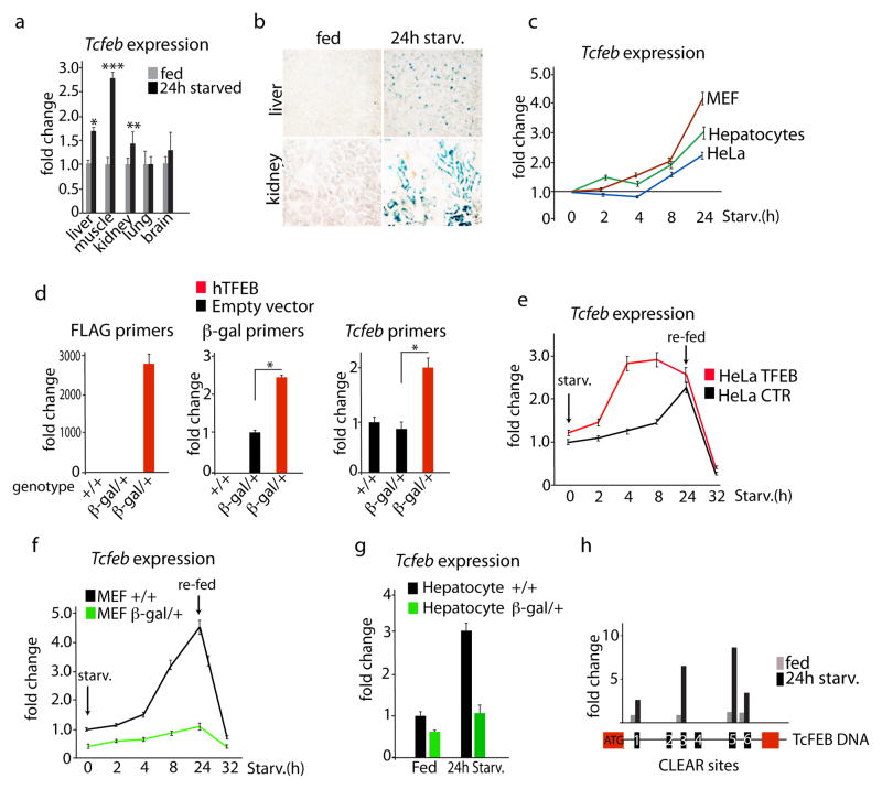
We tested whether starvation activated TFEB transcription. TFEB expression was significantly induced in liver, muscle, and kidney of mice subjected to 24 hours of food deprivation (Fig. 1a). Similar results were observed in vivo using a heterozygous transgenic mouse line (Tcfeb-β-gal) that carries a TFEB gene-trap allele fused with the β-galactosidase gene. In this model we found stronger β-galactosidase staining in the liver and kidney of fasted mice compared to fed mice (Fig. 1b). Starvation time-course studies of HeLa cells, mouse embryonic fibroblasts (MEFs) and hepatocytes revealed a significant and progressive increase of TFEB mRNA and protein expression levels starting as soon as 4 hours after the elimination of nutrients from the culture medium (Fig. 1c and Supplementary Fig. 1a). Notably, the cytoplasm-to-nucleus translocation of TFEB13 occurs at an earlier time point after starvation compared to TFEB transcriptional activation (Supplementary Fig. 1b). These observations led us to hypothesize that TFEB exerted a positive effect on its own transcription.
Fig. 1. Autoregulation of TFEB during starvation.
a) Expression levels of Tcfeb mRNA in tissues isolated from 24h-fasted (starv) 6 week old mice. Values are expressed as fold change relative to Tcfeb expression in mice fed ad libitum (fed). Bars represent mean±s.d. for n=5; *P≤0.05; **P≤0.01; ***P≤0.001. b) Representative β-gal staining of liver and kidney frozen sections isolated from fed and 24h–fasted heterozygous Tcfeb-β-gal mice. c) Time-course expression analysis of TFEB in wild-type HeLa cells, MEFs and hepatocytes after addition of starvation media (time 0). Bars represent mean±s.d. for n=3 independent experiments. d) Expression levels of transfected hTFEB-Flag, Tcfeb-β-gal fusion transcript and endogenous Tcfeb mRNAs in MEFs isolated from control (+/+) and heterozygous Tcfeb-β-gal/+ mice. Bars represent mean±s.d. for n=3 e) Time-course expression analysis of TFEB mRNA in control or TFEB-overexpressing HeLa cells during fasting and re-feeding. Primers specific for the endogenous TFEB were used. Bars represent mean±s.d. for n=3 f) Time-course expression analysis of Tcfeb mRNA in heterozygous Tcfeb-β-gal/+ or control MEFs during fasting and re-feeding. Specific primers for the endogenous Tcfeb were used. Bars represent mean±s.d. for n=3 g) Expression analysis of Tcfeb mRNA in heterozygous Tcfeb-β-gal/+ or control hepatocytes in fed and after 24h fasting. Bars represent mean±s.d. for n=3 h) Chromatin immunoprecipitation (ChIP) analysis from liver of mice fed ad libitum or 24h-fasted. The CLEAR elements in the first intron of Tcfeb genomic DNA are shown as numbered black boxes as indicated in Supplementary Table 1. Red boxes represent exons, and the ATG indicates the first codon (from the mouse Tcfeb isoform b). The histogram shows the amount of immunoprecipitated DNA as detected by qPCR assay. Values were normalized to the input and plotted as relative enrichment over a mock control. Data represent mean ± s.d of three independent experiments.
The overexpression of the human TFEB cDNA in MEFs from heterozygous Tcfeb-β-gal transgenic mice resulted in a significant increase in the transcription of the Tcfeb–β-galactosidase fusion transcript, indicating that the endogenous TFEB gene is positively regulated by the exogenous TFEB (Fig. 1d). This result was also confirmed using a set of primers that specifically amplifies the murine endogenous TFEB transcript, but does not amplify the exogenous human TFEB cDNA (Fig. 1d). These results indicate that exogenous TFEB can induce endogenous Tcfeb expression and suggest the presence of a positive feedback loop.
The positive feedback of TFEB on its own expression was significantly enhanced during starvation and was suppressed by re-adding nutrients to the culture medium (Fig. 1e). In addition, the effect of starvation on TFEB expression was significantly reduced in MEFs and hepatocytes from heterozygous mice carrying a Tcfeb-β-gal allele that lacks the transcriptional transactivation domain, indicating that the positive feedback loop requires a functional TFEB allele and that TFEB induction by starvation is sensitive to TFEB copy number (Fig. 1f,g).
Next, we tested whether TFEB mediates the positive feedback loop on its own expression by directly binding to its promoter. We previously showed that TFEB recognizes E-box-type DNA sequences, named “CLEAR” motifs14. Sequence analysis identified 6 putative CLEAR sites in the promoter region of the TFEB gene (Fig. 1h and Supplementary Table 1). Chromatin immunoprecipitation coupled with quantitative PCR (ChIP-qPCR) of liver samples from starved transgenic mice overexpressing a tagged version of TFEB13 (Supplementary Fig. 2a), showed an enhancement of TFEB binding to CLEAR sites 1, 3,5 and 6 compared to fed controls (Fig. 1h). Thus, in vitro and in vivo data showed that TFEB controls its own expression by virtue of starvation-induced direct binding to CLEAR elements in the TFEB promoter.
TFEB regulates genes involved in lipid metabolism via PGC1α and PPARα
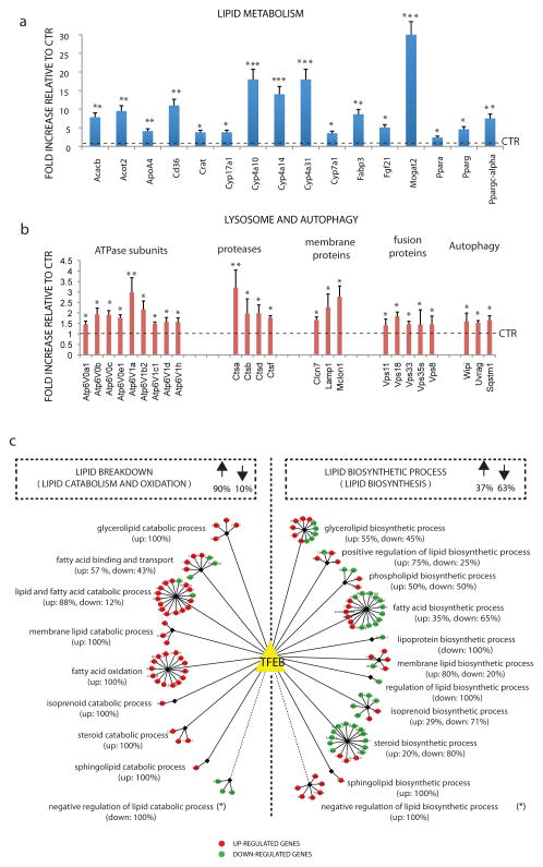
To test whether TFEB is in the metabolic response to starvation we sought to define the complete TFEB-dependent transcriptome in the liver, a primary site for the organismal starvation response. To this end, we injected mice with an adenoviral vector that expresses human TFEB (HDAd-TFEB) under control of a liver-specific promoter (PEPCK) and with a transgeneless control vector. The levels of expression of TFEB protein under these conditions are shown in Supplementary Fig. 2b. Microarray analysis indicated that as a result of TFEB overexpression 773 genes were upregulated and 611 genes were downregulated (GSE35015), using a threshold for statistical significance (FDR<0.05) and further filtering with an absolute Fold Change ≥1.5. Microarray results were validated by quantitative realtime PCR (qRT-PCR) performed on 40 selected genes (Fig. 2a,b). Surprisingly, the gene ontology category15,16 most significantly up-regulated by TFEB overexpression was the cellular lipid metabolic process, which includes monocarboxylic acid, fatty acid, and cellular ketone metabolic processes, among others (Supplementary Table 2). Interestingly, several gene categories related to lipid biosynthesis, such as steroid, lipid and isoprenoid biosynthetic processes were significantly down-regulated (Supplementary Table 3). We also observed that in liver TFEB positively regulates the expression of several genes involved in lysosome organization and autophagy (Supplementary Table 2, Fig. 2b), which is consistent with previous results obtained in cultured cell lines12–14.
Fig. 2. The TFEB lipid metabolism network.
a, b) mRNA levels of the indicated genes involved in (a) lipid metabolism and in (b) autophagy and lysosome pathway were quantified by qRT–PCR in total RNA isolated from liver samples of mice infected with HDAd-TFEB virus. GAPDH was used as a control. Values are mean± s.d for n=3 and are expressed as fold increase compared to control mice (injected with transgeneless viral vector). * p<0.05; **<0.01. Control levels are indicated by dashed line. c) The 124 genes with a known role in the lipid metabolic process, whose expression was perturbed by TFEB overexpression, are represented as coloured circles and assigned to specific lipid breakdown (left) or lipid biosynthesis (right) sub-categories. The percentages of up-regulated (red circles) and down-regulated genes (green circles) are shown both for the two main groups and for each sub-category. *Note that in calculating these percentages genes assigned to the “negative” regulation of lipid catabolic process and to the “negative” regulation of lipid biosynthetic process have been included in the lipid breakdown and in the lipid biosynthesis groups, respectively.
Overall we found that the transcriptional signature of TFEB overexpression in liver was similar to that of starvation (GSE36510; Supplementary Table 4), particularly for genes involved in lipid metabolism (Supplementary Fig. 3 and Supplementary Tables 5 and 6). The global control of lipid metabolism exerted by TFEB is illustrated in Fig. 2c and Supplementary Table 7. These results strongly suggested that TFEB overexpression in fed mice phenocopied the transcriptional effects of nutrient deprivation in vivo, supporting the notion that TFEB is a critical regulator of the response to starvation in the liver.
Interestingly, one of the genes whose expression was significantly upregulated following TFEB overexpression was the peroxisome proliferator-activated receptor γ coactivator 1 α (PGC1α), a known key regulator of liver lipid metabolism that is transcriptionally induced during starvation17,18. To test whether PGC1α is a direct target of TFEB we analysed its promoter and identified three CLEAR sites. ChIP-qPCR from liver extracts showed that TFEB binds to two of these sites in a starvation-dependent manner (Fig. 3a). Furthermore, transactivation of a PGC1α promoter luciferase reporter by TFEB was dependent on the CLEAR sites (Fig. 3b,c) and was enhanced by starvation (Fig. 3d). These results indicated that TFEB directly regulates PGC1α gene expression.
Fig. 3. TFEB directly regulates PGC1α expression during starvation.
a) Chromatin immunoprecipitation (ChIP) analysis from liver of mice fed ad libitum (fed) or 24h-fasted (starved). CLEAR sites in the promoter region of PGC1α are indicated by boxes. Numbers indicate the distance (bp) of the binding element from the start codon. Bar graphs show the amount of immunoprecipitated DNA as detected by qPCR assay. Values were normalized to the input and plotted as relative enrichment over a mock control. Bar graphs represent mean ± s.d of 3 independent experiments *P≤0.05; **P≤0.01. b) Representative diagrams of the constructs containing the promoter region of PGC1α with either intact (PGC1α WT) or deleted (PGC1α DEL) CLEAR elements upstream of the luciferase cDNA. c) Luciferase activity was measured after transfecting increasing amounts of TFEB-Flag in combination with PGC1α WT or PGC1α DEL plasmids. Bar graphs represent mean ± s.d. of n=3 independent experiments *P≤0.05; **P≤0.01; ****P≤0.0001 compared to mock transfected cells.d) Luciferase activity was measured in cells stably overexpressing TFEB cultured in normal and starved media. Bar graphs represent mean ± s.d. of n=3 independent experiments *P≤0.05; **P≤0.01; ****P≤0.0001 compared to mock transfected cells. e) Quantification of mRNA levels of PGC1α in liver and hepatocytes from control, HDAd-TFEB and Tcfeb-LIKO mice treated as indicated. Bar graphs show mean ±s.d. for n=4. *P≤0.05; **P≤0.01; ***P≤0.001. f) Expression analysis of TFEB target genes in liver of mice with indicated genotypes. Bar graphs show mean ±s.d. for n=3. *P≤0.05; **P≤0.01; ***P≤0.001. g) Expression analysis of PGC1α/PPARα target genes in liver from either fasted or fed mice with indicated genotypes. Bar graphs show mean ±s.d. for n=4. *P≤0.05; **P≤0.01; ***P≤0.001 compared to the respective controls (fed or fasted).
These data were confirmed in vivo by measuring and comparing PGC1α expression in mice that overexpress TFEB in the liver and in mice that lack TFEB in the liver (Tcfeb-LiKO) with corresponding control mice. Injection of HDAd-TFEB in the liver of fed animals was sufficient to drive high constitutive PGC1α expression to levels similar as those observed in uninjected starved animals. In contrast, PGC1α induction by starvation was partially blocked by the deletion of TFEB. Furthermore, primary hepatocytes from animals overexpressing TFEB showed high constitutive expression of PGC1α, while TFEB deletion did not affect basal PGC1α expression. In contrast, PGC1α induction was partially blunted in starved hepatocytes lacking TFEB, indicating that starvation induces PGC1α in a TFEB-dependent manner. In starved hepatocytes overexpression of TFEB caused synergistic induction of PGC1α expression beyond the levels obtained by starvation only (Fig. 3e). Together, these results strongly suggested that TFEB directly controls PGC1α induction during liver starvation response, and that the level of TFEB expression is a critical parameter for the magnitude of this response.
To study the role of PGC1α as a mediator of TFEB function, we overexpressed TFEB in a mouse line that lacks PGC1α in the liver, which was generated by injecting PGC1α flox/flox mice with a helper-dependent adenovirus containing the CRE recombinase (HdAD-APOA1-CRE). Fig 3f shows that TFEB-mediated induction of the expression of genes involved in lipid metabolism was severely reduced in the absence of PGC1α, confirming that PGC1α acts downstream of TFEB and mediates TFEB function.
During starvation PGC1α regulates lipid metabolism in the liver via the downstream nuclear receptor peroxisome proliferator activated receptor α (PPARα)17,18, suggesting that PGC1α may mediate TFEB function by controlling the activity of PPARα. To test this hypothesis, we analyzed transcript levels for known targets of PPARα17–20 in Tcfeb-LiKO mice, compared to wild type mice. Starvation caused induction of the PPARα target genes in wild type mice, but not in Tcfeb-LiKO mice (Fig. 3g and Supplementary Fig. 4a), indicating that TFEB is essential for PPARα activation by starvation. Furthermore, the majority (74%) of the genes that were induced by TFEB overexpression in the liver of wild type mice failed to show a transcriptional induction in mice lacking PPARα as measured by microarray analysis of liver tissue (GSE41141; Supplementary Table 8), suggesting that PPARα is an important mediator of TFEB transcriptional activity in the liver during starvation.
TFEB regulates lipid breakdown in the liver
Histological analysis of liver samples did not reveal any significant differences between wild type and Tcfeb-LiKO mice fed with a normal diet. However, after a 24 h fast we observed an accumulation of lipid droplets in Tcfeb-LiKO mice that was not found in wild-type littermates, suggesting a defect in intracellular lipid degradation (Fig. 4a–c). Consistently, we detected an impairment of free fatty acid (FFA) oxidation in cultured hepatocytes (Fig. 4d) and higher levels of circulating FFA (Fig. 4e) and glycerol (Fig. 4f) in Tcfeb-LiKO mice compared to controls. In addition, during starvation Tcfeb-LiKO mice showed decreased plasma levels of circulating ketone bodies, which are mainly produced in the liver from the oxidation of fatty acids (Fig. 4g). These data demonstrate the importance of TFEB in the control of cellular lipid metabolism. In addition, EchoMRI analysis of whole body compositions demonstrated a defective peripheral fat mobilization after a 24 h and a 48 h fast (Fig. 4h). This observation could explain the increased peripheral adiposity in Tcfeb-LiKO mice compared to controls (Fig. 4i), indicating that TFEB activity in liver also affects peripheral fat metabolism.
Fig. 4. Liver fat catabolism in response to starvation is regulated by TFEB.
a) Oil red O staining of liver sections isolated from mice with the indicated genotype fed ad libitum and 24h-fasted. Original magnication 40X. b) Toluidin blue staining of liver sections isolated from fed and 24-h–fasted Tcfeb-LiKO and control mice (Tcfeb flox/flox mice). Arrows indicate lipid droplets. Original magnication 100x. c) Bar graphs show the quantification of the number of lipid droplets/hepatocyte from electron microscopy analysis. Representative image is showed on the right. Values are mean±s.d. of at least 10 cell/mice (n=3 mice/group). *P≤0.05; ***P≤0.001 d) Oxygen consumption rate in primary hepatocytes isolated from control and Tcfeb-LiKO mice was measured with an XF24 analyzer (Seahorse) prior and after addition of palmitic acid (0.2 mM) conjugated with BSA. The vertical red line indicates the time at which palmitate was added to cells. Values are mean±s.d. for 3 independent experiments *P≤0.05. e) Total FFA and f) glycerol in the serum isolated from 6h fasted Tcfeb-LiKO and control mice. Values are mean ± s.d. (n= 5) *P≤0.05; **P≤0.01; ****P≤0.0001 compared to controls. g) Total serum ketones in fed and fasted Tcfeb-LiKO and control mice. Bars are mean±s.d. for n=10. *P≤0.05; **P≤0.01 compared to fed control mice. h) EchoMRI measurement of fat and lean mass in fed and in 24-h and 48-h fasted mice expressed as relative % to fed (100% in the graph). Indicated values are mean±s.d. for n=5. *P≤0.05; **P≤0.01 compared to fed control mice. i) Visceral fat pad mass isolated from 2-month-old-mice with indicated genotypes. Indicated values are mean± s.d. for n=5. *P≤0.05; compared to fed control mice.
Next we addressed the role of TFEB in fat storage and its utilization in animals challenged with a high-fat diet. Liver appearance and lipid content were examined in Tcfeb-LiKO, HDAd-TFEB-injected and in corresponding control mice. Livers from Tcfeb-LiKO mice were large, pale, and filled with lipid vacuoles consistent with an impairment of lipid degradation pathways. Control mice fed with the same diet showed a similar, albeit milder, phenotype, in spite of similar food intake. Conversely, livers from HDAd-TFEB-injected mice displayed normal red color, markedly reduced lipid content compared with wild-type controls, and normal weight in spite of increased food intake compared with control mice, suggesting that TFEB overexpression prevented the effects of the high-fat diet by enhancing lipid degradation (Fig. 5a–c and Supplementary Fig. 4b).
Fig. 5. TFEB regulates lipid catabolism through the autophagic pathway.
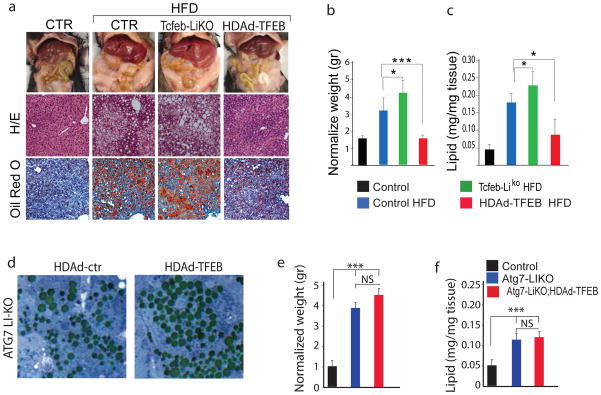
a) Mice with indicated genotype were kept on a high-fat diet for 12 weeks when indicated (HFD). Gross liver morphology (upper panel), H/E (middle panel), and oil red O staining of liver sections (bottom panel). b) Bar graph shows normalized liver weights (mean±s.d. for n=10) and c) total lipid content in mice with indicated genotype (mean±s.d. for n=10). *P≤0.05; **P≤0.01; ***P≤0.001 compared to control. d) Toluidin blue staining of liver sections isolated from ATG7KO mice injected with HDAd-ctr or HDAd-TFEB vector. e) Bar graph shows normalized liver weights (mean±s.d. for n=10 ***P≤0.001) and f) total lipid content in mice with indicated genotype (mean±s.d. for n=5; ***P≤0.001). Mice injected with an empty HDAd virus behaved as wild-type untreated mice, therefore data is not represented in the figure.
Blocking autophagy in the liver leads to hepatomegaly and liver failure21. Recently, a significant increase of lipid droplets, cholesterol and triglycerides was observed in the liver of Atg7 KO mice, suggesting a role of autophagy in lipid degradation9. We analyzed TFEB-mediated lipid degradation in Atg7 liver-KO mice, in which autophagy is blocked21. Liver-specific Atg7 KO mice were generated by injecting Atg7 flox/flox mice with a helper-dependent virus containing the CRE recombinase (HdAD-APOA1-CRE) (Supplementary Fig. 5a). One month after TFEB injection, the mice presented a significant increase in liver size and increased markers of liver damage, as measured by ALT /AST/ALP, which is consistent with previous results21. At the cellular level we observed an accumulation of P6222 (Supplementary Fig. 5b) and lipid droplets (Fig. 5d–f). However, TFEB overexpression failed to decrease lipid droplet number (Fig. 5d), liver weight gain (Fig. 5e) and lipid content (Fig. 5f) in Atg7 liver-KO mice (Supplementary Fig. 5b,c), which is in contrast to the results obtained by TFEB overexpression in wild type mice (Fig. 5a–c). These results indicate that autophagy is required for TFEB-mediated lipid degradation.
TFEB overexpression rescues obesity and metabolic syndrome in mice
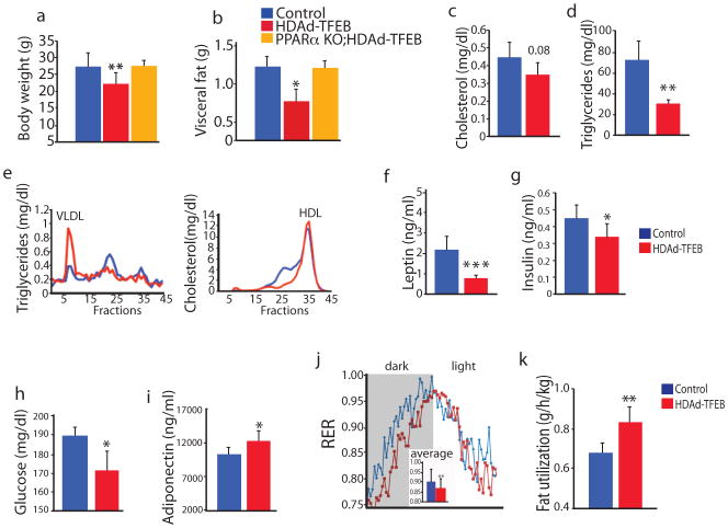
When fed a regular chow diet, HDAd-TFEB-injected mice were significantly leaner than controls (Fig. 6a), with decreased body fat deposition (Fig. 6b), in spite of exhibiting similar food intake. Interestingly, these differences were blunted when HDAd-TFEB was injected in PPARα KO mice (Fig. 6a,b), consistent with our results showing that PPARα is required for transcriptional changes downstream of TFEB. HDAd-TFEB-injected wild type mice also exhibited lower plasma levels of total cholesterol, triglycerides, VLDL, leptin, insulin and glucose (Fig. 6c–i). Indirect calorimetric analysis revealed a decrease in respiratory exchange rate (RER) (Fig. 6j) and an increase in FFA oxidation rate in HDAd-TFEB-injected mice compared with controls (Fig. 6k). Overall, the phenotype observed in HDAd-TFEB-injected mice shared some similarities to those reported for mice under caloric restriction23.
Fig. 6. Metabolic profile of HDAd-TFEB overexpressing mice.
(a) Body weight and (b) visceral fat mass isolated from 2-month-old-mice with the indicated genotypes. n=5. (c–i) Serum metabolic profile in HDad-TFEB mice compared to control mice. (j) Respiratory exchange ratio (RER; VCO2/VO2) and (k) fatty acid utilization calculated from data in (j). Values are mean ± s.d (n = 10) *P_0.05; **P_0.01; compared to controls.
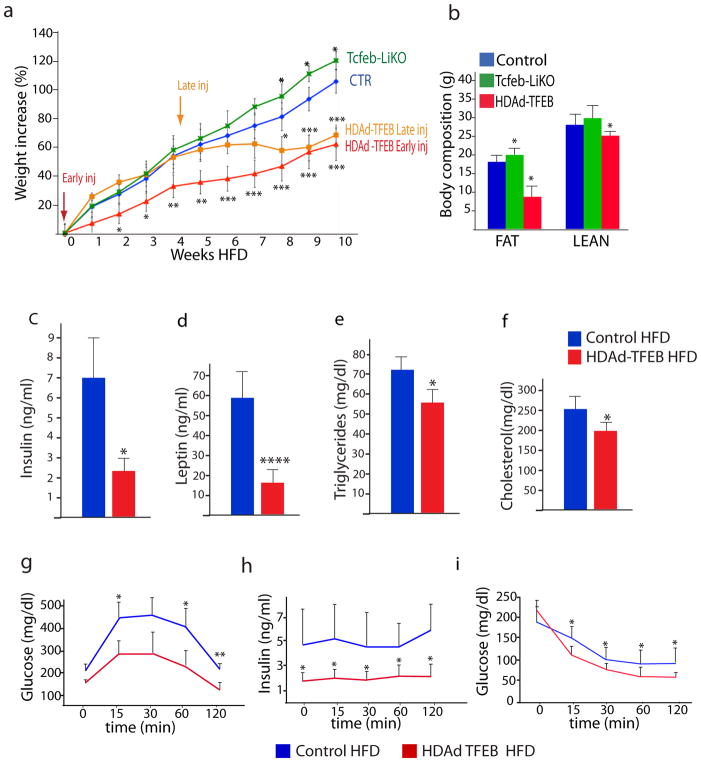
Next we tested the metabolic effects of TFEB gain and loss of function in mice fed with a high-fat diet. We found that in this condition Tcfeb-LiKO mice gained substantially more weight than control littermates (Fig. 7a). Conversely, TFEB overexpression by injection of HDAd-TFEB at the beginning of the high fat diet (“Early inj”) significantly prevented the development of obesity. Furthermore, the injection of HDAd-TFEB after 4 weeks of high fat diet (“Late inj”) completely arrested the development of the obese phenotype. In addition, after 10 weeks of high fat diet the weights of the animals injected at the beginning of the experiment and those injected after 4 weeks were indistinguishable (Fig. 7a). Body composition analysis revealed that the weight difference between Tcfeb-LiKO and HDAd-TFEB-injected mice was largely due to differences in fat accumulation, which was higher in Tcfeb-LiKO and lower in HDAd-TFEB-injected mice compared with controls (Fig. 7b). Of note, the abnormalities in the serum metabolic profile induced by the high-fat diet in wild type mice, which was characterized by the increase of circulating leptin, insulin, triglycerides, and cholesterol, was markedly attenuated in HDAd-TFEB-injected mice (Fig. 7c–f), which also showed improved sugar metabolism, as demonstrated by glucose and insulin-tolerance tests (Fig. 7g–i).
Fig. 7. TFEB prevents diet-induced obesity and metabolic syndrome.
a) Body weight curves of male mice fed with HFD (40% calories from fat) for 10 weeks starting from 5 weeks of age (0 on the x axes). Mice were injected with HDAd-TFEB either 1week before (early inj.), or 4 weeks after, (late inj.) being placed on the HFD, as indicated by the arrows. Values are represented as percentages of weight increase. b) Whole body composition analysis (Echo MRI) of the same mice as in (a) after 10 weeks of HFD. In a and b n=10 mice/group; bars represent mean±s.d. *P≤0.05; **P≤0.01; ***P≤0.001 compared to control HFD group. c–f) Total serum insulin, leptin, triglyceride and cholesterol levels in control and HDAd-TFEB mice kept on high-fat diet (HFD) for 10 weeks. Value are mean ±s.d. n=10. *P≤0.05; ****P≤0.0001 compared to control. g–i) Glucose and insulin tolerance tests in control and HDAd-TFEB mice challenged with HFD for 10 weeks. (g) Glucose and (h) serum insulin levels at the indicated time points after glucose challenge. (i) Glucose levels at the indicated time points after insulin challenge. In g,h,i value are mean ±s.d. n=7 mice/group; *P≤0.05; **P≤0.01; ***P≤0.001 compared to control. Mice injected with an empty HDAd virus behaved as wild-type untreated mice, therefore data is not represented in the figure.
Similar experiments were performed in a genetic model of obesity due to leptin deficiency (Ob/Ob), which is associated with hyperphagia24. Two-month-old Ob/Ob mice, already obese at this early age, were injected with HDAd-TFEB. Six weeks later HDAd-TFEB-injected Ob/Ob mice had significantly reduced levels of circulating triglycerides, cholesterol, glucose and insulin, and an improved glucose tolerance test, compared with untreated Ob/Ob mice, indicating that liver overexpression of TFEB improved the metabolic syndrome phenotype (Supplementary Fig. 6). Together, these observations demonstrate that high TFEB activity can not only prevent the metabolic syndrome, but also revert it once initiated.
Evolutionary conservation of TFEB regulation and function in Caenorhabditis elegans
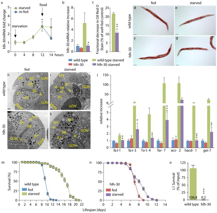
The genetically tractable nematode C. elegans shares approximately 80% of its genes with humans25. The C. elegans genome encodes a single homologue of TFEB, the gene hlh-30 (Supplementary Fig. 7). Its protein product HLH-30 was previously shown to recognize a DNA motif similar to the CLEAR motif in vitro, and to drive transcription of metabolic genes in vivo26. To assess evolutionary conservation of TFEB-mediated starvation responses, we tested whether HLH-30 may act in a similar manner to TFEB during C. elegans starvation. First, we found that hlh-30 mRNA progressively accumulated over a time-course of starvation in wild-type animals, and rapidly decreased after re-introduction of food, similar to mammalian TFEB (Fig. 8a). In contrast, the level of hlh-30 transcript did not increase after 12h of starvation in hlh-30(tm1978) null mutants (Fig. 8b), suggesting that hlh-30 is induced in an hlh-30-dependent manner during starvation by an autoregulatory feedback loop, similar to the mammalian TFEB. C. elegans intestinal cells perform similar metabolic functions to the vertebrate liver, including lipid storage and metabolism. Using semi-quantitative Oil Red O staining, we found that starved wild type animals consumed approximately 20% of their lipid stores, compared to well-fed counterparts (Fig. 8c–g). Starved hlh-30 mutants exhibited a significantly smaller reduction in lipid staining, close to 10%, suggesting that they failed to mobilize lipids as promptly as wild type animals. Consistently, using transmission electron microscopy, we found that intestinal cells in starved wild type animals became depleted of dark-staining lipid droplets (Fig. 8h,i), while those of starved hlh-30 mutants exhibited abundant droplets (Fig. 8j,k). These results suggest that nematodes, similarly to mice, require HLH-30/TFEB to efficiently use lipid stores during periods of starvation. Furthermore, as in liver TFEB-deficient mice, lipid catabolism gene induction is greatly compromised in starved hlh-30 mutants (Fig. 8l). These data suggest that the reason starved hlh-30 mutants fail to mobilize their lipid stores may be because of a severe transcriptional response defect. In wild-type C. elegans, starvation results in lifespan extension27 (Fig. 8m). In contrast, loss of hlh-30 resulted in almost complete abrogation of starvation-induced lifespan extension (Fig. 8n). Additionally, first stage (L1) wild-type larvae arrest in response to food deprivation, resuming development upon food restoration28. In contrast, hlh-30 mutant L1 larvae completely failed to survive starvation-induced arrest, indicating that hlh-30 is not only required in adult animals to survive starvation, but in younger stages as well (Fig. 8o). Considered together, these data demonstrate how hlh-30 expression is induced, that it is required for lipid mobilization, is necessary for a proper transcriptional response, and is required for survival, all during starvation. Therefore, our observations suggest that HLH-30 and murine TFEB share evolutionarily conserved functions in organismal adaptation to starvation.
Fig. 8. Conservation of TFEB-mediated autoregulation and of starvation response in C. elegans.
a) hlh-30 qRT-PCR showing increased expression of C. elegans TFEB gene hlh-30 over a time course of starvation in wild type animals followed by a rapid decrease to basal level following re-feeding of the animals ( mean ± s.e.m. of n=3). *P ≤ 0.05 (t test, compared with wild type at t = 0 h). b) hlh-30 3′ UTR qRT-PCR showing expression of hlh-30 after 12 h starvation in wild type and hlh-30(tm1978) animals (mean ± s.e.m. of n=3). *P ≤ 0.05 (t test, compared with wild type starved). c) Quantification of oil red O stain in starved animals relative to well-fed counterparts (mean ± s.e.m. of n=3).**P ≤ 0.01 (t test, compared with wild type starved). d–g) Representative micrographs of wild type and hlh-30 animals after 8 h starvation and stained with oil red O. h–k) Representative TEMs of wild type and hlh-30 animals after 24 h starvation. IEC, intestinal epithelial cell; EPI, epidermis; BB, brush border; GON, gonad; BWM, body wall muscles, aj, apical junction; ld, lipid droplet. Scale bar is 2 μm. l) qRT-PCR of starvation-induced genes in wild type and hlh-30 animals after 12 h starvation (means ± s.e.m of n=3). *P ≤ 0.05; **P ≤ 0.01; ***P ≤ 0.001 (t test, compared with wild type starved). m–n) hlh-30 is required for starvation-induced lifespan extension. One representative experiment of two independent trials; error bars represents mean ± s.e.m. Median Survival (MS) (wild type fed) = 10 d; MS (wild type starved) = 17 d, P < 0.0001 vs fed using the Log-rank test; MS (hlh-30 fed) = 7 d; MS (hlh-30 starved) = 9 d, P < 0.0001 vs fed. o) L1 arrest assay showing survival of wild-type and hlh-30 starved animals relative to non-starved conditions (mean ± s.e.m. of n=3). ***P ≤ 0.001 (t test, compared with wild type starved).
Discussion
Our study identifies TFEB as a key player in the metabolic response to starvation. TFEB activity is regulated transcriptionally and post-transcriptionally by nutrients, and is required to induce starvation-response genes in both mammals and worms. Most importantly, the absence of TFEB results in an impairment of lipid catabolism and in a more severe metabolic derangement in obese animals, while TFEB overexpression causes the opposite effects and rescues obesity and associated metabolic syndrome.
TFEB mRNA expression is induced by starvation by a post-transcriptional switch that controls TFEB nuclear translocation29–31, which allows TFEB to rapidly respond to nutrient availability, and a positive transcriptional autoregulatory component for a sustained response. Autoregulatory feedback circuits are used by eukaryotic cells to convert a graded input into a binary (ON/OFF) response in eukaryotic gene circuits32–34. Interestingly, the PGC1α gene, a direct target of TFEB, is also subject to an autoregulatory loop35.
Notably, TFEB regulates genes involved in several steps of lipid catabolism, which occur in different cellular compartments, such as the transport of fatty acid chains across the plasma membrane (e.g. CD36 and FABPs), and the β-oxidation of FFA in mitochondria (e.g. Cpt1,Crat, Acadl Acads, Hdad) and in peroxisomes (Cyp4a genes). According to our data most of the effects of TFEB on lipid metabolism appear to be mediated by the direct regulation exerted by TFEB on the PGC1α–PPARα complex. Interestingly, a recent study showed that PGC1α participates in the transcriptional co-activation of TFEB, although the magnitude of this effect appears to be modest36.
The observation that TFEB gain and loss of function in the liver influences whole body energy metabolism suggests that TFEB stimulates the liver secretion of factors that affect the function of other tissues. This is likely to be mediated by PGC1α and PPARα, which are known to regulate the production of secreted hormones37–39.
In previous studies we demonstrated that TFEB controls autophagy by directly regulating lysosomal and autophagy genes12,13. Interestingly, the overexpression of TFEB in mice in which autophagy was genetically suppressed by deletion of Atg7 in the liver did not rescue hepatic steatosis, suggesting that TFEB effects on lipid metabolism require a functional autophagic pathway. Thus, TFEB controls the starvation response by orchestrating the induction of autophagy and PGC1α–PPARα–mediated lipid catabolism. We propose a model (Supplementary Fig. 8) in which adequate nutrition keeps TFEB inactive by cytoplasmic sequestration. During starvation, TFEB translocates to the nucleus where it induces its own expression. This releases the spring for a fast and dramatic metabolic shift to the catabolism of energy stores. Upon nutrient restoration, the feedback loop is quickly interrupted by TFEB nuclear exclusion, restoring the system to baseline. It is likely that global control of lipid metabolism by TFEB arose early during evolution to facilitate organismal adaptation to challenging nutritional conditions, as evidenced by the evolutionary conservation of TFEB autoregulation and by its role in metabolic adaptation to starvation in the invertebrate C. elegans. Finally, the acute beneficial effects of TFEB overexpression in both diet- and genetically-induced obese mice suggest that this regulatory circuit may be an attractive therapeutic target for the modulation of lipid metabolism in obesity-related diseases.
Supplementary Material
Acknowledgments
We thank G. Karsenty, D.Moore, H. Zoghbi, S. Colamarino, E. Abrams, D. Sabatini and R. Zoncu for critical reading of the manuscript. We thank G. Diez-Roux for critical reading of the manuscript, helpful discussions and support in manuscript preparation. We are also grateful to D. Medina, C. Spampanato and F. Annunziata for their contribution. We acknowledge the support of the Italian Telethon Foundation grant numbers TGM11CB6 (C.S. R. DC., A.B) and TGM11SB1 (A.C, and D.dB), the Beyond Batten Disease Foundation (C.S., F.V., T.H., and A.B.); European Research Council Advanced Investigator grant no. 250154 (A.B.); March of Dimes #6-FY11-306 (A.B); US National Institutes of Health (R01-NS078072) (A.B). This work was supported in part by grants from the US National Institutes of Health (R01-HL51586) to L.C. and the Diabetes and Endocrinology Research Center (P30-DK079638, L.C.) and the Mouse Metabolism Core (P.K.S) at Baylor College of Medicine. TJK is in part supported by the Cancer Prevention and Research Institute of Texas (RP110390). O. V. was funded by a Fund for Medical Discovery postdoctoral fellowship from the Massachusetts General Hospital. This study was funded in part by the National Institute of General Medical Sciences of the National Institutes of Health under award number R01GM101056-01 to J. E. I. The content is solely the responsibility of the authors and does not necessarily represent the official views of the National Institutes of Health
Footnotes
Supplementary Information is linked to the online version of the paper. www.nature.com/nature.
Author Contributions C.S., G.M, P.K.S., F.V., O.V., T.H., R.DC., A.C., D.P., T.J.K, A.C.W. performed the experiments. D.d.B. supervised bioinformatic analyses. J.E.I supervised the C. elegans experiments, L.C supervised the metabolic studies, A.B. and C.S. designed the overall study and supervised the work. All authors discussed the results and made substantial contributions to the manuscript.
The authors declare no competing financial interests. Readers are welcome to comment on the online version of this article at www.nature.com/nature.
References
- 1.Bauer M, et al. Starvation response in mouse liver shows strong correlation with lifespan-prolonging processes. Physiol Genomics. 2004;17:230–244. doi: 10.1152/physiolgenomics.00203.2003. [DOI] [PubMed] [Google Scholar]
- 2.Sokolovic M, et al. The transcriptomic signature of fasting murine liver. BMC Genomics. 2008;9:528. doi: 10.1186/1471-2164-9-528. [DOI] [PMC free article] [PubMed] [Google Scholar]
- 3.Hakvoort TB, et al. Interorgan coordination of the murine adaptive response to fasting. J Biol Chem. 2011;286:16332–16343. doi: 10.1074/jbc.M110.216986. [DOI] [PMC free article] [PubMed] [Google Scholar]
- 4.Finn PF, Dice JF. Proteolytic and lipolytic responses to starvation. Nutrition. 2006;22:830–844. doi: 10.1016/j.nut.2006.04.008. [DOI] [PubMed] [Google Scholar]
- 5.Fontana L, Partridge L, Longo VD. Extending healthy life span--from yeast to humans. Science. 2010;328:321–326. doi: 10.1126/science.1172539. [DOI] [PMC free article] [PubMed] [Google Scholar]
- 6.Zinke I, Schutz CS, Katzenberger JD, Bauer M, Pankratz MJ. Nutrient control of gene expression in Drosophila: microarray analysis of starvation and sugar-dependent response. The EMBO journal. 2002;21:6162–6173. doi: 10.1093/emboj/cdf600. [DOI] [PMC free article] [PubMed] [Google Scholar]
- 7.Van Gilst MR, Hadjivassiliou H, Yamamoto KR. A Caenorhabditis elegans nutrient response system partially dependent on nuclear receptor NHR-49. Proc Natl Acad Sci U S A. 2005;102:13496–13501. doi: 10.1073/pnas.0506234102. [DOI] [PMC free article] [PubMed] [Google Scholar]
- 8.Mizushima N. Autophagy: process and function. Genes Dev. 2007;21:2861–2873. doi: 10.1101/gad.1599207. [DOI] [PubMed] [Google Scholar]
- 9.Singh R, et al. Autophagy regulates lipid metabolism. Nature. 2009;458:1131–1135. doi: 10.1038/nature07976. [DOI] [PMC free article] [PubMed] [Google Scholar]
- 10.Singh R, Cuervo AM. Autophagy in the cellular energetic balance. Cell Metab. 2011;13:495–504. doi: 10.1016/j.cmet.2011.04.004. [DOI] [PMC free article] [PubMed] [Google Scholar]
- 11.Yang L, Li P, Fu S, Calay ES, Hotamisligil GS. Defective hepatic autophagy in obesity promotes ER stress and causes insulin resistance. Cell Metab. 2010;11:467–478. doi: 10.1016/j.cmet.2010.04.005. [DOI] [PMC free article] [PubMed] [Google Scholar]
- 12.Sardiello M, et al. A gene network regulating lysosomal biogenesis and function. Science. 2009;325:473–477. doi: 10.1126/science.1174447. [DOI] [PubMed] [Google Scholar]
- 13.Settembre C, et al. TFEB links autophagy to lysosomal biogenesis. Science. 2011;332:1429–1433. doi: 10.1126/science.1204592. [DOI] [PMC free article] [PubMed] [Google Scholar]
- 14.Palmieri M, et al. Characterization of the CLEAR network reveals an integrated control of cellular clearance pathways. Hum Mol Genet. 2011;20:3852–3866. doi: 10.1093/hmg/ddr306. [DOI] [PubMed] [Google Scholar]
- 15.Dennis G, Jr, et al. Database for Annotation, Visualization, and Integrated Discovery. Genome Biol. 2003;4:P3. [PubMed] [Google Scholar]
- 16.Huang da W, Sherman BT, Lempicki RA. Systematic and integrative analysis of large gene lists using DAVID bioinformatics resources. Nat Protoc. 2009;4:44–57. doi: 10.1038/nprot.2008.211. [DOI] [PubMed] [Google Scholar]
- 17.Finck BN, Kelly DP. PGC-1 coactivators: inducible regulators of energy metabolism in health and disease. J Clin Invest. 2006;116:615–622. doi: 10.1172/JCI27794. [DOI] [PMC free article] [PubMed] [Google Scholar]
- 18.Spiegelman BM, Heinrich R. Biological control through regulated transcriptional coactivators. Cell. 2004;119:157–167. doi: 10.1016/j.cell.2004.09.037. [DOI] [PubMed] [Google Scholar]
- 19.Vega RB, Huss JM, Kelly DP. The coactivator PGC-1 cooperates with peroxisome proliferator-activated receptor alpha in transcriptional control of nuclear genes encoding mitochondrial fatty acid oxidation enzymes. Mol Cell Biol. 2000;20:1868–1876. doi: 10.1128/mcb.20.5.1868-1876.2000. [DOI] [PMC free article] [PubMed] [Google Scholar]
- 20.Lin J, Handschin C, Spiegelman BM. Metabolic control through the PGC-1 family of transcription coactivators. Cell Metab. 2005;1:361–370. doi: 10.1016/j.cmet.2005.05.004. [DOI] [PubMed] [Google Scholar]
- 21.Komatsu M, et al. Impairment of starvation-induced and constitutive autophagy in Atg7-deficient mice. J Cell Biol. 2005;169:425–434. doi: 10.1083/jcb.200412022. [DOI] [PMC free article] [PubMed] [Google Scholar]
- 22.Komatsu M, et al. Homeostatic levels of p62 control cytoplasmic inclusion body formation in autophagy-deficient mice. Cell. 2007;131:1149–1163. doi: 10.1016/j.cell.2007.10.035. [DOI] [PubMed] [Google Scholar]
- 23.Bordone L, et al. SIRT1 transgenic mice show phenotypes resembling calorie restriction. Aging Cell. 2007;6:759–767. doi: 10.1111/j.1474-9726.2007.00335.x. [DOI] [PubMed] [Google Scholar]
- 24.Vaisse C, et al. Leptin activation of Stat3 in the hypothalamus of wild-type and ob/ob mice but not db/db mice. Nat Genet. 1996;14:95–97. doi: 10.1038/ng0996-95. [DOI] [PubMed] [Google Scholar]
- 25.Lai CH, Chou CY, Ch’ang LY, Liu CS, Lin W. Identification of novel human genes evolutionarily conserved in Caenorhabditis elegans by comparative proteomics. Genome Res. 2000;10:703–713. doi: 10.1101/gr.10.5.703. [DOI] [PMC free article] [PubMed] [Google Scholar]
- 26.Grove CA, et al. A multiparameter network reveals extensive divergence between C. elegans bHLH transcription factors. Cell. 2009;138:314–327. doi: 10.1016/j.cell.2009.04.058. [DOI] [PMC free article] [PubMed] [Google Scholar]
- 27.Kaeberlein TL, et al. Lifespan extension in Caenorhabditis elegans by complete removal of food. Aging Cell. 2006;5:487–494. doi: 10.1111/j.1474-9726.2006.00238.x. [DOI] [PubMed] [Google Scholar]
- 28.Johnson TE, Mitchell DH, Kline S, Kemal R, Foy J. Arresting development arrests aging in the nematode Caenorhabditis elegans. Mech Ageing Dev. 1984;28:23–40. doi: 10.1016/0047-6374(84)90150-7. [DOI] [PubMed] [Google Scholar]
- 29.Settembre C, et al. A lysosome-to-nucleus signalling mechanism senses and regulates the lysosome via mTOR and TFEB. EMBO J. 2012 doi: 10.1038/emboj.2012.32. [DOI] [PMC free article] [PubMed] [Google Scholar]
- 30.Martina JA, Chen Y, Gucek M, Puertollano R. MTORC1 functions as a transcriptional regulator of autophagy by preventing nuclear transport of TFEB. Autophagy. 2012;8 doi: 10.4161/auto.19653. [DOI] [PMC free article] [PubMed] [Google Scholar]
- 31.Roczniak-Ferguson A, et al. The Transcription Factor TFEB Links mTORC1 Signaling to Transcriptional Control of Lysosome Homeostasis. Sci Signal. 2012;5:ra42. doi: 10.1126/scisignal.2002790. [DOI] [PMC free article] [PubMed] [Google Scholar]
- 32.Becskei A, Seraphin B, Serrano L. Positive feedback in eukaryotic gene networks: cell differentiation by graded to binary response conversion. The EMBO journal. 2001;20:2528–2535. doi: 10.1093/emboj/20.10.2528. [DOI] [PMC free article] [PubMed] [Google Scholar]
- 33.Siciliano V, et al. Construction and modelling of an inducible positive feedback loop stably integrated in a mammalian cell-line. PLoS Comput Biol. 2011;7:e1002074. doi: 10.1371/journal.pcbi.1002074. [DOI] [PMC free article] [PubMed] [Google Scholar]
- 34.Kielbasa SM, Vingron M. Transcriptional autoregulatory loops are highly conserved in vertebrate evolution. PLoS One. 2008;3:e3210. doi: 10.1371/journal.pone.0003210. [DOI] [PMC free article] [PubMed] [Google Scholar]
- 35.Handschin C, Rhee J, Lin J, Tarr PT, Spiegelman BM. An autoregulatory loop controls peroxisome proliferator-activated receptor gamma coactivator 1alpha expression in muscle. Proc Natl Acad Sci U S A. 2003;100:7111–7116. doi: 10.1073/pnas.1232352100. [DOI] [PMC free article] [PubMed] [Google Scholar]
- 36.Tsunemi T, et al. PGC-1alpha rescues Huntington’s disease proteotoxicity by preventing oxidative stress and promoting TFEB function. Sci Transl Med. 2012;4:142ra197. doi: 10.1126/scitranslmed.3003799. [DOI] [PMC free article] [PubMed] [Google Scholar]
- 37.Bostrom P, et al. A PGC1-alpha-dependent myokine that drives brown-fat-like development of white fat and thermogenesis. Nature. 2012;481:463–468. doi: 10.1038/nature10777. [DOI] [PMC free article] [PubMed] [Google Scholar]
- 38.Badman MK, et al. Hepatic fibroblast growth factor 21 is regulated by PPARalpha and is a key mediator of hepatic lipid metabolism in ketotic states. Cell Metab. 2007;5:426–437. doi: 10.1016/j.cmet.2007.05.002. [DOI] [PubMed] [Google Scholar]
- 39.Inagaki T, et al. Endocrine regulation of the fasting response by PPARalpha-mediated induction of fibroblast growth factor 21. Cell Metab. 2007;5:415–425. doi: 10.1016/j.cmet.2007.05.003. [DOI] [PubMed] [Google Scholar]
Associated Data
This section collects any data citations, data availability statements, or supplementary materials included in this article.


