Abstract
Members of the CCp protein family have been previously described to be expressed on gametocytes of apicomplexan Plasmodium parasites. Knocking out Plasmodium CCp genes blocks the development of the parasite in the mosquito vector, making the CCp proteins potential targets for the development of a transmission-blocking vaccine. Apicomplexans Babesia bovis and Babesia bigemina are the causative agents of bovine babesiosis, and apicomplexan Theileria equi causes equine piroplasmosis. Bovine babesiosis and equine piroplasmosis are the most economically important parasite diseases that affect worldwide cattle and equine industries, respectively. The recent sequencing of the B. bovis and T. equi genomes has provided the opportunity to identify novel genes involved in parasite biology. Here we characterize three members of the CCp family, named CCp1, CCp2 and CCp3, in B. bigemina, B. bovis and T. equi. Using B. bigemina as an in vitro model, expression of all three CCp genes and proteins was demonstrated in temperature-induced sexual stages. Transcripts for all three CCp genes were found in vivo in blood stages of T. equi, and transcripts for CCp3 were detected in vivo in blood stages of B. bovis. However, no protein expression was detected in T. equi blood stages or B. bovis blood stages or B. bovis tick stages. Collectively, the data demonstrated a differential pattern of expression of three orthologous genes of the multidomain adhesion CCp family by B. bigemina, B. bovis and T. equi. The novel CCp members represent potential targets for innovative approaches to control bovine babesiosis and equine piroplasmosis.
Introduction
Arthropod-borne protozoan parasites of the phylum Apicomplexa cause major diseases in humans and animals, including malaria, babesiosis, and theileriosis [1], [2], [3]. Apicomplexan parasites have complex life cycle including asexual stages in vertebrate hosts and sexual stages in arthropod vectors [4]. There are currently many knowledge gaps in our understanding of the biology of these parasites, including the molecular events involved in their sexual reproduction. A better comprehension of the sexual events is needed to develop improved approaches to control the diseases caused by apicomplexan parasites. The growing number of available apicomplexan genomes has provided the opportunity to identify novel genes implicated in the parasite life cycle. Genome-based approaches have identified a highly conserved family of six multidomain adhesion proteins, termed CCp family, that are exclusively expressed on the surface of Plasmodium gametocytes [5], [6]. Importantly, knock out of the Plasmodium CCp genes blocks sporozoite formation, thus preventing the development of the parasite in the mosquito vectors, and making the CCp proteins potential targets for the development of transmission-blocking vaccines [6], [7]. The CCp proteins contain at least one signature domain Limulus coagulation factor C (LCCL) and additional adhesion domains, suggesting that these proteins may mediate cell contacts of gametocytes [8]. Members of the CCp family have also been identified in other apicomplexan genera, including Toxoplasma, Cryptosporidium, Eimeria, Theileria and Ascogregarina [9], [10]. Recently three genes of the CCp family have been identified in Babesia divergens [11]. Interestingly, the CCp family has not been identified in any other protozoan except apicomplexan parasites.
Babesia bovis and Babesia bigemina are tick-borne apicomplexan parasites that cause bovine babesiosis, an economically important disease of cattle that threatens the development of the bovine industry globally [12]. Similarly, the tick-borne apicomplexan parasite Theileria equi is the causative agent of equine piroplasmosis, the most economically important parasite disease to the horse industry worldwide [13], [14]. Extensive research efforts have concentrated on the development of approaches to control bovine babesiosis and equine piroplasmosis. However, vaccination with live attenuated parasites is still the most efficient method to prevent bovine babesiosis, and currently there is no vaccine available to control equine piroplasmosis. The recent sequencing of the B. bovis genome and the T. equi genome has provided a crucial tool to investigate the biology of these apicomplexan parasites [15], [16]. Consequently, genome-based studies have identified novel genes and gene families, resulting in a better understanding of parasite biology [17], [18]. Despite these efforts, there is a substantial lack of knowledge concerning the sexual stages of B. bovis, B. bigemina and T. equi. Identification of sexual stage proteins is critical to comprehend parasite biology and develop new immunological and therapeutic interventions to disrupt the parasite life cycle.
In this study we characterized three orthologous genes of the multidomain adhesion CCp family, named CCp1, CCp2 and CCp3, in B. bigemina, B. bovis and T. equi. B. bigemina was used as an in vitro model for sexual stages, and expression of all three CCp genes and proteins was demonstrated in temperature-induced sexual stages. Additionally, the presence of CCp transcripts was shown in vivo in blood stages of B. bovis and T. equi. The newly described orthologous members of the CCp family in B. bigemina, B. bovis and T. equi represent rational targets for the development of novel immunological and therapeutic strategies to control bovine babesiosis and equine piroplasmosis.
Materials and Methods
Babesia Bigemina
B. bigemina isolated in Puerto Rico in 1985 was used for in vitro experiments and parasite culture was performed as described elsewhere [19]. The medium volume of the B. bigemina culture was expanded from 12 ml to 120 ml in a 5-day period, with no addition of extra red blood cells, to increase parasitemia. The expanded culture was used to induce sexual stages as previously described [20]. Briefly, to induce B. bigemina sexual stages, the parasite culture was incubated at 28°C in 5% CO2 for up to 48 h. As a control, a portion of the culture was maintained at 37°C in 5% CO2 for up to 48 h. A Percoll gradient was then used to separate the extracellular from intracellular parasites and the two populations were used to investigate the expression of the CCp genes and proteins. Additionally, bovine sera from B. bigemina-infected animals (Animal Disease Research Unit, USDA, Pullman, WA) were used as controls in immunoblot and immunofluorescence assays.
Babesia Bovis
B. bovis Texas strain [21] was used in this study and parasite culture was performed as described elsewhere [22]. For the B. bovis in vivo studies, a splenectomized calf was intravenously inoculated with approximately 106 B. bovis-infected erythrocytes. For tick-acquisition feeding, 1 gram of larvae (approximately 20,000 larvae) of Rhipicephalus (Boophilus) microplus La Minita strain [23] was placed within a cloth on a splenectomized calf. When approximately 1–2% of the ticks had molted to adult stage, the calf was inoculated with B. bovis as described above and the peak protozoan parasitemia occurred when the adult female ticks were reaching repletion. The calf was monitored daily for the presence of clinical signs of acute bovine babesiosis and maintained throughout the experiment in accordance to protocols approved by the University of Idaho Institutional Animal Care and Use Committee (Permit Number: 2010–54). B. bovis parasitemia in bovine peripheral blood and tick guts was assessed by quantitative real-time PCR (qPCR) using primers to msa-1 gene as previously described [24].
Theileria Equi
T. equi Florida isolate was used in this study and parasite culture was performed as described elsewhere [16]. For the T. equi in vivo studies, a spleen-intact horse was inoculated intravenously with approximately 8×10⁷ infected erythrocytes. The horse was monitored daily for the presence of clinical signs of acute equine piroplasmosis and maintained throughout the experiment in accordance to protocols approved by the University of Idaho Institutional Animal Care and Use Committee (Permit Number: 2010–54). T. equi parasitemia in horse peripheral blood was assessed by qPCR using primers to ema-1 gene as previously described [25].
In silico Gene Identification
In silico gene identification was performed by comparing the amino acid sequences of CCp1 (GenBank ID XP_001348897.1) and CCp3 (GenBank ID XP_001348240) of P. falciparum 3D7 strain, and CCp2 (GenBank ID XP_001615829) of P. vivax Sal-1 strain to the B. bovis and T. equi genomes using BLAST software (http://blast.ncbi.nlm.nih.gov/Blast.cgi). In silico gene identification was also accomplished by comparing the amino acid sequences of B. divergens CCp genes [11] to the B. bovis and T. equi genomes. Amino acid identity ≥ than 30% was used as a criterion for identification of the CCp orthologous genes in B. bovis and T. equi. Multiple alignments of amino acid sequences were generated using the Multiple Alignment Module of LaserGene (http://www.dnastar.com). In silico analysis of the available B. bigemina sequences was performed using BLASTN and TBLAST at the Sanger Institute website (http://www.sanger.ac.uk/cgi-bin/blast/submitblast/b_bigemina). The Simple Modular Architecture Research Tool (SMART) (http://smart.embl-heidelberg.de) and the Transmembrane Hidden Markov Model package 2 (TMHMM2) (http://www.cbs.dtu.dk/services/TMHMM-2.0) were used to predict domains and signal peptides in the CCp protein sequences.
RNA Isolation and Transcript Level Analysis
Reverse transcriptase quantitative real-time PCR (RT-qPCR) was standardized to assess the level of expression of the CCp1-3 genes in B. bovis, B. bigemina and T. equi. Parasite cultures, infected blood and tick guts were collected in RNAlater® (Ambion) and stored at −20°C following the manufacture’s protocol. Total RNA was extracted using the RNAqueous® Kit (Ambion) according to the manufacturer’s protocol. The total RNA samples were analyzed by the Experion™ Automated Electrophoresis System (Bio-Rad) and only samples with an RNA Quality Indicator (RQI) ≥ than 7 were used for cDNA synthesis (Figure S1). Two hundred nanograms of total RNA were utilized for cDNA synthesis using the Superscript® Vilo™ cDNA Synthesis Kit (Invitrogen) following the manufacturer’s protocol. The RT-qPCR were performed in a CFX96™ Real-Time PCR Detection System using the SsoFast™ EvaGreen® Supermix (Bio-Rad). The cycling conditions consisted of an enzyme activation step of 95°C for 30 seconds followed by 40 cycles of 95°C denaturation for 5 seconds and annealing/extension of 60°C for 5 seconds. Reactions were performed in duplicate in 20 µl using 200 nM of each primer and 2 µl of a 1/20 dilution of cDNA as template. The CFX Manager™ Software (Bio-Rad) was used to analyze the RT-qPCR data. Gene expression was normalized to the total amount of RNA used to generate the cDNA, as previously described [24]. The transcription level was then calculated as a relative expression using the formula: Relative expression (sample) = 2 [Cq (control) – Cq (sample)], where the control is the highest Cq value for a given gene of interest, as previously described [24]. Efficiency of amplification and melt curve analyses was performed to evaluate analytical sensitivity and specificity of the RT-qPCR for each gene of interest (Table S1). Standard PCR for bovine actin (primers: gtgtggattggcggct and tactcctgcttgctgat) and horse actin [26] were performed to demonstrate the presence of amplifiable cDNA in all tested samples.
Synthetic Peptides and Polyclonal Antibodies
Synthetic peptides ranging from 15 to 19 amino acids were produced based on the sequence of the B. bovis CCp proteins, as follows: CCp1– KTFESKPSYKEVFK (aa 122–135); CCp2.1 - ESAKKTKDARDKYFLQSV (aa 530–547) and CCp2.2 KRLIRVVNGDPYEIAKIED (aa 1165–1183); and CCp3.1 - PSSLKGTYIYTEDSSI (aa 622–637), CCp3.2 GSYLTFVVEAADVGDVNGI (aa 123–141) and CCp3.3 ASAMFDGVLTPSGGE (aa 254–268) (Figure 1, Table S2, Figure S2, Figure S3 and Figure S4). The percentage of amino acid identity of the peptides and the B. bigemina and T. equi CCp sequences is shown in Table S2. Three 6-week old BALB/c female mice were inoculated with 100 µg of KLH-conjugated CCp1 peptide emulsified in TiterMax® Gold Adjuvant (Sigma) followed by six booster inoculations at 2 weeks interval. Serum titers were assessed by ELISA and the mice exsanguinated 7 days after the last booster inoculation. Two groups of two rabbits were inoculated with either KLH-conjugated CCp2 peptides or KLH-conjugated CCp3 peptides followed by four booster inoculations at 2 weeks interval following the protocol described by Bio-Synthesis, Inc (http://www.biosyn.com). Serum titers were assessed by ELISA and the rabbits exsanguinated 7 days after the last booster inoculation. The rabbit and mouse polyclonal antibodies were used in immunoblot and immunofluorescence assays to investigate the expression of the CCp proteins.
Figure 1. Gene identification, cDNA length, protein length, expected protein size and schematic domain representation of CCp1-3 in Babesia bovis and Theileria equi.
Black lines represent the full-length of the CCp proteins in B. bovis and T. equi. Grey lines represent the regions of the CCp proteins available in B. bigemina. Red lines above CCp1-3 indicate the regions used to design synthetic peptides. R: Ricin B lectin domain. F: F5/F8 type C domain. L: LCCL domain. PL: PLAT domain. SR: Scavenger receptor domain. * In silico data of B. bovis CCp1-3 has been previously shown by Becker et al [11].
Immunoblot Assays
Antigens for immunoblot were prepared from B. bigemina culture, B. bovis-infected bovine blood, B. bovis-infected tick guts, or T. equi-infected horse blood. After adding Laemmli buffer (Bio-Rad), the samples were boiled for 10 min and approximately 10 µg of total protein were separated in 4–20% Mini-PROTEAN® TGX™ Precast Gels (Bio-Rad). The proteins were transferred overnight at 4°C to nitrocellulose membranes. The membranes were blocked for non-specific binding in TNT (10 mM Tris, 150 mM NaCl, and 0.05% Tween 20) containing 5% non-fat milk for 1 hour at room temperature (RT). The membranes were then incubated for 1 hour at RT with an appropriate primary antibody. After washing in TNT, the membranes were incubated with an appropriate HRP conjugated secondary antibody for 30 minutes at RT. After washing in TNT, the immunoblot assays were developed using chemiluminescent HRP antibody detection reagents.
Immunofluorescence Assays
Immunofluorescence assays were performed using antigens from B. bigemina culture, B. bovis-infected bovine blood, B. bovis-infected tick guts, or T. equi-infected blood. B. bigemina culture and parasite-infected blood were adjusted to 20% pack cell volume (PCV) in PBS containing 1% BSA. Ticks fed on a B. bovis-infected calf were dissected and individual tick guts were suspended in PBS containing 1% BSA. Approximately 5 µl of either the 20% PCV solution or the tick gut suspension was used to make a uniform thin film on glass microscope slides. The slides were air dried and fixed for 1 minute in cold acetone. The slides were incubated at 37°C in a humidity chamber for 30 minutes with different concentrations of an appropriate primary antibody. After washing in PBS, the slides were incubated at 37°C in a humidity chamber for 30 minutes with an appropriate FITC conjugated secondary antibody. After washing in PBS, the slides were examined using an epifluorescence microscope equipped with FITC UV excitation and emission filters.
Results
In silico Identification of the CCp Genes
Three orthologous members of the CCp gene family, termed CCp1-3, were identified by comparing the CCp sequences of Plasmodium spp. to the B. bovis and T. equi genomes (Figure 1 and Table S3). The genome screening analysis indicated that orthologous CCp1-3 are single copy genes in the T. equi genome and, as previously described in the B. bovis genome [11]. In the B. bovis genome, CCp1-3 proteins are identified as BBOV_III006360, BBOV_II003700 and BBOV_III008930, respectively, and are annotated as LCCL domain containing proteins. In the T. equi genome, CCp1-3 are identified as BEWA_020400, BEWA_004750 and BEWA_010590, respectively, and annotated as bromodomain containing protein, hypothetical protein and LCCL domain-containing protein, respectively. It was predicted that B. bovis CCp1-3 genes encode for proteins with molecular weight of 177.8, 174.9 and 127.4 KDa, respectively, as previously described [11]. Similarly, it was predicted that T. equi CCp1-3 genes encode for proteins with molecular weight of 174.9, 177.8 and 137.1 KDa, respectively. No orthologous genes for CCp4 and CCp5 were found in the B. bovis and T. equi genomes. Analysis of the available B. bigemina genome sequences also revealed the presence of CCp1-3 in this apicomplexan parasite. However, no full-length sequence of the CCp genes was obtained from B. bigemina, as the complete genome sequence for this parasite is not yet available. Pairwise analysis of the amino acid sequence of CCp1-3 of B. bovis, T. equi, B. divergens and P. falciparum revealed the level of interspecies identity and similarity of these proteins (Table S4). Taken together, the B. bovis CCp1-3 proteins showed greater identity and similarity to B. divergens than to the T. equi and P. falciparum sequences. Likewise, the T. equi CCp1-3 sequences showed a higher level of identity and similarity to B. bovis than to P. falciparum. It is noteworthy that the amino acid identity and similarity was distributed throughout the length of the CCp proteins of B. bovis, T. equi and Plasmodium spp. (Figure S2, Figure S3 and Figure S4). Regarding domain organization, the B. bovis and T. equi CCp proteins presented similar domain distribution when compared to orthologous members in other apicomplexan parasites. CCp1 and CCp2, both in B. bovis and T. equi, contain a similar domain pattern with one LCCL domain, one ricin B lectin domain and one F5/8 type C domain. CCp3 protein, both in B. bovis and T. equi, presents three LCCL domains, two scavenger domains and one PLAT (polycystin-1, lipoxygenase, alpha-toxin) domain (Figure 1).
Expression of CCp1-3 Genes in Babesia bigemina
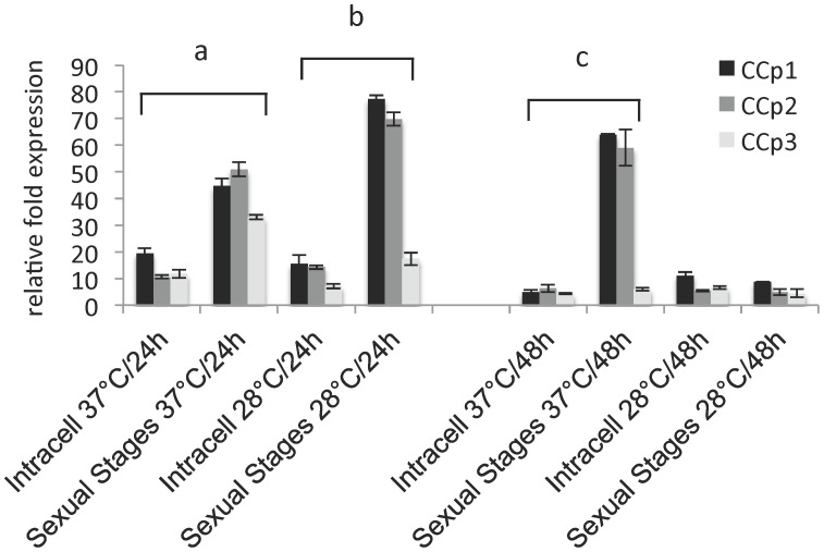
We initially examined the expression of the CCp genes in B. bigemina, since this is the only experimental in vitro system currently available to investigate sexual stages in Babesia spp. and Theileria spp. [4], [20], [27]. B. bigemina sexual stages were induced by incubating the parasite culture for up to 48 h at 28°C in 5% CO2. The extracellular temperature-induced sexual stage parasites were used to investigate the level of expression of the CCp genes and proteins. Extracellular B. bigemina sexual stage parasites were separated from intraerythrocytic parasites by Percoll gradient. Total RNA was extracted from the two parasite populations and relative expression of CCp1-3 was accessed by RT-qPCR (Figure 2). The extracellular population of B. bigemina sexual stages showed a significant up regulation of all three CCp genes when compared to intracellular parasites. Expression of CCp1 and CCp2 peaked at 24 h in sexual stages incubated at 28°C and at 48 h in sexual stages incubated at 37°C. In contrast, CCp3 peaked at 24 h in sexual stages incubated at 37°C. After 48 h of incubation, there was no difference in the expression of CCp3 in sexual stages incubated at either 37°C or 28°C. It is noteworthy that the normalized expression of CCp1 and CCp2 increased (P<0.001) more than 40-fold in sexual stages incubated at 28°C for 24 h when compared to intracellular parasites incubated at the same condition (Figure 2). The results presented in Figure 2 represent the means of three experiments, each containing three technical replicates. It is important to mention that despite the up regulation of CCp-1-3 genes in B. bigemina sexual stages, transcripts for all the three genes were also detected in non-induced parasite cultures kept at 37°C (Figure S5).
Figure 2. Relative expression of B. bigemina CCp1-3 by extracellular sexual stages incubated either at 37°C or 28°C for 24 h and 48 h is compared to gene expression by intracellular parasites (Intracell) incubated under identical conditions for each temperature and time point.
Gene expression was normalized to the total amount of RNA used to generate the cDNA. The transcription level was calculated as a relative expression using the formula: Relative expression (sample) = 2 [Cq (control) – Cq (sample)], where the control is the highest Cq value for a given gene of interest. Results represent the means of three experiments, each containing three technical replicates. Differences in gene expression were determined by the Student’s t-test. “a” indicates the expression of CCp1-3 was significantly higher (P<0.001) by sexual stages incubated for 24 h at 37°C than by Intracell. “b” indicates the expression of CCp1and CCp2 was significantly higher (P<0.001) by sexual stages incubated for 24 h at 28°C than by Intracell. “c” indicates the expression of CCp1and CCp2 was significantly higher (P<0.001) by sexual stages incubated for 37 h at 48°C than by Intracell.
Expression of CCp1-3 Proteins in Babesia bigemina
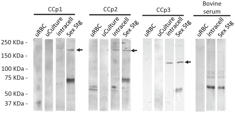
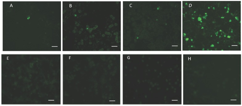
Expression of CCp1-3 proteins was demonstrated in vitro in temperature-induced B. bigemina sexual stages (Figure 3 and 4). Immunoblot analysis using mouse polyclonal anti-CCp1 peptide detected the expression of the CCp1 protein by B. bigemina extracellular sexual stages; however no band with the putative expected size of CCp1 was visualized in intracellular parasites. Interestingly, rabbit polyclonal antibodies against either CCp2 peptides or CCp3 peptides detected the expression of CCp2 and CCp3, respectively, from both extracellular sexual stages and intracellular B. bigemina. No expression of CCp proteins was detected by non-induced B. bigemina cultures using immunoblot (Figure 3). Bovine sera from B. bigemina chronically infected animals reacted primarily to the 58 KDa Rap-1 protein using both intracellular parasites and extracellular sexual stages. The B. bigemina-infected bovine sera also detected other antigens with molecular mass ranging from 75 to 90 KDa. However, the tested B. bigemina-infected bovine sera did not detect antigens with the expected size of the CCp proteins. Figure 3 shows an immunoblot using a representative B. bigemina-infected bovine serum. Immunofluorescence assays using mouse and rabbit polyclonal antibodies generated against CCp synthetic peptides revealed the expression of CCp1-3 in temperature-induced B. bigemina cultures (Figure 4). No expression of CCp proteins was detected by non-induced B. bigemina cultures using immunofluorescence (Figure 4). Immunoblot results showed CCp2 and CCp3 expression by both extracellular and intracellular B. bigemina (Figure 3), and consequently, the immunofluorescence assays were performed using whole culture without Percoll gradient separation. The parasitemia of the B. bigemina culture used for immunofluorescence antigen was approximately 8–10%. With respect to the CCp proteins, only one or two fluorescent parasites were identified on average per microscopic field (100x magnification) indicating that a very low percentage of the temperature-induced B. bigemina parasites were expressing the proteins (Figure 4). Negative controls for the immunofluorescence assay, including pre-immune sera, FITC conjugate, and normal bovine serum are shown in Figure S6.
Figure 3. Immunoblot analysis demonstrating the expression of CCp1-3 by temperature-induced B. bigemina parasites.
Expression of CCp1-3 proteins was investigated in B. bigemina cultures incubated for 24 h at 28°C. Expression of CCp1 was detected only by extracellular sexual stages (Sex Stg) whereas CCp2 and CCp3 were detected in both Sex Stg and intracellular parasites (Intracell) populations. No bands with the expected molecular mass of the CCp proteins were detected in non-induced B. bigemina cultures (uCulture) or in uninfected red blood cells (uRBC). Polyclonal mouse anti-CCp1 peptide, rabbit anti-CCp2-peptides or CCp3-peptides, and bovine serum from an animal chronically infected with B. bigemina were used at a 1∶50 dilution. Anti-mouse, anti-rabbit, or anti-bovine HRP conjugates were used at a 1∶4,000 dilution. Black arrows indicate the expected size of CCp1-3.
Figure 4. Immunofluorescence assays demonstrating the expression of CCp1-3 in temperature-induced B. bigemina cultures.
Antigen for immunofluorescence was prepared from parasite culture adjusted to 20% pack cell volume (PCV). Five µl of the 20% PCV solution was used to make a uniform thin film on glass microscope slides. Expression of CCp1-3 proteins in temperature-induced B. bigemina gametocyte cultures incubated for 24 h at 28°C is shown in panels A, B and C, respectively. CCp expression was not detected in non-induced B. bigemina cultures kept at 37°C as shown in panels E, F, and G. Panels D and H are representative results of immunofluorescence using sera from either B. bigemina-infected or uninfected bovine, respectively. Polyclonal mouse anti-CCp1 peptide, rabbit anti-CCp2 or CCp3 peptides, and bovine sera were used at a 1∶20 dilution. Anti-mouse, anti-rabbit, or anti-bovine FITC conjugates were used at a 1∶80 dilution. The fields shown are representative of the majority of the samples. The panels present a magnification of 100x and white bars indicate 10 µm.
Expression of CCp1-3 in Babesia Bovis
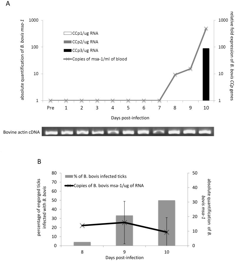
Expression of CCp1-3 genes was evaluated in vitro in B. bovis culture, and in vivo in cattle during acute infection and ticks fed on a calf during acute infection. Transcripts for all three CCp genes were detected in B. bovis culture (Figure S5). During the acute phase of B. bovis infection, the calf temperature ranged from 38.1 to 41.3°C and PVC decreased approximately 40%. B. bovis parasites were first detected at day 8 post-infection and the peak parasitemia (approximately 500 parasites/ml of blood) occurred at day 10 post-infection (Figure 5A). At that time, the B. bovis-infected calf had to be euthanized due to the severity of the disease. Relative expression of the B. bovis CCp genes was evaluated in the calf blood during acute infection. Expression of the B. bovis CCp3 gene was detected at day 10 post-infection. Interestingly, B. bovis CCp1 and CCp2 were not detected during the evaluation period. Standard PCR for bovine actin was performed to demonstrate the presence of amplifiable cDNA in all tested samples (Figure 5A). It is noteworthy that no expression of B. bovis CCp proteins was detected by immunoblot or immunofluorescence either in parasite culture or bovine blood during acute infection. The presence of B. bovis DNA was also examined in engorged R. (B.) microplus female ticks fed on a calf during acute infection. At days 8, 9 and 10 post-B. bovis infection, 24 engorged R. (B.) microplus females were collected per each time point. The ticks were dissected and guts collected in PBS for genomic DNA extraction and in RNAlater® for RNA extraction. A timeframe of less than 2 h passed between tick collection/dissection and tick guts storage in RNAlater®. Genomic DNA and total RNA were extracted from individual guts of ticks fed on the B. bovis-infected calf, and qPCR and RT-qPCR for msa-1 gene were performed. At day 8 of feeding, only one tick gut was positive for B. bovis and the positive sample had 200 parasites per µg of gut DNA. At day 9 of feeding, 33.3% of the tick guts (9 out of 24) were positive for B. bovis and the average number of parasites was 196 (±21) per µg of tick gut DNA. At day 10 of feeding, 50% of the tick guts (12 out of 24) were positive for B. bovis and the average number of parasites was 176 (±26) per µg of gut DNA (Figure 5B). Gene expression was examined in cDNA samples from individual guts of engorged R. (B.) microplus female ticks fed on a calf during B. bovis acute infection. As described for the genomic DNA analysis, at days 8, 9 and 10 post-B. bovis infection, 24 engorged R. (B.) microplus females were collected per each time point and cDNA synthesized from individual ticks guts. Absolute quantification of B. bovis msa-1 transcripts was performed and ranged from 9 to 15 copies in gut samples of engorged female ticks fed on the infected calf (Figure 5B). However, no expression of CCp1-3 genes was detected in the cDNA samples of tick guts. Additionally, it is noteworthy that no expression of the B. bovis CCp proteins was detected by either immunoblot or immunofluorescence in guts of ticks fed on a calf during acute infection.
Figure 5. Expression of CCp1-3 during acute B. bovis infection.
In panel A, relative fold expression of CCp1-3 transcripts in bovine blood during acute B. bovis infection is shown in the right y-axis. Absolute quantification of B. bovis msa-1 DNA in bovine blood is shown in the left y-axis. CCp gene expression was normalized to the total amount of RNA used to generate the cDNA and the transcription level was calculated as a relative expression using the formula: Relative expression (sample) = 2 [Cq (control) – Cq (sample)], where the control is the highest Cq value for a given gene of interest. To confirm the presence of amplifiable cDNA in all tested samples a standard PCR for bovine actin was performed. In panel B, absolute expression of B. bovis msa-1 RNA in bovine blood is shown in the right y-axis and the percentage of engorged ticks infected with B. bovis is shown in the left y-axis.
Expression of CCp1-3 in Theileria Equi
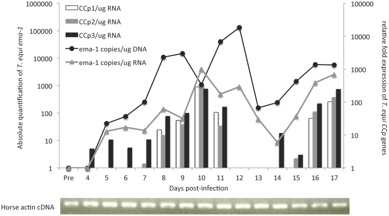
Expression of CCp1-3 genes was evaluated in vitro in T. equi culture, and in vivo in horse blood during acute infection. Transcripts for all three CCp genes were detected in T. equi cultures (Figure S5). During T. equi acute infection, the horse temperature ranged from 37.1 to 40.8°C and PCV decreased 40%. T. equi parasites were first detected at day 5 post-infection and the peak parasitemia (approximately 500 parasites/µg DNA) occurred at day 12 post-infection (Figure 6). Total RNA was extracted from peripheral blood of the infected horse and used to synthesize cDNA. Relative expression of the CCp genes was evaluated in T. equi blood stages during acute infection. Expression of T. equi CCp3 was first detected at day 4 post-infection and was followed by the expression of CCp2 and CCp1 at days 7 and 8 post-infection, respectively. The expression of all T. equi CCp genes peaked at day 10 post-infection and showed an increase of more than 250-fold compared to previous days of infection. Interestingly, the CCp genes were not constitutively expressed during T. equi acute infection. In fact, during the evaluation period, the genes presented two waves of expression that began with the expression of the CCp3 gene followed sequentially by CCp2 and then CCp1 genes. Expression of the T. equi ema-1 gene was also evaluated and shown to be constitutively expressed during the acute phase of the infection. Standard PCR for horse actin was performed to demonstrate the presence of amplifiable cDNA in all tested samples (Figure 6). It is noteworthy that no expression of the T. equi CCp proteins was detected by immunoblot or immunofluorescence either in parasite culture or in horse blood during acute infection.
Figure 6. Expression of CCp1-3 during acute T. equi infection.
Relative fold expression of CCp1-3 transcripts in horse peripheral blood during acute T. equi infection is shown in the right y-axis. Absolute quantification of T. equi emai-1 DNA (black line) and cDNA (grey line) is shown in the left y-axis. CCp gene expression was normalized to the total amount of RNA used to generate the cDNA and the transcription level was calculated as a relative expression using the formula: Relative expression (sample) = 2 [Cq (control) – Cq (sample)], where the control is the highest Cq value for a given gene of interest. To confirm the presence of amplifiable cDNA in all tested samples a standard PCR for horse actin was performed.
Discussion
In this study we investigated the pattern of expression of three newly identified orthologous genes of the multidomain adhesion protein family CCp, named CCp1, CCp2 and CCp3, by the apicomplexan parasites Babesia bovis, Babesia bigemina and Theileria equi. Members of the CCp family have been previously identified in Plasmodium, Toxoplasma, Cryptosporidium, Eimeria, Theileria and Ascogregarina [6], [8], [9]. Recently, three members of the CCp family have been identified in B. divergens [11], [28]; however, no biological data were previously available for CCp genes in B. bovis, B. bigemina or T. equi. Here we have demonstrated that expression of the CCp1-3 genes was significantly up regulated in vitro in B. bigemina sexual stages. We also demonstrated a differential expression of the CCp1-3 proteins in vitro in B. bigemina sexual stages. Additionally, CCp transcripts were detected in vivo in blood stages of B. bovis and T. equi during acute infection.
CCp1-3 were identified as single copy genes in the genomes of B. bovis and T. equi, as previously described for the CCp ortologous genes in Plasmodium spp. and B. divergens [5], [6], [11]. The presence of CCp orthologous genes in several apicomplexan parasites, including Plasmodium, Toxoplasma, Cryptosporidium, Eimeria, Theileria, Ascogregarina and Babesia, suggests a potentially conserved function for this family. It has been described that CCp proteins are expressed on the surface of Plasmodium gametocytes where they play a role in parasite interaction during zygote formation [6], [7], [8]. Therefore, it is tempting to consider that CCp proteins might play a similar function in Babesia and Theileria transmission. Development of mutant Babesia and Theileria lacking the expression of the CCp genes is essential to address this hypothesis. The presence of multi-adhesion domains and the remarkable similarity of domain organization of CCp1-3 among different apicomplexan parasites, including B. bovis, B. bigemina and T. equi described in this manuscript, also prompts us to argue about a potential evolutionary conserved function for these proteins. The similar size and domain organization of CCp1 and CCp2 in numerous apicomplexan parasites, including B. bovis, B. bigemina and T. equi presented in this study, suggests that a gene duplication event may have occurred in the common ancestor of the Apicomplexa phylum, as mentioned elsewhere [6]. Interestingly, neither CCp4 nor CCp5 orthologous genes were found in B. bovis or T. equi. Similar absence of both CCp members was also previously reported in T. parva, T. annulata, and B. divergens [11], [29], [30]. It has been shown in Plasmodium that the other member of the CCp family, termed FNPA gene, does not contain the LCCL signature domain [6], [10], and was not a subject of investigation in this study.
B. bigemina is the only experimental in vitro system currently available to investigate sexual stages in Babesia species and several studies have used this model to study sexual stages [4], [20], [27]. B. bigemina is a particularly important model to study sexual stages in vitro considering the technical limitations to investigate these stages in Babesia and Theileria species. Thus, using B. bigemina, we demonstrated significant up regulation of CCp1-3 genes in temperature-induced sexual stages. We also demonstrated expression of CCp2 and CCp3 proteins both in intracellular and extracellular temperature-induced B. bigemina, and CCp1 expression only in extracellular temperature-induced parasites. Expression of CCp proteins in intracellular and extracellular gametocyte-committed Plasmodium has also been previously described [31]. It remains to be determined if the intracellular B. bigemina parasites expressing CCp proteins were committed to sexual stage.
It has been shown that individuals naturally infected with Plasmodium develop low and inconsistent immune response against the sexual stage antigens Pfs230 and Pfs48/45 [32]. In addition, no immune response against Plasmodium CCp proteins has been reported to date. Here we show that sera from B. bigemina-infected cattle do not recognize antigens in the range of the expected molecular mass of CCp proteins, suggesting low or absence of exposure of these antigens during bovine infection. This result makes the CCp proteins rational targets for the development of immunological approaches to prevent parasite transmission.
Regulation of gene expression and translational repression mechanisms play important roles in sexual differentiation and gametogenesis in eukaryotes. We showed that the CCp1-3 genes were not constitutively expressed in B. bovis and T. equi during acute infection, suggesting the presence of a mechanism to regulate the in vivo expression of these genes. It has been demonstrated that post-transcriptional gene regulation is critical for Plasmodium zygote development [33], [34]. Additionally, a mechanism based on the RNA helicase DOZI (development of zygote inhibited) has been recently shown to repress translation in Plasmodium [35]. Here we document the presence of CCp3 transcripts in B. bovis blood stages and CCp1-3 transcripts in T. equi blood stages; however, no protein products were detected. Considering the molecular similarities among apicomplexan parasites, it is appealing to argue that a DOZI-like mechanism may be involved in translational repression in B. bovis and T. equi. Pradel et al. [31] showed that disruption of the P. falciparum CCp3 gene leads to the loss of the expression of CCp1 and CCp2 proteins, demonstrating co-dependent expression of these genes. Consistent with this observation, we demonstrated that in vivo the expression of the CCp3 gene was followed sequentially by the expression of CCp2 and CCp1, respectively. This was particularly evident in the two waves of expression of the CCp genes during the T. equi acute infection, suggesting some level of co-dependent gene expression.
Control of gene expression and synthesis of stage-specific proteins have been previously demonstrated in Plasmodium [34], [36]. Regulation of the B. bigemina Rap-1b and Rap-1c genes has also been previously demonstrated both at the transcriptional and translational levels [37]. As mentioned above, despite the presence of transcripts, we were not able to demonstrate the expression of the CCp proteins by B. bovis or T. equi during acute infection. Additionally, B. bovis CCp transcripts and protein expression were not detected in guts of ticks fed on a calf during acute infection. The low number of parasites detected in tick guts and the absence of detectable CCp transcripts and proteins may reinforce the hypothesis that a low number of B. bovis parasites survive inside the tick vector and converge to sexual stages, as previously described in different species of Plasmodium [38].
In summary, in this study we investigated the pattern of expression of three orthologous genes of the highly conserved family of multidomain adhesion CCp proteins, named CCp1-3, by B. bovis, B. bigemina and T. equi. Transcripts for CCp1-3 were significantly up regulated in vitro in temperature-induced B. bigemina sexual stages. Expression of all three CCp proteins was demonstrated in vitro in temperature-induced B. bigemina cultures. Gene transcripts were detected in blood stages of B. bovis and T. equi. However, no protein expression was detected in T. equi blood stages or B. bovis blood stages or B. bovis tick stages. It has been demonstrated in Plasmodium that CCp proteins are expressed on the surface of gametocytes and that the knock out of the CCp genes blocks parasite transmission [6], [7], [39]. It was beyond the scope of this study to investigate the function of CCp genes, but considering the in silico, in vivo and in vitro data presented in this paper, it is rational to cogitate that the orthologous members of the CCp family may play a role in sexual stages of B. bovis, B. bigemina and T. equi. Consequently, it is tempting to consider CCp proteins as potential targets to interrupt parasite transmission. Future studies are needed to design novel immunological and therapeutic strategies to address this hypothesis.
Supporting Information
Total RNA samples were analyzed by the Experion Automated Electrophoresis System (Bio-Rad). Panel A shows a typical electropherogram of total RNA samples from Babesia bovis-infected bovine blood (The relative positions of 18S rRNA and 28S rRNA are indicated). Panel B shows a typical microfluidic electrophoresis of four representative RNA samples (1 to 4) and a molecular marker (M). The four representative samples presented RNA Quality Indicator (RQI) ≥ than 7 when analyzed by the Experion software, and in this study, only samples with RQI ≥ than 7 were used for cDNA synthesis.
(TIFF)
Multiple sequence alignments of CCp1 by CLUSTALW2. CCp2 amino acid sequences from Babesia bovis (Texas strain), Theileira equi (Florida isolate), Plasmodium falciparum (3D7 strain) and Plasmodium vivax (Sal-1 strain) were analyzed. Red line indicates the predicted location of the LCCL signature domains of the CCp protein family. Green line indicates the regions used to design synthetic peptides.
(TIF)
Multiple sequence alignments of CCp2 by CLUSTALW2. CCp3 amino acid sequences from Babesia bovis (Texas strain), Theileira equi (Florida isolate), Plasmodium falciparum (3D7 strain) and Plasmodium vivax (Sal-1 strain) were analyzed. Red line indicates the predicted location of the LCCL signature domains of the CCp protein family. Green lines indicate the regions used to design synthetic peptides.
(TIF)
Multiple sequence alignments of CCp2 by CLUSTALW2. CCp3 amino acid sequences from Babesia bovis (Texas strain), Theileira equi (Florida isolate), Plasmodium falciparum (3D7 strain) and Plasmodium vivax (Sal-1 strain) were analyzed. Red lines indicate the predicted location of the LCCL signature domains of the CCp protein family. Green lines indicate the regions used to design synthetic peptides.
(TIF)
Presence of CCp1 - 3 transcripts in cultures of Babesia bovis , Babesia bigemina and Theileria equi kept at 37°C. Results without reverse transcriptase (RT-) are shown for each gene and parasite.
(TIFF)
Immunofluorescence assays of pre-immune sera, normal bovine serum, anti-mouse FITC conjugate, anti-rabbit FITC conjugate, and anti-bovine FITC conjugate. Mouse and rabbit pre-immune sera were used at a 1∶20 dilution. Normal bovine sera were used at a 1∶20 dilution. Anti-mouse, anti-rabbit, or anti-bovine FITC conjugates were used at a 1∶80 dilution. The fields shown here are representative of the majority of the samples. The panels present a magnification of 100x and white bars indicate 10 µm.
(TIFF)
Gene name, primer sequence, amplicon size and parameters of PCR efficiency of the CCp genes in Babesia bovis ( Bb ), Babesia bigemina ( Bbg ) and Theileria equi ( Te ).
(DOC)
Acknowledgments
We appreciate the excellent assistance of Lowell Kappmeyer, David Herndon, Paul Lacy, Ralph Horn and James Allison. We express our gratitude to Dr. Marta da Silva for critically reviewing the manuscript.
Funding Statement
This work was supported by USDA-ARS (United States Department of Agriculture - Agricultural Research Service) CRIS project number. The funders had no role in study design, data collection and analysis, decision to publish, or preparation of the manuscript.
References
- 1. Hay SI, Guerra CA, Tatem AJ, Noor AM, Snow RW (2004) The global distribution and population at risk of malaria: past, present, and future. Lancet Infect Dis 4: 327–336. [DOI] [PMC free article] [PubMed] [Google Scholar]
- 2. Bishop R, Musoke A, Morzaria S, Gardner M, Nene V (2004) Theileria: intracellular protozoan parasites of wild and domestic ruminants transmitted by ixodid ticks. Parasitol 129: S271–S283. [DOI] [PubMed] [Google Scholar]
- 3. Hunfeld KP, Hildebrandt A, Gray JS (2008) Babesiosis: recent insights into an ancient disease. Int J Parasitol 38: 1219–1237. [DOI] [PubMed] [Google Scholar]
- 4. Mehlhorn H, Schein E (1984) The piroplasm: Life cycle and sexual stages. Adv Parasitol 23: 37–103. [DOI] [PubMed] [Google Scholar]
- 5. Delrieu I, Waller CC, Mota MM, Grainger M, Langhorne J, et al. (2002) PSLAP, a protein with multiple adhesive motifs, is expressed in Plasmodium falciparum gametocytes. Mol Biochem Parasitol 121: 11–20. [DOI] [PubMed] [Google Scholar]
- 6. Pradel G, Hayton K, Aravind L, Iyer LM, Abrahamsen MS, et al. (2004) A multidomain adhesion protein family expressed in Plasmodium falciparum is essential for transmission to the mosquito. J Exp Med 199: 1533–1544. [DOI] [PMC free article] [PubMed] [Google Scholar]
- 7. Lavazec C, Moreira CK, Mair GR, Waters AP, Janse CJ, et al. (2009) Analysis of mutant Plasmodium berghei parasites lacking expression of multiple PbCCp genes. Mol Biochem Parasitol 163: 1–7. [DOI] [PubMed] [Google Scholar]
- 8. Simon N, Scholz SM, Moreira CK, Templeton TJ, Kuehn A, et al. (2009) Sexual stage adhesion proteins form multi-protein complexes in the malaria parasite Plasmodium falciparum . J Biol Chem 284: 14537–14546. [DOI] [PMC free article] [PubMed] [Google Scholar]
- 9. Templeton TJ, Enomoto S, Chen WJ, Huang CG, Lancto CA, et al. (2010) A genome-sequence survey for Ascogregarina taiwanensis supports evolutionary affiliation but metabolic diversity between Gregarine and Cryptosporidium . Mol Biol Evol 27: 235–248. [DOI] [PMC free article] [PubMed] [Google Scholar]
- 10. Dessens JT, Sinden RE, Claudianos C (2004) LCCL proteins of apicomplexan parasites. Trends Parasitol 3: 102–108. [DOI] [PubMed] [Google Scholar]
- 11. Becker CA, Malandrin L, Depoix D, Larcher T, David PH, et al. (2010) Identification of three CCp genes in Babesia divergens: novel markers for sexual stages parasites. Mol Biochem Parasitol 174: 36–43. [DOI] [PubMed] [Google Scholar]
- 12. Bock R, Jackson L, de Vos A, Jorgensen W (2004) Babesiosis of cattle. Parasitol 129 Suppl: S247–269 [DOI] [PubMed] [Google Scholar]
- 13. Friedhoff KT, Tenter AM, Müller I (1990) Haemoparasites of equines: impact on international trade of horses. Rev Sci Tech 4: 1187–1194. [PubMed] [Google Scholar]
- 14. Ueti MW, Mealey RH, Kappmeyer LS, White SN, Kumpula-McWhirter N, et al. (2012) Re-emergence of the apicomplexan Theileria equi in the United States: elimination of persistent infection and transmission risk. PLOS One 7: e44713. [DOI] [PMC free article] [PubMed] [Google Scholar]
- 15. Brayton KA, Lau AO, Herndon DR, Hannick L, Kappmeyer LS, et al. (2007) Genome sequence of Babesia bovis and comparative analysis of apicomplexan hemoprotozoa. PLOS Pathog 3: 1401–1413. [DOI] [PMC free article] [PubMed] [Google Scholar]
- 16. Kappmeyer LS, Thiagarajan M, Herndon DR, Ramsay JD, Caler E, et al. (2012) Comparative genomic analysis and phylogenetic position of Theileria equi . BMC Genomic 9: 603. [DOI] [PMC free article] [PubMed] [Google Scholar]
- 17.Suarez CE, Laughery JM, Bastos RG, Johnson WC, Norimine J, et al. (2011) A novel neutralization sensitive and subdominant RAP-1-related antigen (RRA) is expressed by Babesia bovis merozoites. Parasitol: 1–10. [DOI] [PubMed]
- 18. Ferreri LM, Brayton KA, Sondgeroth KS, Lau AO, Suarez CE, et al. (2012) Expression and strain variation of the novel "small open reading frame" (smorf) multigene family in Babesia bovis . Int J Parasitol 42: 131–138. [DOI] [PMC free article] [PubMed] [Google Scholar]
- 19. Vega CA, Buening GM, Green TJ, Carson CA (1985) In vitro cultivation of Babesia bigemina . Am J Vet Res 46: 416–420. [PubMed] [Google Scholar]
- 20. Mosqueda J, Falcon A, Antonio Alvarez J, Alberto Ramos J, Oropeza-Hernandez LF, et al. (2004) Babesia bigemina sexual stages are induced in vitro and are specifically recognized by antibodies in the midgut of infected Boophilus microplus ticks. Int J Parasitol 34: 1229–1236. [DOI] [PubMed] [Google Scholar]
- 21. Goff WL, Johnson WC, Cluff CW (1998) Babesia bovis immunity. In vitro and in vivo evidence for IL-10 regulation of IFN-gamma and iNOS. Ann N Y Acad Sci 849: 161–180. [DOI] [PubMed] [Google Scholar]
- 22. Erp EE, Gravely SM, Smith RD, Ristic M, Osorno BM, et al. (1978) Growth of Babesia bovis in bovine erythrocyte cultures. Am J Trop Med Hyg 5: 1061–1064. [DOI] [PubMed] [Google Scholar]
- 23. Stiller D, Goff WL, Johnson LW, Knowles DP (2002) Dermacentor variabilis and Boophilus microplus (Acari: Ixodidae): experimental vectors of Babesia equi to equids. J Med Entomol 4: 667–670. [DOI] [PubMed] [Google Scholar]
- 24. Bastos RG, Ueti MW, Guerrero FD, Knowles DP, Scoles GA (2009) Silencing of a putative immunophilin gene in the cattle tick Rhipicephalus (Boophilus) microplus increases the infection rate of Babesia bovis in larval progeny. Parasit Vectors 2: 57. [DOI] [PMC free article] [PubMed] [Google Scholar]
- 25. Ueti MW, Palmer GH, Kappmeyer LS, Scoles GA, Knowles DP (2003) Expression of equi merozoite antigen 2 during development of Babesia equi in the midgut and salivary gland of the vector tick Boophilus microplus . J Clin Microbiol 41: 5803–5809. [DOI] [PMC free article] [PubMed] [Google Scholar]
- 26. Schwint ON, Ueti MW, Palmer GH, Kappmeyer LS, Hines MT, et al. (2009) Imidocarb dipropionate clears persistent Babesia caballi infection with elimination of transmission potential. Antimicrob Agents and chemother 53: 4327–4332. [DOI] [PMC free article] [PubMed] [Google Scholar]
- 27. Ribeiro MF, Patarroyo JH (1998) Ultrastructure of Babesia bigemina gametes obtained in "in vitro" erythrocyte cultures. Vet Parasitol 76: 19–25. [DOI] [PubMed] [Google Scholar]
- 28. Becker CA, Malandrin L, Larcher T, Chauvin A, Bischoff E, et al. (2012) Validation of BdCCp2 as a marker for Babesia divergens sexual stages in ticks. Exp Parasitol 133: 51–6. [DOI] [PubMed] [Google Scholar]
- 29. Gardner MJ, Bishop R, Shah T, de Villiers EP, Carlton JM, et al. (2005) Genome sequence of Theileria parva, a bovine pathogen that transforms lymphocytes. Science 309: 134–137. [DOI] [PubMed] [Google Scholar]
- 30. Pain A, Renauld H, Berriman M, Murphy L, Yeats CA, et al. (2005) Genome of the host-cell transforming parasite Theileria annulata compared with T. parva . Science 309: 131–133. [DOI] [PubMed] [Google Scholar]
- 31. Pradel G, Wagner C, Mejia C, Templeton TJ (2006) Plasmodium falciparum: Co-dependent expression and co-localization of the PfCCp multi-adhesion domain proteins. Exp Parasitol 112: 263–268. [DOI] [PubMed] [Google Scholar]
- 32. Bousema JT, Drakeley CJ, Kihonda J, Hendriks JC, Akim NI, et al. (2007) A longitudinal study of immune responses to Plasmodium falciparum sexual stage antigens in Tanzanian adults. Parasite Immunol 29: 309–317. [DOI] [PubMed] [Google Scholar]
- 33. Young JA, Fivelman QL, Blair PL, de la Vega P, Le Roch KG, et al. (2005) The Plasmodium falciparum sexual development transcriptome: a microarray analysis using ontology-based pattern identification. Mol Biochem Parasitol 143: 67–79. [DOI] [PubMed] [Google Scholar]
- 34. Mair GR, Braks JA, Garver LS, Wiegant JC, Hall N, et al. (2006) Regulation of sexual development of Plasmodium by translational repression. Science 313: 667–669. [DOI] [PMC free article] [PubMed] [Google Scholar]
- 35. Mair GR, Lasonder E, Garver LS, Franke-Fayard BM, Carret CK, et al. (2010) Universal features of post-transcriptional gene regulation are critical for Plasmodium zygote development. PLOS Pathogen 6: e1000767. [DOI] [PMC free article] [PubMed] [Google Scholar]
- 36. Dixon MW, Thompson J, Gardiner DL, Trenholme KR (2008) Sex in Plasmodium: a sign of commitment. Trends Parasitol 24: 168–175. [DOI] [PubMed] [Google Scholar]
- 37. Suarez CE, Palmer GH, Florin-Christensen M, Hines SA, Hötzel I, et al. (2003) Organization, transcription, and expression of rhoptry associated protein genes in the Babesia bigemina rap-1 locus. Mol Biochem Parasitol 127: 101–112. [DOI] [PubMed] [Google Scholar]
- 38. Sinden RE, Carter R, Drakeley C, Leroy D (2012) The biology of sexual development of Plasmodium: the design and implementation of transmission-blocking strategies. Malar J 11: 70. [DOI] [PMC free article] [PubMed] [Google Scholar]
- 39. Raine JD, Ecker A, Mendoza J, Tewari R, Stanway RR, et al. (2007) Female inheritance of malarial lap genes is essential for mosquito transmission. PLOS Pathogen 3: e30. [DOI] [PMC free article] [PubMed] [Google Scholar]
Associated Data
This section collects any data citations, data availability statements, or supplementary materials included in this article.
Supplementary Materials
Total RNA samples were analyzed by the Experion Automated Electrophoresis System (Bio-Rad). Panel A shows a typical electropherogram of total RNA samples from Babesia bovis-infected bovine blood (The relative positions of 18S rRNA and 28S rRNA are indicated). Panel B shows a typical microfluidic electrophoresis of four representative RNA samples (1 to 4) and a molecular marker (M). The four representative samples presented RNA Quality Indicator (RQI) ≥ than 7 when analyzed by the Experion software, and in this study, only samples with RQI ≥ than 7 were used for cDNA synthesis.
(TIFF)
Multiple sequence alignments of CCp1 by CLUSTALW2. CCp2 amino acid sequences from Babesia bovis (Texas strain), Theileira equi (Florida isolate), Plasmodium falciparum (3D7 strain) and Plasmodium vivax (Sal-1 strain) were analyzed. Red line indicates the predicted location of the LCCL signature domains of the CCp protein family. Green line indicates the regions used to design synthetic peptides.
(TIF)
Multiple sequence alignments of CCp2 by CLUSTALW2. CCp3 amino acid sequences from Babesia bovis (Texas strain), Theileira equi (Florida isolate), Plasmodium falciparum (3D7 strain) and Plasmodium vivax (Sal-1 strain) were analyzed. Red line indicates the predicted location of the LCCL signature domains of the CCp protein family. Green lines indicate the regions used to design synthetic peptides.
(TIF)
Multiple sequence alignments of CCp2 by CLUSTALW2. CCp3 amino acid sequences from Babesia bovis (Texas strain), Theileira equi (Florida isolate), Plasmodium falciparum (3D7 strain) and Plasmodium vivax (Sal-1 strain) were analyzed. Red lines indicate the predicted location of the LCCL signature domains of the CCp protein family. Green lines indicate the regions used to design synthetic peptides.
(TIF)
Presence of CCp1 - 3 transcripts in cultures of Babesia bovis , Babesia bigemina and Theileria equi kept at 37°C. Results without reverse transcriptase (RT-) are shown for each gene and parasite.
(TIFF)
Immunofluorescence assays of pre-immune sera, normal bovine serum, anti-mouse FITC conjugate, anti-rabbit FITC conjugate, and anti-bovine FITC conjugate. Mouse and rabbit pre-immune sera were used at a 1∶20 dilution. Normal bovine sera were used at a 1∶20 dilution. Anti-mouse, anti-rabbit, or anti-bovine FITC conjugates were used at a 1∶80 dilution. The fields shown here are representative of the majority of the samples. The panels present a magnification of 100x and white bars indicate 10 µm.
(TIFF)
Gene name, primer sequence, amplicon size and parameters of PCR efficiency of the CCp genes in Babesia bovis ( Bb ), Babesia bigemina ( Bbg ) and Theileria equi ( Te ).
(DOC)


