Abstract
A ribozyme based gene control element enabled the spatio-temporal regulation of gene function in mammalian cell culture with light.
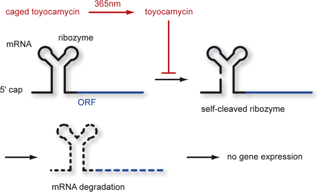
The involvement of RNA in the regulation of gene function is becoming increasingly prominent.1 Recently, a variety of natural riboswitches have been discovered in either the 3’ or 5’ untranslated region (UTR) of genes.2 Moreover, one natural riboswitch based on an a cis-acting allosteric ribozyme, which is activated by a small molecule ligand (glucosamine-6-phosphate), is known.3 Cleavage of this ribozyme results in mRNA degradation and gene silencing. One of the most prominent ribozymes is the hammerhead ribozyme which contains a central core of 15 conserved nucleotides.4 This RNA enzyme and its site-specific cleavage of RNA strands has been intensively studied in vitro.4, 5 However, in vivo applications of the minimal hammerhead ribozyme have been hampered by the necessity of high salt concentrations for sufficient catalytic activity.6 Recently, the Mulligan lab reported a mutated version of the full-length hammerhead ribozyme which was active under physiological conditions and led to transgene inactivation in mammalian cell culture and mice when inserted into the 5’ UTR of a gene.7 The active ribozyme removes the 5’ cap from the mRNA through self-cleavage leading to an inhibition of translation, mRNA degradation, and thus gene silencing (Scheme 1). Subsequent screening of compound collections for inhibitors of ribozyme activity delivered the natural product toyocamycin (1).8 This antibiotic efficiently inhibits ribozyme function in vivo at a micromolar concentration and thus induces gene expression.
Scheme 1.
A self-cleaving hammerhead ribozyme located in the 5’ untranslated region of a transgene leads to gene silencing. Toyocamycin (1) inhibits ribozyme function and induces expression of an open reading frame (ORF). Using a caged toyocamycin (2), this process can be controlled with light.
Due to the modular nature and the simplicity of this gene expression system (in comparison to transcription factors, operators, promoters, etc.), and its function in mammalian cell culture and mice, we adapted it to achieve photochemical gene regulation. Light-activation of gene function has the advantage of being precisely controllable in a spatial and temporal fashion.9 Moreover, light irradiation is minimally invasive inducing only low perturbations to the system under study.10 All previously reported eukaryotic light-inducible gene expression systems are based on transcriptional activation, thus the implemenation of a ribozyme photoregulatory system is novel.11 In order to achieve photochemical control over translation we decided to install a light-removable photocaging group on 1.
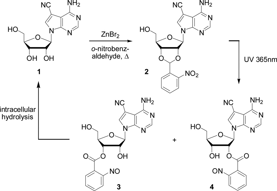
The precise mechanism of action of how 1 inhibits hammerhead ribozyme function is still unknown; however, preliminary evidence suggests the incorporation of 1 into the ribozyme by RNA polymerase II.8 Thus, we hypothesized that an inactive analog of 1 can be generated by blocking either the 3’ or the 5’ position with a caging group. Initial experiments of synthesizing a 2-nitrobenzyl or 6-nitropiperonyl ether specifically at the 5’ hydroxyl group of 1 were unsuccessful. A synthesized ortho-nitroveratryl carbonate at the 5’ position (as a mixture together with 3’ and 2’ carbonates) was not stable under physiological conditions. Finally, we synthesized the dioxolane caged 2 in one step from 1 through treatment with zinc bromide12 in neat ortho-nitrobenzaldehyde at 60 °C for 24 h (67% yield, Scheme 2).13 All attempts to conduct the same reaction with 6-nitropiperonal in order to obtain a favorable bathochromic shift in the absorption maximum of the caged compound failed.
Scheme 2.
Conversion of toyocamycin (1) into caged toyocamycin 2. UV light irradiation generates the esters 3 and 4 which are hydrolyzed intracellularly to generate toyocamycin (1).
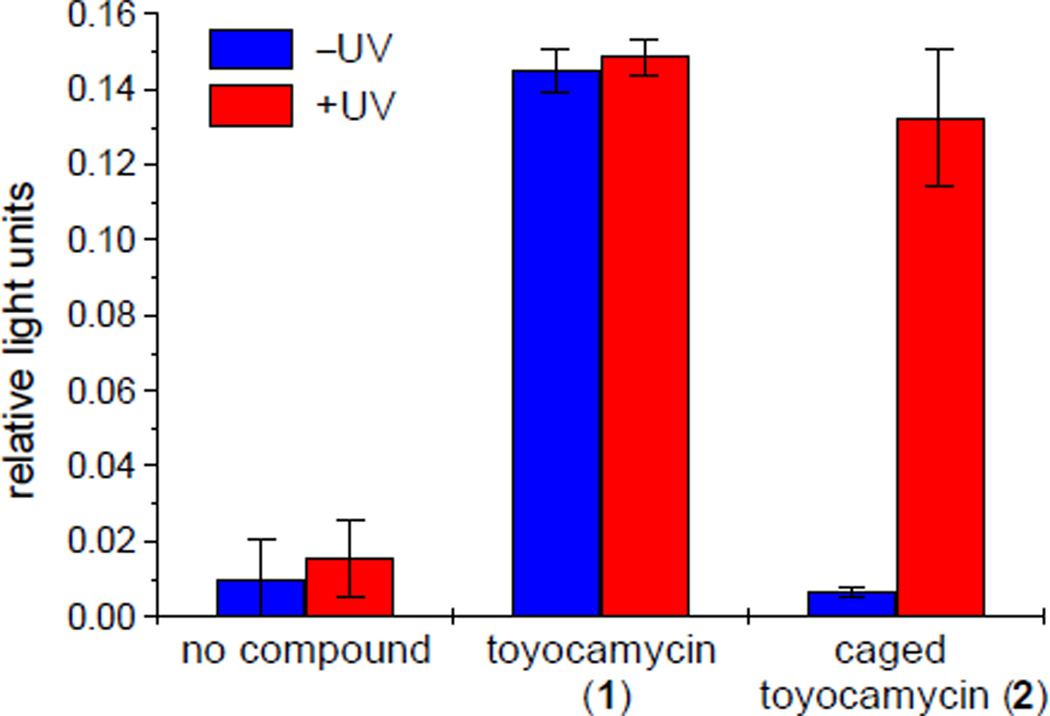
We speculated that 2 is inactive as a repressor of ribozyme function, which was verified by a luciferase assay in HEK293T cells (Fig. 1). Transcripts encoded by the N117-Luc plasmid contain the self-cleaving ribozyme upstream of the firefly luciferase open reading frame (Scheme 1, ORF = luciferase).7 Here, the caged toyocamycin 2 (10 µM) leads to a low luciferase signal which is within the error margin of the signal obtained when the cells harboring the reporter construct are not exposed to any small molecule, demonstrating the inactivity and cellular stability of 2 towards decaging (Fig. 1). An initial experiment irradiating 2 in the absence of cells with UV light of 365 nm for 10 min (25 W hand-held UV lamp) revealed a complete photochemical conversion into the benzoic esters 3 and 4 in a ratio of 1:1 as determined by 1H NMR and GC (Scheme 2 and see the Supplementary Information). We hypothesized that the esters 3 and 4 will be enzymatically hydrolyzed to active toyocamycin (1) intracellularly. Thus, HEK293T cells transfected with the N117-Luc construct were exposed to 2 (10 µM) for 48 h followed by a change to media not containing 2 and a brief UV irradiation (365 nm, 5 min, 25 W hand-held UV lamp). A 20-fold activation of luciferase activity was detected profiding expression levels virtually identical with the induction using regular toyocamycin. Within the error margin, UV irradiation itself had no effect on luciferase activity in any of these experiments (Fig. 1).
Fig. 1.
Luciferase assay demonstrating the induction of gene expression with toyocamycin (1), the inactivity of caged toyocamycin (2) in the absence of UV light, and the restoration of gene activity through UV irradiation. All experiments were conducted in triplicate and the average ratio of luciferase light units (firefly/Renilla) with its standard deviation is reported.
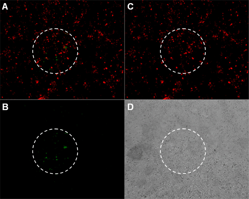
In order to apply the developed photochemical deactivation of ribozyme function in the spatial control of gene activity, we generated the N117-GFP plasmid encoding a green fluorescent protein (GFP) transcript with the self-cleaving ribozyme located in the 5' UTR (see Supplementary Information). A monolayer of 293T cells was transfected with N117-GFP (and a DsRed transfection control) and incubated with growth media containing 2 (10 µM) for 48h. The media containing 2 was removed, the cells were washed with PBS buffer, and subsequently irradiated for 30 sec through an inverted compound microscope equipped with a 100 W Xe/Hg lamp using a DAPI fluorescence filter (340–380 nm excitation). The cells were imaged after 24 h (to provide GFP expression and maturation) with a Leica DM5000B compound microscope revealing precise spatial control of gene expression since GFP activity was only observed in the irradiated area (Fig. 2). No diffusion and gene activation by the decaged toyocamycin in neighboring cells was observed. Thus, a highly stringent spatially and temporally regulated gene expression system was discovered.
Fig. 2.
Spatial control of gene expression in 293T cells harboring N117-GFP. The area within the white circle was briefly irradiated with UV light. A) Montage of the GFP and DsRed images. B) GFP expression is only visible in the irradiated area. C) DsRed transfection control. D) Brightfield image of the cell monolayer.
In summary, we developed a light-activated gene expression system for mammalian cells based on a self-cleaving hammerhead ribozyme and a photocaged small molecule inhibitor of ribozyme function. Excellent induction of gene expression after UV irradiation was observed, and spatial control of gene expression was achieved through UV irradiation of a cell monolayer through the optics of a regular fluorescent microscope. We expect that this light-activation methodology will find widespread application in the spatial-temporal investigation of gene function, especially since this ribozyme gene regulation system is structurally and conceptually much simpler than previously described photochemical gene regulation systems, and since it has shown to be active in vertebrate species.7
DDY acknowledges a graduate research fellowship from the ACS Medicinal Chemistry Division. AD is a Beckman Young Investigator and a Cottrell Scholar. We thank Prof. Richard C. Mulligan (Harvard University) for generously providing the N117-Luc plasmid, Xin Xiong for technical support, and Berry & Associates, Inc. for 1. This research was supported in part by research grant no. 5-FY05-1215 from the March of Dimes Birth Defects Foundation and the NIH (GM079114).
Supplementary Material
Footnotes
Electronic Supplementary Information (ESI) available: Experimental protocols for the synthesis and UV irradiation of 2, analytical data and 1H NMR spectrum of 2, luciferase assay protocol, construction of N117-GFP, and a protocol for the spatially controlled GFP activation. See http://dx.doi.org/10.1039/b000000x/
References
- 1.a) Win MN, Smolke CD. Biotechnol. Genet. Eng. Rev. 2007;24:311–346. doi: 10.1080/02648725.2007.10648106. [DOI] [PubMed] [Google Scholar]; b) Kawasaki H, Wadhwa R, Taira K. Differentiation. 2004;72:58–64. doi: 10.1111/j.1432-0436.2004.07202006.x. [DOI] [PubMed] [Google Scholar]
- 2.a) Winkler WC, Breaker RR. Chembiochem. 2003;4:1024–1032. doi: 10.1002/cbic.200300685. [DOI] [PubMed] [Google Scholar]; b) Tucker BJ, Breaker RR. Curr. Opin. Struct. Biol. 2005 doi: 10.1016/j.sbi.2005.05.003. [DOI] [PubMed] [Google Scholar]; c) Mandal M, Breaker RR. Nat. Rev. Mol. Cell. Biol. 2004;5:451–463. doi: 10.1038/nrm1403. [DOI] [PubMed] [Google Scholar]; d) Brantl S. Trends Microbiol. 2004;12:473–475. doi: 10.1016/j.tim.2004.09.008. [DOI] [PubMed] [Google Scholar]
- 3.Winkler WC, Nahvi A, Roth A, Collins JA, Breaker RR. Nature. 2004;428:281–286. doi: 10.1038/nature02362. [DOI] [PubMed] [Google Scholar]
- 4.Doudna JA, Cech TR. Nature. 2002;418:222–228. doi: 10.1038/418222a. [DOI] [PubMed] [Google Scholar]
- 5.a) Strobel SA, Cochrane JC. Curr. Opin. Chem. Biol. 2007;11:636–643. doi: 10.1016/j.cbpa.2007.09.010. [DOI] [PMC free article] [PubMed] [Google Scholar]; b) Silverman SK. RNA. 2003;9:377–383. doi: 10.1261/rna.2200903. [DOI] [PMC free article] [PubMed] [Google Scholar]; c) Khan AU, Lal SK. J. Biomed. Sci. 2003;10:457–467. doi: 10.1007/BF02256107. [DOI] [PubMed] [Google Scholar]; d) Blount KF, Uhlenbeck OC. Biochem. Soc. Trans. 2002;30:1119–1122. doi: 10.1042/bst0301119. [DOI] [PubMed] [Google Scholar]
- 6.a) Breaker RR. Nature. 2004;432:838–845. doi: 10.1038/nature03195. [DOI] [PubMed] [Google Scholar]; b) Rossi JJ. Trends Biotechnol. 1995;13:301–306. doi: 10.1016/S0167-7799(00)88969-6. [DOI] [PubMed] [Google Scholar]
- 7.Yen L, Svendsen J, Lee JS, Gray JT, Magnier M, Baba T, D'Amato RJ, Mulligan RC. Nature. 2004;431:471–476. doi: 10.1038/nature02844. [DOI] [PubMed] [Google Scholar]
- 8.Yen L, Magnier M, Weissleder R, Stockwell BR, Mulligan RC. Rna. 2006;12:797–806. doi: 10.1261/rna.2300406. [DOI] [PMC free article] [PubMed] [Google Scholar]
- 9.a) Young DD, Deiters A. Org. Biomol. Chem. 2007;5:999–1005. doi: 10.1039/b616410m. [DOI] [PubMed] [Google Scholar]; b) Tang X, Dmochowski IJ. Mol. Biosyst. 2007;3:100–110. doi: 10.1039/b614349k. [DOI] [PubMed] [Google Scholar]; c) Mayer G, Heckel A. Angew. Chem. Int. Ed. Engl. 2006;45:4900–4921. doi: 10.1002/anie.200600387. [DOI] [PubMed] [Google Scholar]; d) Curley K, Lawrence DS. Pharmacol. Ther. 1999;82:347–354. doi: 10.1016/s0163-7258(98)00055-2. [DOI] [PubMed] [Google Scholar]; e) Adams SR, Tsien RY. Annu. Rev. Physiol. 1993;55:755–784. doi: 10.1146/annurev.ph.55.030193.003543. [DOI] [PubMed] [Google Scholar]
- 10.a) Robert C, Muel B, Benoit A, Dubertret L, Sarasin A, Stary A. J. Invest. Dermatol. 1996;106:721–728. doi: 10.1111/1523-1747.ep12345616. [DOI] [PubMed] [Google Scholar]; b) Schindl A, Klosner G, Honigsmann H, Jori G, Calzavara-Pinton PC, Trautinger F. J. Photochem. Photobiol. B. 1998;44:97–106. doi: 10.1016/s1011-1344(98)00127-4. [DOI] [PubMed] [Google Scholar]
- 11.a) Cruz FG, Koh JT, Link KH. J. Am. Chem. Soc. 2000;122:8777–8778. [Google Scholar]; b) Lin W, Albanese C, Pestell RG, Lawrence DS. Chem. Biol. 2002;9:1347–1353. doi: 10.1016/s1074-5521(02)00288-0. [DOI] [PubMed] [Google Scholar]; c) Cambridge SB, Geissler D, Keller S, Curten B. Angew. Chem. Int. Ed. Engl. 2006;45:2229–2231. doi: 10.1002/anie.200503339. [DOI] [PubMed] [Google Scholar]
- 12.Lin WY, Lawrence DS. J. Org. Chem. 2002;67:2723–2726. doi: 10.1021/jo0163851. [DOI] [PubMed] [Google Scholar]
- 13.Yousefi-Salakdeh E, Murtola M, Zetterberg A, Yeheskiely E, Stromberg R. Bioorg. Med. Chem. 2006;14:2653–2659. doi: 10.1016/j.bmc.2005.11.049. [DOI] [PubMed] [Google Scholar]
Associated Data
This section collects any data citations, data availability statements, or supplementary materials included in this article.


