



Abstract
Sports injuries involving the hand and wrist are common and, as a result, many different practitioners (athletic trainers, physical therapists, primary care physicians, general orthopedic surgeons) will encounter these injuries. In addition to thorough evaluation, an understanding of typical pathologies seen in the athlete enhances diagnosis and facilitates appropriate, expedient management. These injuries are complex because they can be either acute traumatic or repetitive in origin and often involve both the bony skeleton and soft tissues. This article provides a review of athletic injuries to the wrist with particular focus on physical evaluation and management of the most common and challenging fractures and soft tissue injuries.
Keywords: Sports injuries, Wrist, Scaphoid fracture, Scapholunate ligament tear, Triangular fibrocartilaginous complex (TFCC) tear, Hamate hook fracture, Pisiform fracture, Extensor carpi ulnaris (ECU)
Introduction
Hand and wrist injuries are common in the athletic population. Between 3 % and 9 % of all sports injuries involve the hand and wrist [1]. A recent study surveying sports- related injuries in high school athletes found that fractures of the hand and wrist were the most common body site, making up nearly 40 % of all such fractures [2•]. Hand surgeons routinely treat traumatic injuries sustained during contact sports such as basketball and football, but also treat as many, if not more, injuries due to repetitive or overuse from racquet sports, golf, and gymnastics. The increasing rate of competitive athletic participation in children and adolescents, combined with an increasingly active aging population, make an understanding of these injuries critical for a host of practitioners. Before referral to a hand surgeon, these injuries will routinely be evaluated by athletic trainers, physical therapists, primary care physicians or general orthopedic surgeons (Table 1).
Table 1.
Summary of common wrist injuries in the athlete, diagnosis and management
| Diagnostic keys | Preferred initial management in the athlete | |
|---|---|---|
| Acute injuries | ||
| Scaphoid fracture | • Persistent tenderness to palpation palmarly or dorsally | • Non-displaced waist or distal fracture – cast immobilization or immediate surgical fixation |
| • Careful scrutiny of plain radiographs, serial imaging and use of CT or MRI | • Displaced waist or distal fracture – immediate surgical fixation | |
| • Proximal pole fracture – immediate surgical fixation | ||
| Scapholunate ligament tear | • Dorsal wrist swelling | • Confirmation of suspected diagnosis by MRI |
| • Tenderness to palpation 1 cm distal to dorsoradial tubercle | • Early (<6 weeks) direct ligament repair and k-wire fixation | |
| • SL widening compared with contralateral side on standard or stress radiographs | ||
| TFCC tear (traumatic) | • Point tenderness to palpation in fovea | • Unstable DRUJ – immediate surgical repair |
| • Pain reproduced by wrist ulnar deviation | • Stable DRUJ – immobilization versus early MRI and surgical repair/debridement | |
| • DRUJ instability when compared with contralateral side | ||
| Hamate fracture | • At-risk sport including baseball, golf, tennis | • Non-displaced – cast immobilization versus surgical fragment excision |
| • Direct tenderness to palpation palmarly | • Displaced – surgical fragment excision | |
| Pisiform fracture | • Direct tenderness to palpation palmarly | • Symptomatic immobilization |
| ECU subluxation | • Athlete reports: clicking, popping, instability | • Immobilization versus surgical repair or reconstruction |
| • Confirmation of instability by physical exam or dynamic ultrasonography | ||
| Chronic and repetitive injuries | ||
| TFCC tear (degenerative) | • Point tenderness to palpation in fovea | • Immobilization, activity modification, ± corticosteroid injection |
| • Pain reproduced by wrist ulnar deviation | • MRI if symptoms persist >6 weeks | |
| Hamate nonunion | • At-risk sport including baseball, golf, tennis | • Surgical fragment excision |
| • Direct tenderness to palpation palmarly | ||
| ECU tendinitis | • Tenderness to palpation over distal ulnar | • Immobilization, activity modification |
| • Pain with resisted wrist extension in ulnar deviation | • ± corticosteroid injection if symptoms persist >6 weeks | |
Evaluation of the injured wrist begins with thorough inspection. Any swelling is an indication of injury and merits further investigation. Swelling can be subtle and the contralateral, uninjured extremity is used to assess symmetry. While the anatomy of the wrist is complex, the superficial location of the majority of structures makes direct palpation possible. Throughout this article, we will emphasize specific elements of physical examination that facilitate clinical diagnosis of these common injuries. The purpose of this review is to provide a comprehensive outline of common injuries sustained to the wrist due to participation in sports. We emphasize physical examination diagnosis and provide in depth discussion of the management of the most common injuries and those that are particularly challenging to diagnose and treat.
Scaphoid fracture
The scaphoid is the most commonly fractured carpal bone, accounting for up to 70 % of all carpal fractures [1, 3, 4]. Timely diagnosis and treatment of these fractures is critical due to its tenuous retrograde vascularity and well-documented risk for nonunion with progression to wrist arthritis [3–7]. Appropriate physical examination, clinical suspicion and radiographic evaluation prevent the common misdiagnosis of “wrist sprain”. Injury is most commonly sustained from a fall onto an outstretched hand and swelling is often subtle and generalized about the wrist. Palpation of the “anatomic snuffbox” corresponds to the waist of the scaphoid. The distal pole is actually more superficial and easily palpable volarly as the most proximal bony prominence on the radial aspect of the palm at the level of the wrist crease. Pain with palpation in either of these locations or with axial loading to the thumb should raise suspicion for scaphoid fracture.
Radiographic diagnosis of scaphoid fracture can also be challenging. Standard wrist radiographs including posteroanterior (PA), lateral, and an oblique view can be augmented by a PA ulnar deviation view, which extends the scaphoid, allowing for better visualization. Subtle fractures may not become apparent on routine radiographs until 1–2 weeks following injury. Classic management includes thumb spica immobilization of suspected scaphoid fractures during this time period [3, 8]. This may not be ideal in high level athletes and some authors have advocated early use of advanced imaging modalities, computed tomography (CT), or magnetic resonance imaging (MRI), to expedite diagnosis and treatment [1, 8–10].
Treatment of the radiographically confirmed acute scaphoid fracture varies from cast immobilization to operative management with internal fixation based on the fracture location and displacement. Classically, nondisplaced or minimally displaced fractures of the scaphoid waist or distal pole, where vascularity is more robust, have been managed with thumb spica cast immobilization, while even minimally displaced fractures of the less vascular proximal pole are managed with internal fixation. Debates in the management of scaphoid fractures have included: the use of above or below the elbow thumb spica casting in conservative treatment, internal fixation through the palmar or dorsal approach, optimal screw placement in operative fixation, and the treatment of minimally displaced scaphoid waist fractures. Currently, the most controversial topic is operative vs nonoperative management of minimally displaced scaphoid waist fractures with some authors suggesting that early operative management can speed time to union providing benefits of return to work and function [11, 12, 13•]. These factors become increasingly pertinent as they relate to athletes and return to play [14].
Our preferred management of acute scaphoid fractures of non- or minimally-displaced fractures of the distal pole of the scaphoid includes below-elbow thumb spica cast immobilization for 8–10 weeks, or until there is radiographic evidence of healing as demonstrated either by plain radiography or CT. We are more aggressive with surgical management of proximal pole fractures, even if minimally displaced due to limited vascularity. In surgery, care is taken to avoid exposure of the dorsal ridge where the critical blood supply to the scaphoid is located. The optimal screw trajectory is in a center-center position in line with the long axis of the scaphoid. We use intraoperative fluoroscopy to determine screw trajectory. We treat displaced scaphoid waist fractures with internal fixation and approach these using either a minimal dorsal or palmar approach based on the degree of displacement (more displacement is best approached through a palmar approach) and surgeon preference. In management of non- or minimally-displaced fractures of the scaphoid, we present the risks and benefits of surgical management to the athlete so that a collaborative, informed decision can be made regarding management of these controversial fractures. For many athletes, cast immobilization for 2–3 months is unacceptable, making surgical management appealing.
The take home point on scaphoid fractures in athletes is clinical suspicion and early diagnosis. Management of scaphoid nonunion or malunion for missed scaphoid fractures is beyond the scope of this article, but involves more complex surgical management and limited ability to restore pre-injury level of function.
Scapholunate interosseous ligament tear
Injuries to the scapholunate (SL) interosseous are the most common ligamentous injuries in the wrist [1]. The SL ligament is a key stabilizer of the proximal carpal row of the wrist and its dorsal component is the most important [15]. Similar to scaphoid fracture, injury most commonly results from a fall on an outstretched hand. While sometimes the diagnosis is straightforward, it can be challenging with poorer outcomes associated with management of chronic injuries. This makes early recognition of injury paramount. Untreated complete ligament injuries progress to a predictable pattern of wrist arthritis called scapholunate advanced collapse, or SLAC wrist.
Physical findings of a scapholunate interosseous ligament injury can be nonspecific and include generalized wrist swelling and tenderness to palpation over the dorsal wrist. The examiner should make an effort to localize tenderness to the SL ligament which can be directly palpated 1 cm distal to the dorsoradial tubercle (Lister’s tubercle). In an uninjured wrist, this space is normally concave. While difficult in the setting of acute injury due to pain and guarding, the scaphoid shift test, or Watson’s maneuver, is another sign of SL laxity. To perform this test, the examiner places a thumb on the distal pole of the scaphoid. The examiner uses his/her other hand to axially load the wrist and bring the wrist from ulnar to radial deviation. In a positive test, dorsally directed pressure on the scaphoid results in dorsal wrist pain and a palpable “clunk” [16]. Examination of the contralateral side helps to differentiate normal vs pathologic laxity.
Radiographic evaluation of suspected SL tear should begin with standard wrist views. Complete SL discontinuity will manifest as diastasis of the SL interval, which is normally < 3 mm or < 2 mm different from the contralateral side [17]. Incomplete injuries, so called pre-dynamic or dynamic, will not show any abnormality on standard imaging. Instead, stress views, such as a clenched fist in combination with wrist supination and ulnar deviation, which drives the capitate into the scapholunate interval, can reveal dynamic scapholunate diastasis. Definitive diagnosis is usually confirmed with MRI (with or without arthrography). The use of wrist arthroscopy for diagnosis and treatment of partial tears of the SL ligament is a current topic of research interest [18–20].
We treat acute, partial tears of the SL ligament in athletes with time off from sport and splint or cast immobilization for 4–6 weeks until clinical examination becomes pain-free. Gradual return to sport is based on symptomatology. Complete acute tears of the SL ligament should be managed early with surgical repair to avoid static changes in carpal alignment [15, 19, 21]. While several techniques for ligament repair and reconstruction have been described, we favor direct ligament repair through a dorsal approach using a suture anchor with 2 supplementary Kirschner wires transfixing the SL interval and an additional wire from the scaphoid to the capitate. In more chronic cases, once signs of carpal malalignment are visible radiographically (scaphoid flexion), we prefer a Szabo-style capsulodesis to augment primary repair [21]. Kirschner wires remain in place with cast immobilization for up to 10 weeks with regular clinical evaluation and radiographs to assess for pin complications such as infection and migration.
Scapholunate ligament injuries represent a spectrum of pathology and occur in combination with injury to multiple other supporting wrist ligaments. Early identification of complete tears that are amenable to repair maximize outcome in this difficult injury, especially if done prior to development of carpal malalignment. Athletes should be counseled that such injuries in athletes will necessitate significant time off from sport.
Triangular fibrocartilaginous complex (TFCC) tear
The TFCC is composed of composed of a relatively avascular central disc, dorsal, and palmar radioulnar ligaments, the subsheath of the extensor carpi ulnaris and the ulnar collateral ligament. The poor vascularity of the central disc influences the inherent healing potential of the TFCC and determines surgical management. Sports-related injuries of the TFCC occur both due to acute trauma as well as overuse and are a common cause of ulnar-sided wrist pain in the athlete. Acute traumatic injuries result from an axial load through the wrist. While the radiocarpal joint bears the majority of the force transmitted through the wrist, the force through the ulnocarpal joint is linked to ulnar variance. Athletes with an ulna that is relatively long compared with the radius (positive ulnar variance) are at increased risk for injury due to increased forces transmitted through the ulnocarpal joint in this setting. Repetitive injuries to the TFCC are most common in sports requiring forceful wrist flexion and rotation such as golf, tennis, and baseball. In addition to ulnar-sided wrist pain, the athlete may notice popping or clicking with wrist motion. Direct pressure over the TFCC, through direct palpation palmar to the ulnar styloid, recreates symptoms. Ulnar deviation combined with axial load, supination, and pronation may also reproduce pain. As the TFCC is a major stabilizer to the distal radioulnar joint (DRUJ), its stability should be checked on examination by assessing motion of the ulna relative to the radius. DRUJ motion should be compared with that of the contralateral wrist in a similar position of forearm rotation to differentiate normal from pathologic laxity.
Radiographic evaluation of a purely soft tissue TFCC injury will not demonstrate a pathognomonic abnormality. It can, however, demonstrate secondary findings, such as positive ulnar variance or ulnar styloid fracture, which may aid in diagnosis. Fractures of the ulnar styloid base may destabilize the TFCC and DRUJ. MRI evaluation, with or without arthrography, is sensitive for detection of TFCC pathology. Recent studies suggest that MRI will detect abnormalities in many asymptomatic wrists [22, 23•]. The practitioner should always be aware of false positives and use physical examination to corroborate diagnostic imaging. Arthroscopy remains the gold standard for diagnosis of TFCC pathology [17].
In the classification system of Palmer, tears of the TFCC are classified as either traumatic or degenerative [24]. Blood supply to the TFCC is more robust peripherally and relatively avascular centrally [17, 24, 25]. This anatomy guides surgical treatment. In the absence of mechanical symptoms (distal radioulnar joint (DRUJ) instability, catching, locking, both acute and degenerative tears are most commonly managed initially with a period of conservative treatment. Our preferred management of suspected TFCC injuries in the non-elite athlete includes a 4–6 week period of sport avoidance or modification, immobilization with an off-the-shelf or fabricated orthoplast wrist splint, and a course of anti-inflammatories followed by gradual return to full participation. If pain that limits athletic participation persists, we will at this point obtain an MRI. Because MRI is highly sensitive for detecting TFCC abnormality, we find MR arthrogram to be more specific in detecting clinically relevant tears in the TFCC by the leakage of contrast from the ulnocarpal joint into the DRUJ. There is little literature to either support or refute the utility of ulnocarpal corticosteroid injection in the treatment of TFCC pathology and we reserve use of steroid injection to aid in diagnosis or to treat acute pain that has failed oral anti-inflammatories.
Acute tears that are at the well-vascularized periphery of the TFCC are most amenable to repair. In contradistinction, chronic tears and those of the central disc are less likely to heal even with repair and are most commonly treated with arthroscopic debridement. Open and arthroscopically-assisted mini-open approaches to TFCC tear have been most commonly described [26]. As techniques in wrist arthroscopy have advanced, all-inside repair techniques can also be employed with good clinical results [27, 28]. Athletes who undergo TFCC repair can expect a 6–12 week period of immobilization and slow return to activities involving forceful pronosupination. Due to the lengthy recovery following repair of an acute tear, our management of the elite athlete may differ in an attempt to minimize or strategically place time away from sport. In this case, MRI diagnosis may be obtained earlier to facilitate definitive management.
Hook of hamate and pisiform fracture
While comprising less that 3 % of all carpal fractures, fractures of the hook of the hamate and pisiform should be considered in athletes with persistent ulnar sided wrist pain, whether from a traumatic fall onto an outstretched hand [29] or with an insidious onset. Fractures of the hamate and pisiform can be difficult to diagnose on routine radiographic views of the wrist and an appropriate index of suspicion combined with directed physical exam aid in early diagnosis. The pisiform is a sesamoid bone and the attachment of the flexor carpi ulnaris (FCU). It is a palpable prominence proximally in the hypothenar eminence at the wrist crease. Distal and radial to the pisiform, the hook of the hamate is also palpable deep to the hypothenar eminence. This superficial anatomy predisposes both to traumatic injury. Baseball, golf, hockey, and tennis athletes sustain repetitive trauma to the hypothenar eminence through use of a bat, club, stick, and racquet and these activities may lead to fracture of the hamate hook.
After directed physical examination, work-up of athletes with suspected hamate or pisiform fracture includes specific radiographic views of the wrist as conventional views are usually negative. The carpal tunnel radiographic view can demonstrate pathology at either the pisiform or hamate hook (Fig. 1). To further evaluate the pisiform, oblique lateral views can show the pisotriquetral articulation. We utilize CT and MRI to detect occult hamate fractures when suspicion is high and plain radiography is negative.
Fig. 1.
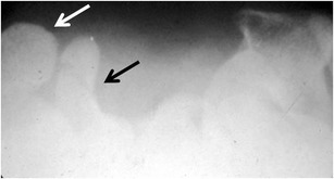
Carpal tunnel view is obtained with maximal wrist extension and x-ray beam oriented 15 degrees from the palm. The white arrow shows a normal pisiform and black arrow shows a minimally displaced hook of the hamate fracture. (Reproduced with permission of Dr. Fufa, personal files)
Pisiform fractures are most commonly acute injuries and can be treated non-operatively with symptomatic immobilization in a wrist brace, padding and activity modification for a period of 4–6 weeks. Occasionally, corticosteroid injection to the pisotriquetral joint may be indicated for pain that persists after immobilization. Less commonly, complete pisiform excision is used for cases of nonunion or refractory pain secondary to pisotriquetral arthritis.
If diagnosed acutely, nondisplaced hamate hook fractures may heal uneventfully with cast immobilization [30]. Due to propensity for the hamate hook to go on to nonunion, careful follow-up is needed. While internal fixation both through formal open palmar approach or percutaneous dorsal approach [31, 32•] has been described, we do not routinely utilize this technique. In most cases, we favor fragment excision through a palmar approach taking care to identify and protect the motor branch of the ulnar nerve. Fragment excision avoids the potential complications of flexor tendon or ulnar nerve irritation by fracture fragments or hardware. Athletes can expect return to sport quickly based on symptomatology, estimated at 7–10 weeks following excision [1].
Extensor carpi ulnaris tendonitis and instability
Extensor carpi ulnaris (ECU) tendonitis is the second most common tendonitis of the wrist (next to DeQuervain’s tenosynovitis) and is seen commonly in baseball and other sports requiring excessive ulnar deviation such as in hitting a baseball or swinging a golf club. Tendonitis is differentiated from ECU tendon instability that results from an acute injury that is exacerbated by repetitive movements in stick athletes such as baseball players, but also in golf and racket athletes. Instability is due to disruption of the fibro-osseous tunnel stabilizing the tendon at the distal ulna. Athletes may complain of pain on the dorso-ulnar wrist which may be associated with painful snapping or clicking during pronosupination.
The ECU muscle and tendon can be palpated along its course from its origin at the lateral epicondyle to its insertion in the dorsal base of the 5th metacarpal. Direct palpation may elicit symptoms in cases of tendonitis and sensitivity can be increased with resisted wrist extension in ulnar deviation and supination. It is important to also perform dynamic examination through direct palpation of the ECU over the distal ulna during passive and active pronosupination as this may demonstrate subluxation.
Typically, ECU tendonitis and instability are accurately diagnosed with physical examination. Radiographs do not typically demonstrate specific abnormality, but may show evidence of prior trauma, such as ulnar styloid fracture, to suggest etiology for disruption of the fibro-osseous sheath. When needed to confirm a diagnosis of tendonitis, MRI may reveal typical patterns including an enlarged tendon, fluid signal within the tendon sheath, and intrasubstance high signal intensity. Dynamic imaging with ultrasound to document subluxation has also been described [33] and can diagnose more subtle cases of instability that can’t be voluntarily reproduced or elicited on physical exam.
Most ECU tendonitis can be effectively managed with conservative therapies. Immobilization (with the forearm in neutral or slight pronation and the wrist neutral), medical management with anti-inflammatories, and activity modification are the mainstays of nonoperative treatment. Injection of corticosteroid can also be used in refractory cases while surgery is reserved for cases in which all the above modalities have failed. There is less consensus on the appropriate timing or treatment for ECU instability. It can be managed initially with similar conservative measures as used for ECU tendonitis. Some authors have suggested that acute injuries can be treated with a period of cast immobilization with the forearm pronated and wrist radially deviated [34]. However, improved understanding of the anatomy of the 6th extensor compartment and of the tear patterns seen in ECU instability suggests that surgical management is often needed in cases that have established symptoms of recurrent instability and pain. Particularly in cases in which the fibro-osseous sheath is disrupted from the radial wall of the tunnel (as diagnosed by MRI), it is felt that the position of the tendon may inhibit the ability for primary healing of the sheath. In the young, active athlete, acute repair is most often indicated for reliably good result and return of pre-injury function. When indicated, we perform stabilization using either direct repair (if tissue quality is sufficient) or reconstruction (using local retinacular tissue) through a dorsal approach. Rehabilitation following reconstruction must progress slowly under the direction of a hand therapist and athletes may not return to full activity for 3 to 4 months.
Conclusions
Wrist injuries are common in athletes and can result from either trauma or repetitive injury. For the clinician performing initial evaluation, knowledge of the most common injuries and directed physical examination aid in early diagnosis and treatment.
Acknowledgments
Disclosure
DT Fufa: none; CA Goldfarb: Receives royalties from Wolters Kluwer.
References
Papers of particular interest, published recently, have been highlighted as: • Of importance
- 1.Rettig AC. Athletic injuries of the wrist and hand. Part I: traumatic injuries of the wrist. Am J Sports Med. 2003;31:1038–48. doi: 10.1177/03635465030310060801. [DOI] [PubMed] [Google Scholar]
- 2.Swenson DM, Yard EE, Collins CL, et al. Epidemiology of US high school sports-related fractures, 2005–2009. Clin J Sport Med. 2010;20:293–9. doi: 10.1097/JSM.0b013e3181e8fae8. [DOI] [PubMed] [Google Scholar]
- 3.Gelberman RH, Wolock BS, Siegel DB. Fractures and non-unions of the carpal scaphoid. J Bone Joint Surg Am. 1989;71:1560–5. [PubMed] [Google Scholar]
- 4.Hove LM. Epidemiology of scaphoid fractures in Bergen, Norway. Scand J Plast Reconstr Surg Hand Surg. 1999;33:423–6. doi: 10.1080/02844319950159145. [DOI] [PubMed] [Google Scholar]
- 5.Gelberman RH, Menon J. The vascularity of the scaphoid bone. J Hand Surg Am. 1980;5:508–13. doi: 10.1016/s0363-5023(80)80087-6. [DOI] [PubMed] [Google Scholar]
- 6.Mack GR, Bosse MJ, Gelberman RH, YU E, et al. The natural history of scaphoid non-union. J Bone Joint Surg Am. 1984;66:504–9. [PubMed] [Google Scholar]
- 7.Lindstrom G, Nystrom A. Natural history of scaphoid non-union, with special reference to "asymptomatic" cases. J Hand Surg Br. 1992;17:697–700. doi: 10.1016/0266-7681(92)90204-F. [DOI] [PubMed] [Google Scholar]
- 8.Sjolin SU, Andersen JC. Clinical fracture of the carpal scaphoid–supportive bandage or plaster cast immobilization? J Hand Surg Br. 1988;13:75–6. doi: 10.1016/0266-7681_88_90057-5. [DOI] [PubMed] [Google Scholar]
- 9.Amadio PC, Berquist TH, Smith DK, Ilstrup DM, Cooney WP 3rd, Linscheid RL, et al. Scaphoid malunion. J Hand Surg Am. 1989;14:679–87. doi: 10.1016/0363-5023(89)90191-3. [DOI] [PubMed] [Google Scholar]
- 10.Kumar S, O'Connor A, Despois M, Galloway H, et al. Use of early magnetic resonance imaging in the diagnosis of occult scaphoid fractures. the CAST Study (Canberra Area Scaphoid Trial) N Z Med J. 2005;118:U1296. [PubMed] [Google Scholar]
- 11.Bond CD, Shin AY, McBride MT, Dao KD. Percutaneous screw fixation or cast immobilization for nondisplaced scaphoid fractures. J Bone Joint Surg. 2001;83–A:483–8. doi: 10.2106/00004623-200104000-00001. [DOI] [PubMed] [Google Scholar]
- 12.Ram AN, Chung KC. Evidence-based management of acute nondisplaced scaphoid waist fractures. J Hand Surg Am. 2009;34:735–8. doi: 10.1016/j.jhsa.2008.12.028. [DOI] [PMC free article] [PubMed] [Google Scholar]
- 13.Ibrahim T, Qureshi A, Sutton AJ, Dias JJ, et al. Surgical vs nonsurgical treatment of acute minimally displaced and undisplaced scaphoid waist fractures: pairwise and network meta-analyses of randomized controlled trials. J Bone Joint Surg Am. 2011;36:1759–68. doi: 10.1016/j.jhsa.2011.08.033. [DOI] [PubMed] [Google Scholar]
- 14.Rettig AC, Kollias SC. Internal fixation of acute stable scaphoid fractures in the athlete. Am J Sports Med. 1996;24:182–6. doi: 10.1177/036354659602400211. [DOI] [PubMed] [Google Scholar]
- 15.Kuo K, Wolfe SW. Scapholunate instability: current concepts in diagnosis and management. J Hand Surg Am. 2008;33:998–1013. doi: 10.1016/j.jhsa.2008.04.027. [DOI] [PubMed] [Google Scholar]
- 16.Hwang JJ, Goldfarb CA, Gelberman RH, Boyer MI. The effect of dorsal carpal ganglion excision on the scaphoid shift test. J Hand Surg Br. 1999;24:106–8. doi: 10.1016/S0266-7681(99)90053-0. [DOI] [PubMed] [Google Scholar]
- 17.Rosner JL, Zlatkin MB, Clifford P, Ouellette EA, Awh MH, et al. Imaging of athletic wrist and hand injuries. Semin Musculoskelet Radiol. 2004;8:57–79. doi: 10.1055/s-2004-823015. [DOI] [PubMed] [Google Scholar]
- 18.Darlis NA, Kaufmann RA, Giannoulis F, Sotereanos DG, et al. Arthroscopic debridement and closed pinning for chronic dynamic scapholunate instability. J Hand Surg Am. 2006;31:418–24. doi: 10.1016/j.jhsa.2005.12.015. [DOI] [PubMed] [Google Scholar]
- 19.Manuel J, Moran SL. The diagnosis and treatment of scapholunate instability. Hand Clin. 2010;26:129–44. doi: 10.1016/j.hcl.2009.08.006. [DOI] [PubMed] [Google Scholar]
- 20.Dohi Y, Omokawa S, Ono H, Aoki M, Akahane M, Wada T, et al. Arthroscopic gap distance can predict the degree of scapholunate ligament tears: a cadaver study. J Orthop Sci. 2012;17:64–9. doi: 10.1007/s00776-011-0172-x. [DOI] [PubMed] [Google Scholar]
- 21.Szabo RM. Scapholunate ligament repair with capsulodesis reinforcement. J Hand Surg Am. 2008;33:1645–54. doi: 10.1016/j.jhsa.2008.08.016. [DOI] [PubMed] [Google Scholar]
- 22.Blazar PE, Chan PS, Kneeland JB, Leatherwood D, Bozentka DJ, Kowalchick R, et al. The effect of observer experience on magnetic resonance imaging interpretation and localization of triangular fibrocartilage complex lesions. J Hand Surg Am. 2001;26:742–8. doi: 10.1053/jhsu.2001.24966. [DOI] [PubMed] [Google Scholar]
- 23.Iordache SD, Rowan R, Garvin GJ, Osman S, Grewal R, Faber KJ, et al. Prevalence of triangular fibrocartilage complex abnormalities on MRI scans of asymptomatic wrists. J Hand Surg Am. 2012;37:98–103. doi: 10.1016/j.jhsa.2011.10.006. [DOI] [PubMed] [Google Scholar]
- 24.Palmer AK. Triangular fibrocartilage complex lesions: a classification. J Hand Surg Am. 1989;14:594–606. doi: 10.1016/0363-5023(89)90174-3. [DOI] [PubMed] [Google Scholar]
- 25.Nagle DJ. Triangular fibrocartilage complex tears in the athlete. Clin Sports Med. 2001;20:155–66. doi: 10.1016/S0278-5919(05)70253-2. [DOI] [PubMed] [Google Scholar]
- 26.Chloros GD, Wiesler ER, Poehling GG. Current concepts in wrist arthroscopy. Arthroscopy. 2008;24:343–54. doi: 10.1016/j.arthro.2007.10.006. [DOI] [PubMed] [Google Scholar]
- 27.Yao J, Dantuluri P, Osterman AL. A novel technique of all-inside arthroscopic triangular fibrocartilage complex repair. Arthroscopy. 2007;23:1357. doi: 10.1016/j.arthro.2007.02.010. [DOI] [PubMed] [Google Scholar]
- 28.Yao J, Lee AT. All-arthroscopic repair of Palmer 1B triangular fibrocartilage complex tears using the FasT-Fix device. J Hand Surg Am. 2011;36:836–42. doi: 10.1016/j.jhsa.2011.01.005. [DOI] [PubMed] [Google Scholar]
- 29.Vigler M, Aviles A, Lee SK. Carpal fractures excluding the scaphoid. Hand Clin. 2006;22:501–16. doi: 10.1016/j.hcl.2006.07.007. [DOI] [PubMed] [Google Scholar]
- 30.Whalen JL, Bishop AT, Linscheid RL. Nonoperative treatment of acute hamate hook fractures. J Hand Surg Am. 1992;17:507–11. doi: 10.1016/0363-5023(92)90363-T. [DOI] [PubMed] [Google Scholar]
- 31.Saint-Cyr M, Oni G, Wong C, Sen MK, LaJoie AS, Gupta A, et al. Dorsal percutaneous cannulated screw fixation for delayed union and nonunion of the scaphoid. Plast Reconstr Surg. 2011;128:467–73. doi: 10.1097/PRS.0b013e31821e703b. [DOI] [PubMed] [Google Scholar]
- 32.Scheufler O, Radmer S, Andresen R. Dorsal percutaneous cannulated mini-screw fixation for fractures of the hamate hook. Hand Surg. 2012;17:287–93. doi: 10.1142/S0218810412970039. [DOI] [PubMed] [Google Scholar]
- 33.MacLennan AJ, Nemechek NM, Waitayawinyu T, Trumble TE, et al. Diagnosis and anatomic reconstruction of extensor carpi ulnaris subluxation. J Hand Surg Am. 2008;33:59–64. doi: 10.1016/j.jhsa.2007.10.002. [DOI] [PubMed] [Google Scholar]
- 34.Inoue G, Tamura Y. Recurrent dislocation of the extensor carpi ulnaris tendon. Br J Sports Med. 1998;32:172–4. doi: 10.1136/bjsm.32.2.172. [DOI] [PMC free article] [PubMed] [Google Scholar]