

Abstract
Colonoscopy is a widely used diagnostic and therapeutic procedure. While it is a relatively safe procedure, there is a risk of some complications. Splenic injury after colonoscopy is a very rare but a life-threatening complication; around 105 cases have been reported in the literature so far. Owing to the rarity of this complication, no management standards were defined. In the literature, most of the patients were managed with operative intervention and less frequently with observation. We report a case of splenic injury and massive hemoperitoneum due to colonoscopy treated non-operatively.
Background
Colonoscopy is a widely used procedure for the screening and surveillance of gastrointestinal disease and the use of the colonoscopy has increased significantly over the past decade. While colonoscopy is a relatively safe procedure, there is a risk of some complications such as haemorrhage and perforation, with incidence rates of 0.1–0.6% and 0.1–0.3%, respectively.1 Splenic injury (SI) after colonoscopy is a very rare but a life-threatening complication; more than 100 cases have been reported in the literature so far.2 Owing to the rarity of this complication, no management standards were defined. In the literature, most of the patients were managed with operative intervention (68–77%) and less frequently with observation or angioembolisation.3 4 We report a case of SI after colonoscopy treated non-operatively.
Case presentation
A 53-year-old man underwent diagnostic colonoscopy for evaluation of abdominal pain and chronic constipation. Colonoscopy was performed by an experienced endoscopist (>600 procedures/year) after the administration of the intravenous sedation. The procedure was uncomplicated and the entire colon was visualised without difficulty (no biopsy was taken and no abdominal pressure was performed). The patient tolerated the procedure well and was discharged from hospital after 1 h. His medical history was remarkable for asthma and the history of distal gastrectomy 15 years ago due to ulcers.
Investigations
Five hours after the procedure, the patient started to feel abdominal pain, sweating, weakness and dizziness and he was brought to the emergency department. His initial examination showed mild abdominal tenderness without guarding. Blood pressure was 100/60 mm Hg and pulse was 122 bpm. Haemoglobin level was 8.2 g/dL, haematocrit was 26%, white cell count was 9600/mm3 and platelet count was 380 000/mm3. Coagulation studies were normal. The upright abdominal x-ray did not show any evidence of free air (figure 1). Aggressive fluid resuscitation was initiated to the patient, in whom intra-abdominal free fluid and splenic haematoma was detected in the ultrasonography. Shortly afterwards, the patient’s blood pressure was measured as 130/70 mm Hg and the pulse as 96 bpm. With a prediagnosis of SI, contrast-enhanced abdominal CT was performed, which revealed free fluid in the perihepatic and perisplenic areas and hypodense areas (subcapsular haematoma) in the spleen (figure 2). Since the patient’s vital signs were stable, the decision was taken to follow non-operatively.
Figure 1.
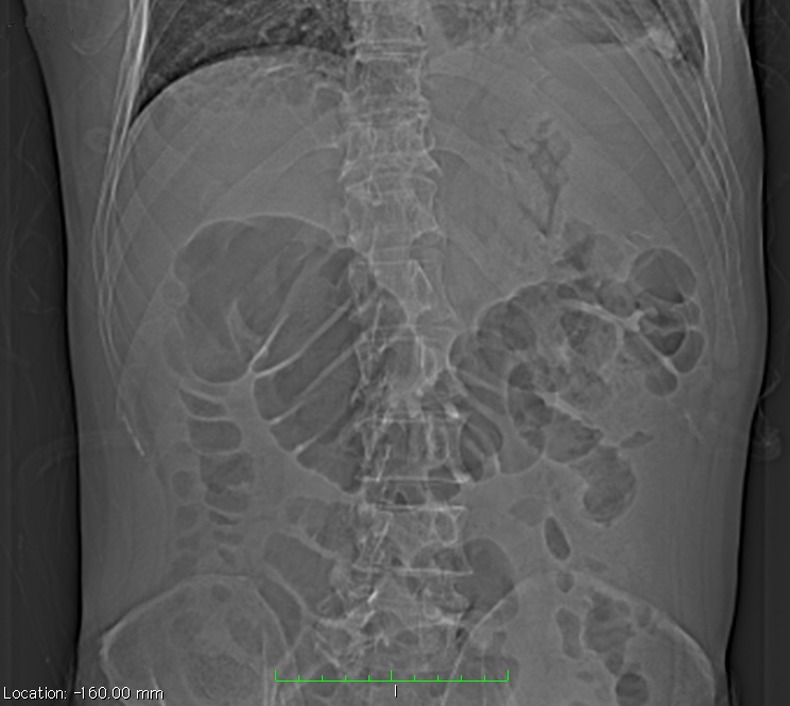
X-Ray radiograph shows dilated transverse colon and no intra-abdominal free air.
Figure 2.

Two sections of CT scan show massive intra-abdominal fluid and subcapsular splenic haematoma.
Treatment
The patient was followed with bed rest, blood transfusion and close clinical and laboratory monitoring. There were no signs of hypovolaemia, and the patient remained stable during the 5-day follow-up. The patient was discharged with a haemoglobin value of 12 g/dL. One month later, the patient's abdominal CT revealed resorption of the free fluid and persisting subcapsular haematoma in the spleen (figure 3A).
Figure 3.

CT scan views of the patient 1 month (A) and 6 month (B) later.
Outcome and follow-up
The patient had an uneventful course in the 6 months of the follow-up with the same CT findings (figure 3B).
Discussion
In the large series reported in the literature, SI is an uncommon complication of colonoscopy. Since it was first defined by Wherry and Zehner in 1974, it has remained a rare complication that is only reported in case reports or case series.5 6 However, it is likely that the true incidence of this complication is underestimated due to asymptomatic or unreported cases. Interestingly, although colonoscopy was introduced in the 1960s, 77% of cases of SI due to colonoscopy have been reported in the period after the year 2000.2 It will probably be encountered more frequently in the coming years, with increasing and widespread use of colonoscopy.
Different mechanisms for the occurrence of this complication have been described. The common aspect of factors such as difficulties in intubation, looping, traction on the splenocolic ligaments, adhesions cause the traction of the splenic ligament as well as the avulsion and the laceration of the splenic capsule.4 In particular, adhesions between the colon and spleen due to previous abdominal surgery tend to increase this effect. In the reported series, 48–64% of patients have a history of previous abdominal surgery.2 7 In the presented case, the patient had a history of gastric surgery, and despite the uncomplicated procedure, the spleen's vulnerability had increased leading to avulsion and subcapsular haematoma. In addition, this patient did not have other risk factors, such as inflammatory bowel disease, previous colonoscopies, splenomegaly, malaria or coagulopathy.8
Symptoms at the time of admission will vary depending on the severity of the injury. Hypovolaemic shock and haemodynamic instability because of massive intra-abdominal bleeding can be observed, as well as only signs of irritation of the diaphragm or the peritoneum. Although it is known that some patients can have a delayed admission, most of the patients seek medical attention within the first 24 h.3 In addition to the clinical and the laboratory evaluations during admission, radiological examination is also important. Plain radiography will assist ruling out suspicions of perforation, but its diagnostic value is low for bleeding. Ultrasonography (also focused assessment ultrasonography for trauma—FAST), CT and diagnostic peritoneal lavage are the methods that can be applied. CT is considered to be the most valuable diagnostic tool for SI because of its advantages such as the quantitative evaluation of haemoperitoneum, grading of the injury and to exclude other organ injuries.9 The presented case was primarily evaluated with plain radiography and ultrasound, and CT was used for evaluation of SI.
The treatment approach is determined depending on the patient’s clinical presentation and the CT findings. The high success rates of non-operative management (NOM), first in children, and later in adults has engendered a significant change in the management of the SI treated with splenectomy for many years. However, it is arguable whether the NOM applied to patients with trauma is suitable for SI following colonoscopy. In a recent review of SI after colonoscopy, splenectomy was recommended due to high incidence of failure rates observed with NOM, in particular for patients with haemodynamic instability or persisting abdominal pain and with large subcapsular haematoma. However, in the same study, splenectomy was performed in more than half of the patients because they were admitted in the delayed fashion, and the failure rate for intentional NOM was reported as 31% (6/15).3 In the literature, there are varying rates of failure for NOM. These rates depend on the severity of injury, as well as the trauma approach algorithm and the emergency surgical capabilities of the referred centre. NOM for SI has been reported at rates of up to 70%. Failure rates vary between 10% and 33%.10 11 Although the increased failure rates are reported associated with high grade of SI, haemoglobin drop greater than 3 g/dL, amount of fluid on CT of greater than 300 mL and need for blood transfusion; the most important issue for NOM is haemodynamic stability.2 12 As stated in the guideline of the Eastern Association for the Surgery of Trauma, haemodynamic stability is the key point in making this decision irrespective of the grade of injury, and in the presence of haemodynamic stability, the patient could be managed with bed rest, serial haematocrit measurement and physical examinations.13 There are various definitions of haemodynamic instability and it may be defined as a state where the circulatory system is not able to provide for perfusion of the tissues and ongoing hypotension despite fluid resuscitation.14 In haemodynamically stable patients with ongoing bleeding (especially in case of detection of extravasation or pseudoaneurysm in the CT), angiographic embolisation is another treatment option. However, one should be aware of the risk of splenic infarct or lack of haemorrhage control due to the arterial circulation from short gastric arteries. It is known that the circulation of the gastric remnant is provided through the splenic artery in individuals with previous distal gastrectomy. Therefore, in the presented case, the preservation of the splenic circulation was more essential and the patient, who remained stable throughout the follow-up period, was discharged uneventfully despite the massive intraperitoneal haemorrhage and intraperitoneal fluid was resolved in the short-term follow-up.
Learning points.
Splenic injury (SI) after colonoscopy, which has been previously reported on very rare occasions, is encountered more frequently due to the increasing use of colonoscopy.
Diagnosis and treatment algorithms should be established for this complication, which is also a medicolegal issue, and the patients should be informed about complications that might develop after endoscopic procedures.
Treatment algorithm of SI after colonoscopy should be similar with traumatic SI.
Non-operative management should be assessed for haemodynamically stable patients.
Footnotes
Competing interests None.
Provenance and peer review: Not commissioned; externally peer reviewed.
References
- 1.Fisher DA, Maple JT, Ben-Menachem T, et al. ASGE Standards of Practice Committee Complications of colonoscopy. Gastrointest Endosc 2011;2013:745–52 [DOI] [PubMed] [Google Scholar]
- 2.Singla S, Keller D, Thirunavukarasu P, et al. Splenic injury during colonoscopy—a complication that warrants urgent attention. J Gastrointest Surg 2012;2013:1225–34 [DOI] [PubMed] [Google Scholar]
- 3.Michetti CP, Smeltzer E, Fakhry SM. Splenic injury due to colonoscopy: analysis of the world literature, a new case report, and recommendations for management. Am Surg 2010;2013:1198–204 [DOI] [PubMed] [Google Scholar]
- 4.Shankar S, Rowe S. Splenic injury after colonoscopy: case report and review of literature. Ochsner J 2011;2013:276–81 [PMC free article] [PubMed] [Google Scholar]
- 5.Crispin A, Birkner B, Munte A, et al. Process quality and incidence of acute complications in a series of more than 230,000 outpatient colonoscopies. Endoscopy 2009;2013:1018–25 [DOI] [PubMed] [Google Scholar]
- 6.Zandonà C, Turrina S, Pasin N, et al. Medico-legal considerations in a case of splenic injury that occurred during colonoscopy. J Forensic Leg Med 2012;2013:229–33 [DOI] [PubMed] [Google Scholar]
- 7.Saad A, Rex DK. Colonoscopy-induced splenic injury: report of 3 cases and literature review. Dig Dis Sci 2008;2013:892–8 [DOI] [PubMed] [Google Scholar]
- 8.Duarte CG. Splenic rupture after colonoscopy. Am J Emerg Med 2008;2013:117.e1–3 [DOI] [PubMed] [Google Scholar]
- 9.Fishback SJ, Pickhardt PJ, Bhalla S, et al. Delayed presentation of splenic rupture following colonoscopy: clinical and CT findings. Emerg Radiol 2011;2013:539–44 [DOI] [PubMed] [Google Scholar]
- 10.Requarth JA, D'Agostino RB, Jr, Miller PR. Nonoperative management of adult blunt splenic injury with and without splenic artery embolotherapy: a meta-analysis. J Trauma 2011;2013:898–903 [DOI] [PubMed] [Google Scholar]
- 11.Peitzman AB, Heil B, Rivera L, et al. Blunt splenic injury in adults: Multi-institutional Study of the Eastern Association for the Surgery of Trauma. J Trauma 2000;2013:177–87 [DOI] [PubMed] [Google Scholar]
- 12.Velmahos GC, Toutouzas KG, Radin R, et al. Nonoperative management of blunt injury to solid abdominal organs: a prospective study. Arch Surg 2003;2013:844–51 [DOI] [PubMed] [Google Scholar]
- 13.EAST Practice Management Guidelines Work Group Practice management guidelines for the nonoperative management of blunt injury to the liver and spleen. East Assoc Surg Trauma http://www.east.org [Google Scholar]
- 14.Moore FA, Davis JW, Moore EE, et al. WTA critical decisions in trauma: management of adult blunt splenic injury. J Trauma 2008;2013:1007–11 [DOI] [PubMed] [Google Scholar]