

Description
The comet-tail artefact appears as a dense tapering trail of echoes just distal to a strongly reflecting structure. This reverberation type of artefact occurs when there is a marked difference in acoustic impedances between an object and its surrounding.1 A 12-year-old boy presented with a short history of acute abdominal pain without vomiting and fever was studied with an abdominal ultrasound showing the presence of multiple small echogenic foci with comet-tail artefact (figure 1). On subsequent MRI, these lesions were hyperintense on T2-weighted and hypointense on T1-weighted images, respectively (figure 2). A cause of abdominal pain was identified in a concomitant acute adenomesenteritis. Comet-tail artefact arises from reverberation of the US beam within a small cyst or gas bubble. The multiple echoes generated register as a trail on the image.
Figure 1.
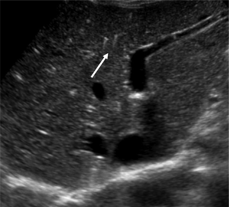
Abdominal ultrasound showing the presence of multiple small echogenic foci with comet-tail artefact (white arrow).
Figure 2.
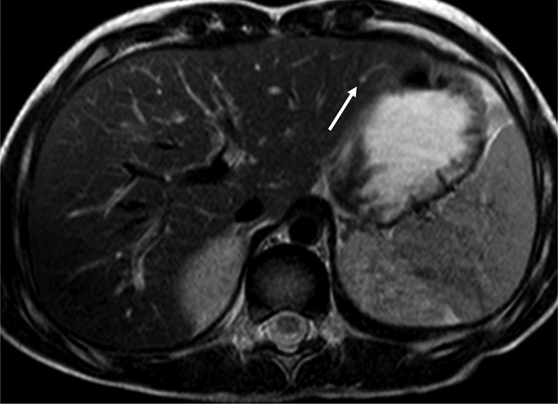
Abdominal MRI showing how these lesions were hyperintenses on T2 (white arrow).
The appearance may also be seen with von Meyenburg complexes, a benign condition found in up to 5% of the population.
Comet-tail artefact must be differentiated from the ring-down artefact. In the past, it has been thought that the ring-down artefact was a variant of comet-tail artefact because of a similar US appearance. Despite this, these two artefacts have separate mechanisms. In ring-down artefact, the transmitted ultrasound energy causes resonant vibrations within fluid trapped between a tetrahedron of air bubbles. These vibrations create a continuous sound wave. This phenomenon is displayed as a line or series of parallel bands extending posterior to a gas collection.2
The comet-tail artefact is a grey-scale US finding seen when small calcific/crystalline highly reflective objections are found, and is believed to be a special form of reverberation artefact.3 The artefact often is helpful in situations in which grey-scale imaging does not provide adequate information for a conclusive diagnosis.
Learning points.
The presence of comet-tail artefacts could avoid unnecessary invasive (ie, liver biopsy) or non-invasive (ie, CT) approaches.
The artefact often is helpful in situations in which grey-scale imaging does not provide adequate information for a conclusive diagnosis.
Footnotes
Contributors: PM and FDP followed the patient during the hospital stay; EC performed the follow-up and OA was the supervisor.
Competing interests: None.
Patient consent: Obtained.
Provenance and peer review: Not commissioned; externally peer reviewed.
References
- 1.Ziskin MC, Thickman DI, Goldenberg NJ, et al. The comet tail artifact. J Ultrasound Med 1982;2013:1–7 [DOI] [PubMed] [Google Scholar]
- 2.Feldman MK, Katyal S, Blackwood MS. US artifacts. Radiographics 2009;2013:1179–89 [DOI] [PubMed] [Google Scholar]
- 3.Tchelepi H, Ralls PW. Color comet-tail artifact: clinical applications. AJR Am J Roentgenol 2009;2013:11–18 [DOI] [PubMed] [Google Scholar]