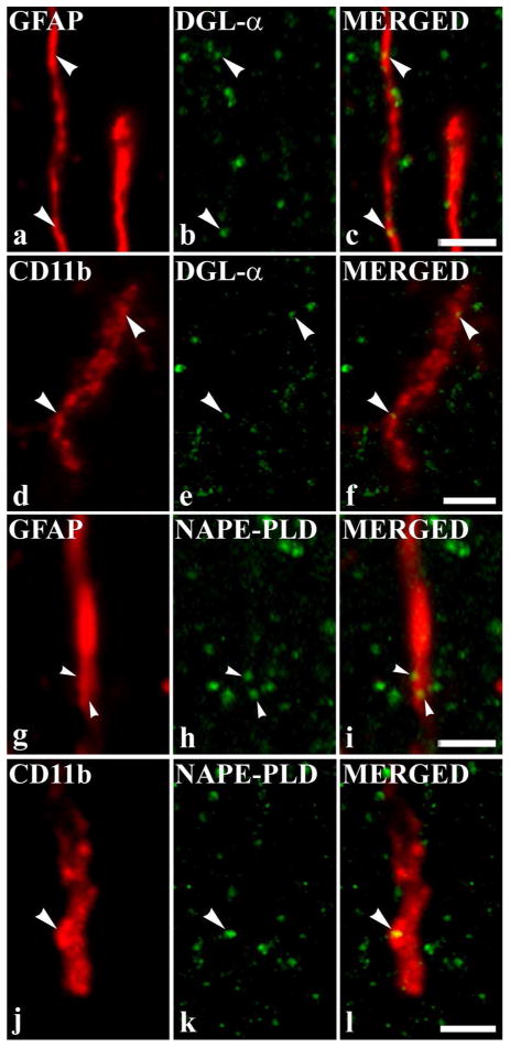
Figure 4.
Co-localization of DGLα and NAPE-PLD with glial markers. Micrographs of single 1.6 μm thick laser scanning confocal optical section (compressed images of three consecutive 1 μm thick optical sections with 0.3 μm separation in the Z axis) illustrating the co-localization between immunolabeling for DGLα (green; b) and immunoreactivity for a marker which is specific for astrocytes (GFAP, red; a); between immunolabeling for DGLα (green; e) and immunoreactivity for a marker which is specific for microglial cells (CD11b, red; d); between immunolabeling for NAPE-PLD (green; h) and immunoreactivity for a marker which is specific for astrocytes (GFAP, red; g); between immunolabeling for NAPE-PLD (green; k) and immunoreactivity for a marker which is specific for microglial cells (CD11b, red; j) in the superficial spinal dorsal horn. Mixed colors (yellow) on the superimposed image (c, f, i, l) indicate double labeled spots within the illustrated glial processes. Puncta immunoreactive for DGLα or NAPE-PLD which are also stained for the glial marker are marked with arrowheads. Scale bar: 2 μm.

