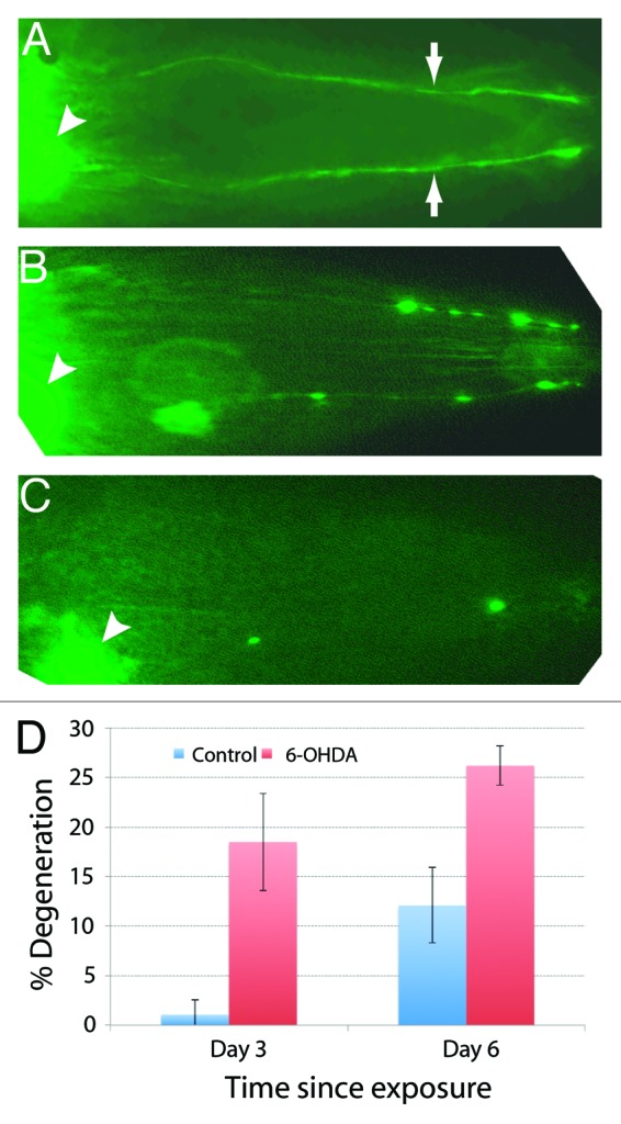
Figure 7. Degeneration of DA neurons in 6-OHDA-exposed worms visualized by dat-1p::YFP expression. In all cases anterior is toward the right. (A) In wild-type bhEx120 animals, YFP fluorescence can be observed in CEP cell bodies (arrowhead) and their processes (arrows). (B) Exposure to 6-OHDA causes spotty appearance of CEP neuronal processes indicating degeneration of neurons. (C) Another 6-OHDA-treated animal. The neuronal processes are almost completely missing. (D) Quantification of neuronal defects in 3-d- and 6-d-old control (untreated) and 6-OHDA-treated animals (sample size: 81 6-OHDA and 88 controls for day 3 set; 42 6-OHDA and 33 controls for day 6 set).
