Abstract
Digital pathology has grown dramatically in the last 10 years and has created opportunities to not only support the triaging of difficult cases among specialists within an organization, but also enable remote pathology consultations with external organizations across the world. This study investigated one organization's need for a vendor agnostic Digital Pathology Consultation workflow solution that overcomes the challenges associated with the transfer of large studies across a local area network or across the Internet. The organization investigated is a large multifacility healthcare organization that consists of 20 hospitals spread across a wide geographical area. The organization has one of the largest academic pathology departments in the USA, with more than 100 diagnostic anatomic pathologists. This organization developed a set of web-based tools to support the workflow of digital pathology consultations and allow the viewing of whole slide images. The challenges and practical implementations of two different use cases are addressed: the occasional end user (professional or patient) requesting a second opinion and the external laboratory or hospital looking for an established consultative relationship with a large volume of cases. The solution presented in this study addresses the challenges associated with the distribution of large images and the lack of established imaging standards, while providing for a convenient and secure portal for pathologist report entry and distribution.
Keywords: Digital pathology, Whole slide imaging, Telepathology, Pathology consultations, Imaging streaming, Asynchronous imaging transfer, Image size
Background
The organization investigated is a large multifacility healthcare organization that consists of 20 hospitals and 400 outpatient sites spread across a worldwide geographical area and that has one of the largest academic pathology departments in the USA, with more than 100 diagnostic anatomic pathologists. This organization has been using digitized pathology slides, including whole slide images (WSI), for over a decade to support the internal triaging of difficult pathology cases to appropriate experts and to provide remote pathology consultations to external organizations across the world [1–3]. WSI are accurate, high-resolution images captured from traditionally prepared histopathology and cytopathology specimens (glass slides), where many individual microscopic image fields are quilted together to form a seamless image product. WSI are similar in structure and size to geospatial imagery, and a corollary can be drawn to products such as Google Earth™.
Due to the size of the data involved, the organization was faced with many challenges such as the ability to transfer these large imaging studies across the Internet in a timely manner. Also critical was the ability to integrate the diversity of imaging solutions available on the market, as different vendors offer unique benefits to support various pathology subspecialties. A common viewing solution was desired in order to maximize user software familiarity and promote peer acceptance of the workflow.
As the telepathology business was expanding, both for internal use and to support external clients, there was a growing need to create a set of user-friendly tools that would facilitate standardized implementations and clinical workflow. These software tools needed to be flexible with respect to vendor integration so as not to restrict an organization's imaging equipment choices for specific vendor solutions. Pathology facilities are not always “rich” in terms of information technology (IT) expertise or infrastructure, so the ultimate solution had to be practical and easy to deploy and manage. The solution presented in this study addresses the challenges associated with the distribution of large images and the lack of established imaging standards, while providing for a convenient and secure portal for pathologist report entry and distribution.
Whole Slide Imaging
Current generation WSI scanning systems are comprised of software-controlled robotic hardware platforms that work according to similar principles as traditional compound microscopes. A glass pathology slide is loaded into the scanner and moved continuously at high speed under a microscope objective lens. As the histopathology specimen has a tissue section of known thickness, real-time focusing adjustments determined by software algorithms are utilized to keep the resulting images in focus. The objective lens determines the output resolution of the image and also the overall speed of capture, with higher resolution resulting in smaller fields of view and longer scan times [4].
The resulting image is represented in full color (typically 24 bits per pixel) with a dimension of many thousands of pixels in both width and height. Due to the data size, the image is often stored in a pyramid-like structure organized into layers, each of different resolution, to facilitate efficient viewing via specialized vendor-supplied software. A typical pathology case may contain multiple WSI images, where each image can be in the hundreds or thousands of megabytes on disk [4].
Lack of Established Standards
The market of digital pathology is dominated by a handful of vendors who have each developed proprietary imaging formats to optimize certain properties or functionality of the image acquisition and viewing solutions that each sells. Many options for both transmitted light as well as fluorescence imaging exist, as well as slide handling capabilities and value-added imaging features to optimize the resulting WSI product. Vendors offer supplemental software solutions for viewing and image distribution, and may include rich workflow functionality and specific analysis applications for image quantification.
Table 1 illustrates a selection of vendors currently active in the WSI imaging market. This table is not intended as a complete list and is based only on the organization's experience to date. As this field is in rapid evolution, this tabulated information is subject to change.
Table 1.
Selection and summary of wsi vendors
| Vendor | Imaging modality | Software solutions |
|---|---|---|
| TL = transmitted light | ||
| FL = epifluorescence | ||
| A = additional modalities | ||
| Aperio www.aperio.com | ✓ TL/FL/A | ✓ Local and web-based viewing |
| Optical configuration options | ✓ Pathology workflow, image management and collaboration | |
| ✓ Image analysis solutions | ||
| 3DHISTECH www.3dhistech.com | ✓ TL/FL | ✓ Local and web-based viewing |
| Optical/camera configuration options | ✓ Pathology workflow, image management and collaboration | |
| ✓ Image analysis solutions | ||
| ✓ Pathology education solutions | ||
| Ventana Digital Pathology http://www.ventanadigitalpathology.com/ | ✓ TL | ✓ Local and web-based viewing |
| Optical configuration options | ✓ Pathology workflow, image management and collaboration | |
| ✓ Image analysis solutions | ||
| Hamamatsu Photonics www.hamamatsu.com | ✓ TL/FL | ✓ Local and web-based viewing |
| Optical configuration options | ✓ Image management, metadata association and collaborative viewing | |
| Leica Microsystems www.leica.com | ✓ TL/FL | ✓ Local and web-based viewing |
| Optical configuration options | ✓ Pathology workflow, image management and collaboration | |
| Additional modalities supported with alternate devices | ||
| ✓ Image analysis solutions | ||
| ✓ Pathology education solutions | ||
| Philips http://www.research.philips.com/initiatives/digitalpathology/index.html | ✓ TL | ✓ Enterprise pathology workflow solution |
| Large-scale lab automation design | ||
| Omnyx www.omnyx.com | ✓ TL | ✓ Enterprise pathology workflow solution |
| Large-scale lab automation design | ||
| Olympus www.olympus.com | ✓ TL/FL/A | ✓ Image viewing |
| Optical/camera configuration options | ✓ Analysis software tools | |
| MikroScan Technologies www.mikronet.com | ✓ TL | ✓ Image viewing software |
| Optical configuration options | ✓ Remote collaboration supported by desktop sharing |
While it is openly accepted that WSI solutions offer unique value to enable pathologists to collaborate over large geographical distances [1, 4, 5], there are also challenges encountered while attempting to use such solutions for routine second opinion consultation. A typically configured scanning system, using a ×20 objective lens, has a good combination of imaging speed and resolution. However, at times, higher resolution is needed to visualize the subtle pathology expressed by a difficult case. Additional imagery data obtained by scanning multiple focal planes may be needed to improve visualization, or additional microscopy modalities such as immunofluorescence may be required to support specialty staining protocols.
Given these factors, a comprehensive one-vendor solution may not be possible to meet all WSI requirements to fully support consultation requests. This heterogeneity inherently forces vendors to develop specific imaging formats to support, optimize, and integrate various device capabilities. However, it is important to have a common viewing platform to minimize end user software training and maximize application familiarity.
Even though there are currently no widely accepted standards, the recent publication of the Digital Imaging and Communications in Medicine (DICOM) supplement 145 provides technical specifications for storage of whole slide imaging data [6]. DICOM provides a data framework that enables the integration of diverse scanners, archiving solutions, diagnostic viewing tools, and printers from multiple manufacturers. The introduction of the DICOM standard in pathology is expected to enable clinical laboratories and pathology groups to store digital pathology images in a format that is compatible across diverse systems. In addition, it should enable more flexibility in the selection of solutions available on the market and more interoperability between digital pathology systems and diagnostic tools, as well as other systems such as Picture Archiving and Communication System and Vendor-Neutral Archive solutions already in use in different domains like radiology and cardiology [4, 6].
Today, however, the lack of an established standard is a reality, and it will take some time for all of the vendors to converge on adopting a standard, updating their software and supporting a standardized imaging solution. Efforts are underway by some groups to build open source tools for digital pathology, such as the “OpenDP” consortium [7] and eSlide[8]. These tools are maturing quickly, but are not yet stable enough for mainstream production use.
Imaging Study Size
Another significant challenge in digital pathology is the average imaging study file size. For a WSI, the file size can range on average from 50 Mb to over 6 GB, and this depends on the magnification at which the slide is scanned. In addition, the number of slides in a pathology imaging study can typically range between 2 slides and 60. Therefore, WSI studies can be in the magnitude of thousands of megabytes [4] in contrast to other specialties, such as radiology, where the average size of a digital imaging study is usually tens or hundreds of megabytes. Tables 2 and 3 compare the average study on-disk file size (compressed) between different radiology modalities and different pathology studies in the organization examined. These tables are not intended as complete lists and are based only on the organization's experience to date. As these imaging domains are in continuous evolution, the tabulated information is subject to change.
Table 2.
Average size per radiology modality
| Radiology modality | Avg. size (MB) |
|---|---|
| CT scan | 153.4 |
| Ultrasound | 69.2 |
| X-ray angiography | 157.5 |
| Computerized radiography | 14.1 |
| MRI | 98.6 |
| Mammography | 83.5 |
| PET | 365.9 |
| Digital radiography | 17.4 |
| Radiofluoroscopy | 30.1 |
| Nuclear medicine | 7.0 |
| Radiographic image (conventional plain films) | 36.7 |
| Endoscopy image | 6.8 |
| Radiotherapy | 30.6 |
| Panoramic X-ray | 5.1 |
| Breast imaging | 38.8 |
| Magnetic resonance angiography | 55.6 |
Table 3.
Average size per pathology study type
| Biopsy type | Slides per case study (typical) | Average size per case study (MB) | |
|---|---|---|---|
| ×20 resolution compressed | ×40 resolution compressed | ||
| Dermatopathology | 6 | 1,392 | |
| GU | 9 | 1,701 | |
| Head and neck | 3 | 1,965 | |
| Hematopathology | 31 | 40,300 | |
| Neuropathology | 9 | 1,872 | |
| Thoracic pathology | 12 | 3,240 | |
| Transplant pathology, needle | 2–3 | 250–375 | 700–1050 |
| Transplant pathology, wedge | 2–3 | 600–900 | 1,500–2,250 |
For efficient storage, WSIs are stored in compressed format. The compression method utilized is typically JPEG or JPEG 2000 encoding, with a high quality factor to preserve image detail. A typical WSI captured with a ×20 objective lens would potentially represent 20+ Gb of storage if uncompressed, but after compression, the size is reduced to an average range of 200–650 Mb [4].
A whole slide imaging study of a single pathology case, which may include several slides, can result in the compressed imaging study being an average size of 2–3 GB. Typically, the image sets are consolidated on a server, with sufficient storage allocation, connected to the scanning platform either via direct connection or a high-speed network (Gigabit). To view the images, the end user must have native file access to the image via network resource sharing or utilize a vendor-supplied web-based viewing system to allow for thin-client connectivity. While direct file access would appear the most straightforward method, often, IT security issues and firewalls prevent this type of connectivity, especially between disparate institutions. As such, this often blocks the adoption of digital pathology when all parties are not on the same LAN [4].
The process of transferring a set of digital slides over the Internet can take a significant amount of time, tying up a workstation for the duration of the transfer. Brief interruptions in the connectivity over a long upload time may cause the data transfer to fail, forcing a human operator to monitor the transfer and restart if necessary. Table 4 shows transfer times for different study sizes and for different available bandwidths under ideal conditions.
Table 4.
Image file transfer times per available bandwidth
| Transfer times (hours/minutes/seconds) | ||||||
|---|---|---|---|---|---|---|
| Network speed (Mbps) | Imaging study size (MB) | |||||
| 100 | 500 | 1,024 | 2,048 | 3,072 | ||
| LAN Hi | 100 | 00:00:14 | 00:01:10 | 00:02:28 | 00:04:42 | 00:07:16 |
| WiFi | 54 | 00:00:40 | 00:03:31 | 00:06:11 | 00:14:58 | 00:20:16 |
| Fiber Std | 20 | 00:01:40 | 00:08:22 | 00:17:09 | 00:33:44 | 00:52:14 |
| LAN Low | 10 | 00:02:07 | 00:10:24 | 00:21:29 | 00:44:10 | 01:07:04 |
| Cable | 1.5 | 00:22:38 | 01:53:16 | 03:52:46 | 07:38:51 | 11:38:10 |
Connectivity was tested between a commercial pathology laboratory in China and the organization in the USA which is the subject of this study. Both the commercial laboratory in China and the US Hospital have high-speed access to the Internet. However, when setting up a connection between these two locations, for example through a virtual private network, the actual connection speed was very variable, and on average, the connection speed achieved was 2 Mbps. As a result, if the commercial laboratory in China was to transfer a typical whole slide imaging study of 2 Gb, it would take more than 18 h before a pathologist could view the case and start rendering a consultation to the requesting physician in China. In addition, short network interruptions over such an extended amount of time could cause the data transfer to halt and fail. Such extended transfer times and potential interruptions can severely impact the turnaround time of remote pathology consultations.
Methods
A case study analysis was performed to investigate the organization's digital pathology system which was built upon data and experiences collected over a 10-year period. The investigators are also members of the information system team responsible for developing, implementing, and maintaining the solution. Informal interviews with the information system designers and the end user pathologists were performed, information system documentation was reviewed, and quantitative data in the form of system performance were collected. Descriptive statistics were used to analyze the data, the findings were presented, and a discussion concluded the investigation.
Results
To be effective, pathology consultation tools based on WSI must allow the client requesting a consultation to share imaging data with remote pathologist(s). The organization set out to develop a robust and format-agnostic consultation platform that could accept large WSI data sets. The submission platform also needed to capture all necessary clinical information associated with cases for consultation, allow secure and reliable WSI transmission, and be user friendly. Slide labeling, as well as the resulting WSI filename, was automated using bar coding to remove chances of human error in naming data files. The organization emphasized the adoption of connection security measures in order to protect the privacy of patients treated over a public network. A streamlined workflow needed to be implemented to ensure prompt turnaround time as well as obtain buy-in from pathologists.
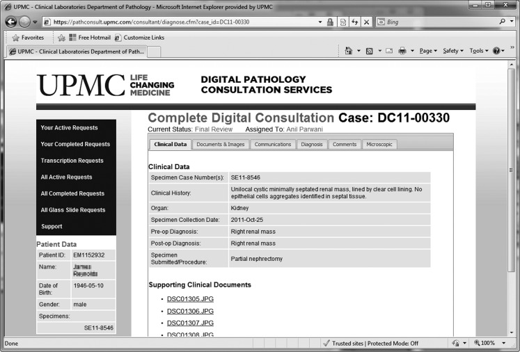
The end result was a telepathology portal that offers diagnostic consultations to both peripheral community pathologists within the organization and external users seeking expert second opinion consultation (Fig. 1). This web-based solution can accept all forms of digital images including static microscopic images, digitized whole slides, or even snapshots from a handheld digital camera. The unique attribute regarding the solution presented in this article is that it is vendor agnostic and allows the asynchronous upload of WSIs, as well as the streaming of imaging data directly from the source storage location.
Fig. 1.
Telepathology user interface
The implementation utilized a mix of client–server technology, combining a web front-end application with fillable forms to allow for user registration, case information, and submission as well as overall workflow logic, including label printing, reporting, and billing tasks. The application is tied to a database, with separate table space for each client to maintain security. The database, currently SQL Server 2008 R2, manages the workflow events and links each case with the associated WSI data. The WSI data themselves are stored on a traditional file system (with redundancy) due to size. WSI viewing is supported by a Java client–server application, designed to be image format agnostic, that enables more advanced programmatic control of system resources (such as multiple image access threads) to maximize performance and support multiple platforms. The client–server design allows the organization to deploy the same technology footprint for both internal uses and external clients. Clients may choose from two options for image sharing: a simpler and more basic asynchronous transfer or a client–server viewing which requires more advanced network connectivity between the institutions.
The web tool features the following:
Client submission and request tracking: offers Secure Sockets Layer (https) secure login for client personnel to submit patient data, upload whole slide and static images, view the current status, and view and print second opinion reports
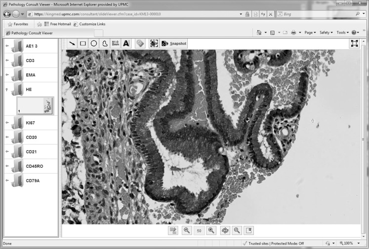
Host pathologist: hosts the diagnosis tool where pathologists can view WSIs, annotate and capture image snapshots, view clinical data, and submit a diagnosis for second opinion reports (Fig. 2)
Host consultation services: allow a manager to triage requests, assign them to pathologists, monitor workflow, and maintain personnel data
Host transcriptionist: lists the current transcription queue with data entry fields for diagnoses dictated by pathologists through a voice dictation system
Fig. 2.
Vendor-neutral viewer ‐ is showing a viewer of a whole slide image, snapshots and annotation tools
Asynchronous Transfer
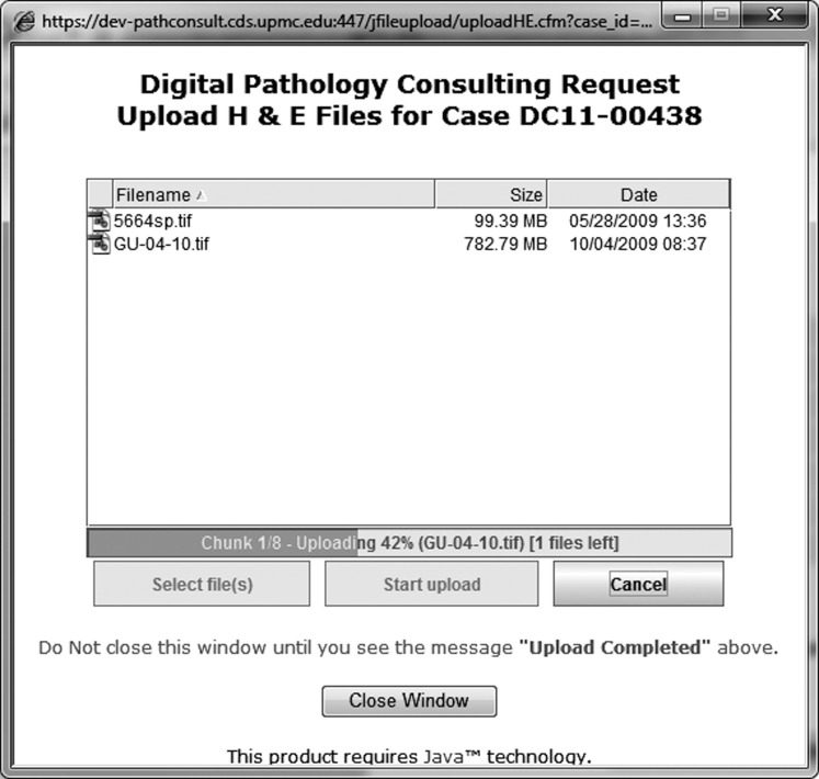
For those users with lower consult volumes, such as less than ten per month, the asynchronous transfer method developed by the organization leverages technology that frees up the user and the remote workstation during the transmission of large datasets. This enables the users to continue to upload cases or to perform any other type of work on their workstation while the transfer occurs in the background. In addition, the asynchronous transfer method automatically monitors and resumes transmission from any point of failure. The web portal features an asynchronous upload tool (Fig. 3) supporting transfers of multigigabyte WSI files reliably over the Internet ensuring delivery of files 100 % of the time unless interrupted by the operator.
Fig. 3.
Asynchronous upload tool ‐ is showing a window with bu;ons to upload images and an uploading progress bar
Real-Time Imaging Viewing
When supporting an external organization like a laboratory or a hospital requesting a high volume of consultations and fast turnaround time, the transmission of entire digital pathology studies via the Internet may be labor intensive, extremely time consuming, or very expensive, even when using a dedicated connection with high bandwidth. The solution developed by the organization offers the option to install a software service on the client's remote storage location linking the appropriate imaging data set and streaming the information in real time to a connected client viewer where the pathologist is providing the consultation. A sophisticated algorithm only transmits the information that fits in the screen of the pathologist's workstation display, therefore limiting the amount of data that needs to be transmitted and enabling quick turnaround of the pathology consultations. Algorithmic methods are also utilized to asynchronously prefetch surrounding image areas and to cache imagery already received to minimize wait time for high latency connections. The software service also provides separate access paths for the slide imagery and the label/identification area enhancing electronic patient information security.
A common challenge with thin-client connectivity to a large data source revolves around maximizing performance over a wide area network. Bandwidth is the measure of how much data can be transmitted over a given period of time, and latency is the amount of time that a network data request takes to propagate the network and notify of delivery. As WSI data delivery to a thin-client viewer is synchronous by design (data cannot be lost and must be acknowledged by the receiver), minimizing latency is a foremost design consideration especially with larger geographical distances. Java™ is a functionally rich environment, which allowed developers to make use of multiple threads to create simultaneous requests for image data, reducing the effects of latency. The areas to be refreshed are queued, and as the requests are filled, the queue is emptied.
Vendor Agnostic
The solution used by this organization leverages a vendor-neutral viewer that is compatible with the most common imaging pathology formats available in the market. The WSI viewer is a Java applet that uses SlideViewer framework in conjunction with a slide selector and has the ability to capture, upload, and view screenshots of the slide. SlideViewer is an internally developed high-level Java application programming interface (API) that describes a vendor agnostic digital slide viewer [9]. The SlideViewer API supports multiple WSI formats by either connecting directly to vendors' proprietary image servers or by connecting to an open source OpenSlide [10] Image Server that, in turn, supports multiple WSI formats from pathology glass slide scanner vendors such as Aperio, Hamamatsu, Trestle and Mirax, as well as radiology DICOM images and JPG/GIF/PNG files. Additional image support can be added by integrating a third-party Java WSI client into the SlideViewer API to provide a seamless viewing experience. As of May 20, 2012, SlideViewer can connect to Aperio Image Server, Hammamatsu NanoZoomer Server, Zeiss Tile Server, and OpenSlide Server.
The SlideViewer API offers many additional features that seamlessly work across multiple virtual slide vendors, including full slide annotation framework, as well as recording and playback framework. Virtual slides can be cropped, rotated, and flipped in real time without modifying original files. SlideViewer uses tile caching, multithreaded tile fetching, and many other features that increase the speed and performance of viewing WSI.
SlideViewer is an integral part of SlideTutor ITS, an Intelligent Tutoring System in the domain of dermatological pathology. As a part of SlideTutor ITS, SlideViewer has been undergoing improvements through many studies involving training pathology residents and community pathologists [11]. SlideViewer for the organization investigated has proven to be an effective tool that is scalable, responsive, and highly customizable to the vendor's needs.
Practical Applications
In addition to using this solution within its 20-hospital health system, the organization has expanded the use of this telepathology system internationally. In September 2011, the Telepathology Web Portal went live for one of the largest commercial diagnostic laboratories in China. This exciting endeavor provided the organization with a unique opportunity to tap into a market which seeks valued US-based expert opinions in the field of anatomic pathology. The pathologists in the USA have the ability to remotely view WSI scanned on a whole slide scanner and hosted on an imaging server at a facility in Guangzhou, China. The Web Portal viewer affords the pathologists the ability to remotely manipulate, annotate, and take snapshots of areas of interest on the WSI, which can also be included in the final report.
Web-based pathology workflow solutions also create opportunities to combine advancements in diagnostic tools, such as incorporating fluorescence-based techniques to improve both accuracy and specificity of information available about a specimen. WSI techniques allow for collaboration with such specimens, whereas before, any consultation would have to be done at the same location as the physical slides due to imaging requirements. By incorporating WSI viewing into the solution, the organization enables external collaborators to take advantage of such techniques that may not be easily available or practical within their local histology facility [4, 12].
Discussion
With all complex IT solutions, institutional-level challenges may be present that constrict how such a solution may be deployed. Such challenges are related to network connectivity and firewall configurations when linking two separate organizations, and the related IT infrastructure rules that must be established and followed. While a direct link to a client to support remote WSI viewing may be the most practical, oftentimes, it takes much time and relationship building to facilitate such a link. Therefore, to overcome this challenge, the solution was to provide both a “general” tool for the casual user, offering upload capability, as well as a custom implementation linking institutions. Table 5 summarizes the advantages and disadvantages of the general telepathology portal using the aforementioned asynchronous transfer method and the custom pathology portal that leverages client software on the remote storage hardware to allow streaming of imaging data.
Table 5.
Telepathology portal access comparisons
| General telepathology portal occasional user (asynchronous upload) | Custom/client telepathology portal large volume (imaging streaming) | |
|---|---|---|
| Pro | Simple, easy to use; readily available to submit consultation requests | Support of large consult volumes |
| Real-time consultations; no need for image transfers | ||
| Does not require any setup at the client site | Custom logic (case routing, historical data) and custom data for specialty pathology areas | |
| No specific IT-level engagement needed | Bar coding, specimen identification automation to minimize risk of mislabeling | |
| Workflow and hardware optimization | ||
| No direct cost to implement | ||
| With web-based collaboration tools, a highly interactive experience is possible | ||
| Con | Limited automation; requires more user intervention | Implementation cost is higher |
| Requires project level engagement between clients to resolve IT connectivity issues and installation of software at the remote site | ||
| Potential long file upload times for whole slide images | ||
| Limited ability to organize information; no custom data |
The implementation of an agnostic viewing solution also provides several benefits, such as allowing the use of the same viewing architecture to support several clients, all with different vendor WSI scanning solutions. Minimization of end user (pathologist) training was also a recognized benefit of a single, common viewing solution.
However, most prevalent is the ability of this overall solution to minimize patient wait time for conclusive results and to facilitate critical diagnostic decisions when a procedure requires pathology interpretation. Many specialized pathology areas often do not have sufficient volume in smaller institutions, such as transplantation or neuropathology, and when a complex case does arise, the actual glass slides are conventionally sent via standard courier for a second opinion. This process can take days and risks specimen loss and/or damage. When reviewing the material, the consulting party often may request additional tissue preparations, which would involve additional shipping expense and time. As a practical example, consultative second opinions between a specialty hospital located in Italy and the organization described here would take on average 3–4 days with significant courier costs due to the distance involved.
Once the organization transitioned over to the telepathology system, turnaround time for second opinion was reduced to less than one working day (or same day with the time zone advantage from CET to EST for biopsies consulted on in the morning hours). Additionally, both pathology users could collaborate on the same physical specimen by viewing the same image. Specimen rejection, where the imaging system resolution was not sufficient or there was some other imaging issue, required the glass slide to be sent, but occurred in less than 2 % of the telepathology consultations.
Conclusions
The telepathology portal enables pathologists from near and far to gain immediate access to an expert team of diagnostic anatomic pathologists who are available to render a diagnosis in subspecialty areas.
The organization highlighted in this study has one of the largest academic pathology departments in the USA with expertise in all areas of anatomic pathology. Many of the pathologists available for consultation are leaders in their subspecialty fields. By leveraging the Digital Pathology Consultation Portal, overnight shipping and mailing charges are avoided, loss of slides are no longer a concern, difficulties regarding the transfer of human tissues across international borders are circumvented, and results are delivered much more quickly and via a secure website than using traditional consultation methods, thus expediting the client's satisfaction and ultimately the patient's treatment.
The security of this application is handled with multiple methods. All data transmissions are encrypted using the “https” communication protocol. Sensitive patient identifiers are encrypted in the database, and when required by international regulations, no demographic patient information is shared with the pathologist providing the consultation except for gender and age. Every web page validates against the user ID, and all information including also reports is generated dynamically. No static reports and patient or clinical data are stored on the web servers, and users can only generate and view reports associated with their user ID.
In summary, this organization has implemented a comprehensive solution to support a variety of local, regional, and international remote telepathology consultations overcoming the challenges associated with the large size of digital pathology studies and the lack of established imaging standards. The web interface and the easy setup of the infrastructure streamline access and opportunities for pathology consultations. The organization has enabled their client's digital imaging systems and whole slide scanners to become an easy gateway to real-time telepathology consulting and simplified the mechanism to obtain second opinions. The organization's solution presented herein has also overcome the significant challenges inherent in web-based telepathology, making this solution viable and scalable for second opinion consultations.
References
- 1.Minervini MI, Yagi Y, Marino IR, Lawson A, Nalesnik M, Randhawa P, Wu T, Fung JJ, Demetris A. Development and experience with an integrated system for transplantation telepathology. Hum Pathol. 2001;32:1334–1343. doi: 10.1053/hupa.2001.29655. [DOI] [PubMed] [Google Scholar]
- 2.Horbinski C, Wiley CA. Comparison of telepathology systems in neuropathological intraoperative consultations. Neuropathology. 2009;29:655–663. doi: 10.1111/j.1440-1789.2009.01022.x. [DOI] [PubMed] [Google Scholar]
- 3.Wiley CA, Murdoch G, Parwani A, Cudahy T, Wilson D, Payner T, Springer K, Lewis T. Interinstitutional and interstate teleneuropathology. J Pathol Inform. 2011;2:21. doi: 10.4103/2153-3539.80717. [DOI] [PMC free article] [PubMed] [Google Scholar]
- 4.Parwani AV, Feldman M, Balis U, Pantanowitz L. Digital imaging. In: Pantanowitz L, Balis U, Tuthill M, editors. Pathology Informatics: Modern Practice and Theory for Clinical Laboratory Computing. Chicago: ASCP Press; 2012. pp. 231–256. [Google Scholar]
- 5.Thrall M, Pantanowitz L, Khalbuss W. Telecytology: clinical applications, current challenges, and future benefits. J Pathol Inform. 2011;2:51. doi: 10.4103/2153-3539.91129. [DOI] [PMC free article] [PubMed] [Google Scholar]
- 6.Singh R, Chubb L, Pantanowitz L, Parwani A. Standardization in digital pathology: supplement 145 of the DICOM standards. J Pathol Inform. 2011;2:23. doi: 10.4103/2153-3539.80719. [DOI] [PMC free article] [PubMed] [Google Scholar]
- 7.Open Digital Pathology Consortium. http://www.opendp.org/. Accessed 22 Jan 2013
- 8.Medical Informatics & Telemedicine Lab, University of Udine, Italy. http://www.eslide.net/
- 9.Crowley RS, Legowski E, Medvedeva OM, Tseytlin E, Roh E, Jukic D. Evaluation of an intelligent tutoring system in pathology: effects of external representation on performance gains, metacognition, and acceptance. JAMIA. 2007;14(2):182–190. doi: 10.1197/jamia.M2241. [DOI] [PMC free article] [PubMed] [Google Scholar]
- 10.Goode A, Satyanarayanan M: A vendor-neutral library and viewer for whole-slide images. Technical Report CMU-CS-08-136, Computer Science Department, Carnegie Mellon University, 2008
- 11.Mello-Thoms C, Mello CA, Medvedeva O, Castine M, Legowski E, Gardner G, Tseytlin E, Crowley R. Perceptual analysis of the reading of dermatopathology virtual slides by pathology residents. Arch Pathol Lab Med. 2012;36(5):551–562. doi: 10.5858/arpa.2010-0697-OA. [DOI] [PubMed] [Google Scholar]
- 12.Pantanowitz L, Valenstein PN, Evans AJ, Kaplan KJ, Pfeifer JD, Wilbur DC, Collins LC, Colgan TJ. Review of the current state of whole slide imaging in pathology. J Pathol Inform. 2011;2:36. doi: 10.4103/2153-3539.83746. [DOI] [PMC free article] [PubMed] [Google Scholar]


