“The real voyage of discovery consists not in seeking new landscapes, but in having new eyes,” Marcel Proust said.
On May 4, 1962, Masaki Watanabe MD opened new eyes. On that day, he performed the first arthroscopic menisectomy using the arthroscopic instruments he developed [7, 14, 22]. The patient was a 17-year-old boy who twisted his knee playing basketball. Watanabe débrided the flap tear of the medial meniscus. The patient went home the same day. In 6 weeks, he was back playing basketball [22].
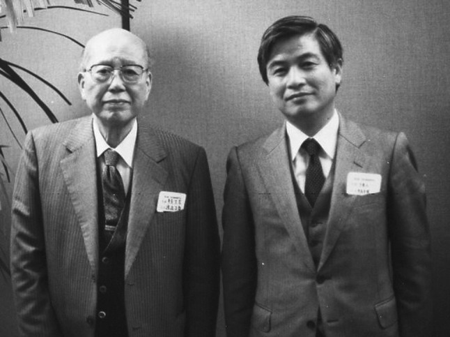
A pioneer in the development of the modern arthroscope with fiberoptic illumination, Watanabe is widely considered the “father of arthroscopy.” (Fig. 1) [8] His arthroscope was a specialized endoscope, derived from a rudimentary cystoscope. The humble orthopaedic surgeon from Japan rarely spoke of his own personal achievements, so his peers did the talking for him, championing his instruments and techniques at international orthopaedic meetings.
Fig. 1.

Watanabe (left) with his former student Prof. Hideshige Moriya (right) in June 1980. Watanabe is widely considered the “father of arthroscopy”
Arthroscopic surgery is now commonplace—an indispensible outpatient procedure in many parts of the world. The benefits of minimally invasive surgery and early mobilization afforded by arthroscopic surgery are accepted as the standard of care.
It was a long road to commonplace. The development of the arthroscope is a story of mentorship, dedication, and passion. Watanabe, among many others, revolutionized orthopaedic surgery. The quest to look into body cavities with small incisions dates to Pompeii, but the modern version of the arthroscope emerged from Japan.
Early Development
The first report of a diagnostic knee arthroscopy was in 1912 by the Danish surgeon, Severin Nordentoft, who called the approach “arthroscopia genu.” [1, 9–11] He used a “trochar endoscope” of his own design and described seeing the patellofemoral joint and the anterior horns of the meniscii in his paper, “On the Endoscopy of Closed Cavities Using My Trocar Instrument.” [1, 9, 11] He believed the device would be useful for the diagnosis of synovial disorders, specifically tuberculosis and syphilis [18]. He also stressed the use of sterile irrigation fluid [3, 17]. Six years later, this critical observation became a reality in Japan.
In 1918, Kenji Takagi professor and orthopaedic surgeon from Tokyo University visualized the knee joint of a cadaver [8, 12, 18, 20] using a 7.3 mm cystoscope [19], known as the Charriere No. 22 [14, 22]. In 1922, Takagi evaluated the effects of tuberculosis in joints, examining the early stages of arthritis and obtaining biopsies for diagnosis [14].
That same year, using a modified Jacobean cytoscope independently, Eugen Bircher reported his findings on 20 knees [4]. He found that the arthroscopic appearance of the knee did not correlate with radiographs. Bircher identified nine torn meniscii at arthroscopy [15]. From 1921–1926, his publications described 60 arthroscopies and were the first to detail knee arthroscopy in actual patients [1–3, 15, 18, 20].
Takagi’s quest to devise a functional knee arthroscope resulted in many different prototypes, each with a different model number [12, 15, 22]. The first model, created in 1931, was 3.5 mm in diameter (Table 1) [8, 9, 12]. In 1936, he began taking color photographs and movies of the procedure [12]. Takagi successfully visualized the knee, shoulder, ankle, and spine [14]. He continued his work with the arthroscope and the instrumentation until the demands of World War II prevented further exploration [8]. Takagi remained committed to the arthroscope and was certain that it would make a significant contribution to modern surgery. His student, Masaki Watanabe [13] shared his vision [14, 22].
Table 1.
The development of the Watanabe arthroscope
| Year | Major event |
|---|---|
| 1931 | Kenji Takagi devises his No. 1 arthroscope, the 3.5 mm prototype of the modern arthroscope. |
| 1936 | Takagi takes color photographs and movies of knee arthroscopy. |
| 1938 | Takagi produces his No. 12 arthroscope. |
| 1949 | Masaki Watanabe transfers to Tokyo Teisin Hospital. |
| 1954 | Watanabe removes a xanthoma from the knee. |
| 1957 | Watanabe and colleagues publish “The Atlas of Arthroscopy.” |
| 1959 | The Watanabe No. 21 becomes a production model. |
| 1962 | Watanabe performs the first arthroscopic partial meniscectomy. |
| 1964 | Robert Jackson learns knee arthroscopy from Watanabe. |
| 1965 | Jackson brings knee arthroscopy to North America at the Toronto General Hospital using the Watanabe No. 21 arthroscope. |
| 1967 | Jackson presents his experience with the Watanabe No. 21 arthroscope at the Orthopaedic Research and Education Foundation in Atlanta. |
| 1968 | Jackson and Isao Abe present an instructional lecture on knee arthroscopy at the American Academy of Orthopaedic Surgeons annual meeting. |
| 1968 | Leonard Peltier orders the first Watanabe No.21 arthroscope in the US. |
| 1969 | Richard O’Connor visits Watanabe to learn knee arthroscopy. |
| 1972 | John Joyce organizes the first course on arthroscopy in Philadelphia. |
| 1974 | Richard O’Connor reports on the first partial meniscectomies performed in North America. |
| 1974 | The International Arthroscopy Association is established. |
| 1982 | The Arthroscopy Association of North America is founded. |
The Watanabe Arthroscope
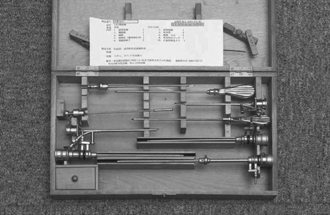
Watanabe studied with Takagi and later joined the surgical staff at Tokyo University until World War II, serving as an intelligence officer [9]. Watanabe returned after the war and transferred to Tokyo Teishin (Postal Services Ministry) Hospital where he became the director of the Department of Orthopaedic Surgery [9, 14, 22]. He resumed his study of the arthroscope and systematically modified it for knee arthroscopy. His first version was a scope with a small field of view [22]. In 1951, he produced the 4 mm No. 13 arthroscope by modifying a pediatric cystoscope (Fig. 2) [22]. He continued to improve the device, numbering each successive model. Watanabe transitioned the arthroscope from purely a diagnostic tool to a diagnostic and therapeutic device.
Fig. 2.

In 1951, Watanabe produced the 4 mm No. 13 arthroscope by modifying a pediatric cystoscope
Tuberculosis had become much less common in Japan after World War II, but knee injuries from industrial and traffic accidents and sports were on the rise [14]. To Watanabe, this resulted in a natural transition. He performed his first live arthroscopy on a horse’s ankle to determine the cause of a talar chondral defect thought to be congenital by one of the horse societies [14, 21]. Watanabe found that the horse ankle resembled a human knee “upside down.” [14] In 1955, he arthroscopically removed a xanthomatous giant cell tumor from the suprapatellar pouch [22] and recorded it. This footage is widely considered as the first recorded arthroscopy on a patient [9].
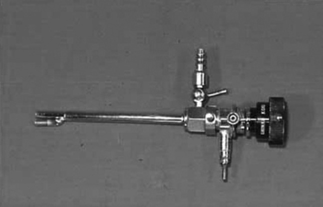
In 1958, Watanabe produced the No. 21 arthroscope, which is remarkably similar to the current models [18] (Fig. 3). The No. 21 design became the world’s first production model and the last version with an incandescent light source [8, 9, 23]. In all versions, the surgeon had to look directly into the eyepiece of the arthroscope. The sheath was 6 mm in diameter while the telescope was 4.9 mm in diameter and 16.5 mm long [6]. The wide angle lens had an astounding field of view of 100° and a depth of focus from 1 mm to infinity [6, 9]. In 1967, Watanabe exchanged the incandescent light source for the new fiber light (cold light) producing the No. 22 arthroscope [9]. In 1970, he produced the first ultrathin fiberoptic arthroscope, called the No. 25. The ultrathin fiberoptic arthroscope had a 2 mm sheath and was the basis for the Dyonics™ (Smith & Nephew, Memphis, TN, USA) “needlescope” [9, 22].
Fig. 3.

In 1958, Watanabe produced the No. 21 arthroscope, which is remarkably similar to current models
Introduction to the Western World
Watanabe dedicated his life to the development of arthroscopy. He meticulously documented his surgical findings, trying to improve the arthroscope and its instruments. But it would take an international collaboration for the arthroscope to gain acceptance and popularity outside of Japan.
In 1964, Robert W. Jackson, then of the Toronto General Hospital, traveled to Japan to study tissue cultures [7, 8]. He sought out Watanabe at the Tokyo Teishin Hospital to learn about arthroscopy [7], the first foreigner to do so [9]. Jackson observed Watanabe or his assistant perform cases twice a week for 6 months. Dr. Hiroshi Ikeuchi, his second assistant, translated [7, 9].
Surprisingly, Watanabe did not recommend that Jackson introduce arthroscopy into his practice [22] because it had yet to be accepted. Yet, Jackson began knee arthroscopy with the Watanabe No. 21 when he returned to Toronto in 1965 [7]. In 1967, Jackson presented his experience with the Watanabe No. 21 at two different national meetings: the Orthopaedic Research and Education Foundation in Atlanta [7] and the Association of Academic Surgeons in Toronto [7, 8]. Jackson and his new Japanese colleague, Dr. Isao Abe, presented the first Instructional Course Lecture on arthroscopy of the knee at the 1970 annual meeting of the American Academy of Orthopaedic Surgeons [7, 8]. They published their experience of their first 200 consecutive cases on a variety of knee conditions with acceptable examinations in 179 cases (89.5%) [6].
Richard O’Connor MD, another pioneer in the development of arthroscopy, visited Watanabe in 1971 and learned the principles and techniques of knee arthroscopy [9, 16, 22]. His 1974 publication of 21 patients undergoing knee arthroscopy with the No. 21 after an acute twisting injury, focused on the description of meniscal and ACL injury [15]. O’Connor performed the first partial arthroscopic meniscectomies in North America [8, 9, 14].
S. Ward Cascells MD possessed a keen interest in chondromalacia patella [9]. He read in a journal that Watanabe performed an arthroscopy [9]. To learn the technique, he observed Jackson in Toronto and then wrote to Watanabe [9]. He was the first to use the No. 21 in the United States [17]. In 1971, he published the first paper in English on operative arthroscopy in patients in the American edition of the Journal of Bone and Joint Surgery.
“I am convinced that the Watanabe arthroscope is a good diagnostic tool which gives the examiner information not available by any other means,” Cascells wrote [5, 9].
Jackson’s dogged promotion of the Watanabe technique, documentation of the procedures and their results, as well as training of surgeons, helped introduce arthroscopy to the western world (7, 13, 15, 18].
Pioneer
Prof. Hideshige Moriya trained under Watanabe from July 1973 to December 1975. In a conversation (March 2013), Moriya spoke of Watanabe’s commitment to the arthroscope.
“In the 1970s, Tokyo Teisin Hospital had a shortage of doctors, particularly in the Department of Orthopaedic Surgery,” Moriya said. “This was because most young orthopaedic surgeons at Tokyo Teisin University were not interested in arthroscopy, whereas it was the focus and passion of Watanabe. He was always thinking of further development of arthroscopy. He loved the No. 21 arthroscope as if it were one of his grandchildren. In the operating theater, he handled it with skill and love.”
Watanabe, who died on October 15, 1995 of complications following treatment of a femoral neck fracture [13], was a meticulous innovator with a passionate commitment to a device initially used to treat tuberculosis. He envisioned the utility of the device as it applied to knee surgery, not just for infection. He also incorporated the new technologies of each era to improve the device. The knee was well suited to his focus because of its anatomy, having width and a potential space for better visualization, Moriya explained.
“Watanabe did the first arthroscopic meniscectomy in the world in 1962,” Moriya said. “However, for the one and a half years while I was trained under him, he did not speak even one word concerning this procedure. It is what we now consider a major event in arthroscopic surgery. I do not think he expected the extensive use of endoscopy, including general surgery. However, I think Watanabe would be happy to see the application of endoscopy. For his contributions, I believe Watanabe is happy in Heaven.”
Footnotes
Note from the Editor-in-Chief: In “Giants in Orthopaedic Surgery,” a columnist explores the life and achievements of an orthopaedic surgeon who changed our profession, by interviewing other surgeons whose lives the “Giant” touched through mentorship or collaboration. We welcome reader feedback on all of our columns and articles; please send your comments to eic@clinorthop.org.
Each author certifies that he or she, or a member of his or her immediate family, has no commercial associations (eg, consultancies, stock ownership, equity interest, patent/licensing arrangements, etc) that might pose a conflict of interest in connection with the submitted article.
All ICMJE Conflict of Interest Forms for authors and Clinical Orthopaedics and Related Research® editors and board members are on file with the publication and can be viewed on request.
Clinical Orthopaedics and Related Research® neither advocates nor endorses the use of any treatment, drug, or device. Readers are encouraged to always seek additional information, including FDA approval status, of any drug or device before clinical use.
The views expressed in this article are those of the author(s) and do not necessarily reflect the official policy or position of the Department of the Navy, Department of Defense, or the United States Government.
17 U.S.C. 105 provides that ‘Copyright protection under this title is not available for any work of the United States Government.’ Title 17 U.S.C. 101 defines a United States Government work as a work prepared by a military service member or employee of the United States Government as part of that person’s official duties.
References
- 1.Bigony L. Arthroscopic surgery: a historical perspective. Orthop Nurs. 2008;27:349–354. doi: 10.1097/01.NOR.0000342421.67207.68. [DOI] [PubMed] [Google Scholar]
- 2.Bircher E. Bietrag zur pathologie (arthritis deformans) und diagnose der meniscus-verletzungen (arthoendoskopie) Bruns’Bietr Klin Chir. 1922;127:239–250.
- 3.Bircher E. Die arthroendoskopie. Zentralbl Chir. 1921;48:1460–1461. [Google Scholar]
- 4.Burman MS. Arthroscopy or the direct visualization of joints: an experimental cadaver study. J Bone Joint Surg Am. 1931;13:669–695. doi: 10.1097/00003086-200109000-00003. [DOI] [PubMed] [Google Scholar]
- 5.Cascells SW. Arthroscopy of the knee joint: a review of 150 cases. J Bone Joint Surg Am. 1971;53:287–298. [PubMed] [Google Scholar]
- 6.Jackson RW, Abe I. The role of arthroscopy in the management of disorders of the knee. An analysis of 200 consecutive examinations. J Bone Joint Surg Br. 1972;54:310–322. [PubMed] [Google Scholar]
- 7.Jackson RW. Memories of the early days of arthroscopy: 1965–1975. The formative years. Arthroscopy. 1987;3:1–3. doi: 10.1016/S0749-8063(87)80002-6. [DOI] [PubMed] [Google Scholar]
- 8.Jackson RW. The introduction of arthroscopy to North America. Clin Orthop Relat Res. 2000;374:183–186. doi: 10.1097/00003086-200005000-00016. [DOI] [PubMed] [Google Scholar]
- 9.Jackson RW. A history of arthroscopy. Arthroscopy. 2010;26:91–103. doi: 10.1016/j.arthro.2009.10.005. [DOI] [PubMed] [Google Scholar]
- 10.Keiser CW, Jackson RW. Eugen Bircher (1882–1956) the first knee surgeon to use diagnostic arthroscopy. Arthroscopy. 2003;19:771–776. doi: 10.1016/S0749-8063(03)00693-5. [DOI] [PubMed] [Google Scholar]
- 11.Kieser CW, Jackson RW. Severin Nordentoft: the first arthroscopist. Arthroscopy. 2001;17:532–535. doi: 10.1053/jars.2001.24058. [DOI] [PubMed] [Google Scholar]
- 12.Kim SJ, Shin SJ. Technical evolution of arthroscopic knee surgery. Yonsei Med J. 1999;40:569–577. doi: 10.3349/ymj.1999.40.6.569. [DOI] [PubMed] [Google Scholar]
- 13.Masaki Watanabe 1911–1995. J Bone Joint Surg Br. 1995;77B:662.
- 14.O’Connor RL. Arthroscopy in the diagnosis and treatment of acute ligament injuries of the knee. J Bone Joint Surg Am. 1974;56:333–337. [PubMed] [Google Scholar]
- 15.Peltier LF. Orthopedics: A History and Iconography. San Francisco, CA: Norman Publishing. 1993:241–263.
- 16.Richard L. O’Connor 1933–1980. J Bone Joint Surg Am. 1982;64:315. [Google Scholar]
- 17.S. Ward Cascells MD. 1915–1996. J Bone Joint Surg Am. 1996;78:1289.
- 18.Sherk HH. Getting It Straight: A History of American Orthopaedics. Rosemont, IL: American Academy of Orthopaedic Surgeons. 2008:201–240.
- 19.Takagi K. Practical experience using Takagi’s arthroscope. J Jpn Orthop Assoc. 1933;8:132. [Google Scholar]
- 20.Takagi K. The arthroscope. J Jpn Orthop Assoc. 1939;14:359–441. [Google Scholar]
- 21.Watanabe M. Arthroscopy of the ankle joint of the horse. J Jpn Orthop Assoc. 1949;22:59. [Google Scholar]
- 22.Watanabe M. Memories of the early days of arthroscopy. Arthroscopy. 1986;2:209–214. doi: 10.1016/S0749-8063(86)80073-1. [DOI] [PubMed] [Google Scholar]
- 23.Watanabe M, Takeda S. The number 21 arthroscope. J Jpn Orthop Assoc. 1960;34:1041. [Google Scholar]


