Abstract
Purpose
Leptin is a potent adipokine that plays a significant role in tumor development and the progression of breast cancer. The aim of this study was to evaluate whether leptin affects the response to tamoxifen treatment in estrogen receptor (ER)-positive breast cancer cells.
Methods
Leptin, leptin receptor (Ob-R), and activation of signaling pathways were studied by Western immunoblotting. The effects of leptin on tamoxifen-dependent growth inhibition were studied in MCF-7 and T-47D cells.
Results
Leptin was expressed in MCF-7 and T-47D and had a proliferative effect on MCF-7 cells. Leptin significantly inhibited the antiestrogenic effect of tamoxifen in both cells only under β-estradiol (E2) (20 nM) conditions. In MCF-7, the inhibitory effect against tamoxifen was a result from the activation of the ERK1/2 and STAT3 signal transduction pathway.
Conclusion
Leptin interferes with the effects of tamoxifen under E2 stimulated conditions in ER-positive breast cancer cells. These results imply that inhibition of leptin is expected to enhance the response to tamoxifen in ER-positive breast cancer cells, and, therefore, could be a promising way to overcome endocrine resistance.
Keywords: Breast neoplasms, Hormone resistance, Leptin
INTRODUCTION
The incidence of obesity and breast cancer has been continuously increasing worldwide. Obesity is an important and modifiable risk factor for the development and progression of postmenopausal breast cancer [1]. An increase in breast cancer risk associated with higher body weight among postmenopausal women has been attributed to elevations in circulating estrogen levels [2]. Although it is clear that estrogen plays an important role in various aspects of breast cancer development, other factors which have been interested in proteins synthesized in adipose tissue, which may influence breast cancer development, can be involved [3,4]. Leptin is one of these proteins and a 16-kDa protein hormone product of the obese (ob) gene as well as a potent adipocytokine [5]. Leptin binds to the leptin receptor (Ob-R), belonging to the class I cytokine receptor superfamily, which activates the janus kinase signal transducer, an activator of transcription (JAK/STAT) and the Ras/extracellular signal-regulated kinase (ERK1/2) signal transduction pathways [6]. A number of isoforms of Ob-R have been identified, but the long isoform (Ob-Rb) has been noted to contain an active intracellular signaling domain and to have the ability to activate the intracellular JAK/STAT, Ras/ERK1/2, and PI3K-Akt-GSK3 pathways [7]. Short isoforms (Ob-Ra) lacks major domains and mainly activate MAPK and have little effect on STAT activation [8]. In the breast, leptin is required for normal mammary gland development and lactation, however, leptin may also contribute to tumorigenesis in mammary tissue [9]. Several actions of leptin, including stimulation of normal and tumor cell growth, cellular migration and invasion, and enhancement of angiogenesis, suggest that this hormone could promote an aggressive estrogen-independent breast cancer phenotype [10]. Recent studies have suggested that the leptin and estrogen systems are involved in functional cross-talk. For instance, leptin has been shown to modulate aromatase activity either in a positive or negative fashion [11-14]. Furthermore, 17β-estradiol (E2) has been shown to up-regulate the synthesis of leptin mRNA and protein in adipocytes [15]. E2 can also modulate Ob-R expression [16], possibly through a putative estrogen responsive element in the Ob-R gene promoter [17]. In MCF-7 cells, leptin has been shown to induce aromatase gene expression, elevating aromatase activity and increasing estrogen synthesis [11]. Leptin has also been shown to enhance estrogen receptor α (ERα)-dependent transcription by decreasing ERα ubiquitination and degradation, especially in the presence of the antiestrogen ICI 182,780 [18]. Furthermore, leptin has been shown to transactivate ERα via the ERK1/2 pathway [19]. Therefore, we hypothesized that circulating leptin plays an important role in tumor development especially in ER-positive breast cancer cells, which are known to have significant resistance to hormone therapy in ER-positive breast cancer. We studied the effect of leptin on the response to tamoxifen in ER-positive breast cancer cells and investigated changes in the activation of multiple leptin stimulated Ob-R signal transduction pathways.
METHODS
Cell culture and therapeutic agents
MCF-10A, MCF-7, MDA-MB231, MDA-MB453, and MDA-MB468 cell lines were obtained from the American Type Culture Collection (Manassas, USA); T-47D, HCC-70, SK-BR3, HCC-1954, Hs578T, and HCC-38 cell lines were obtained from Korea Cell Bank (Seoul, Korea). MCF-10A normal breast cell line was grown in DMEM/F12 (Gibco-BRL, Gaithersburg, USA) media with 5% horse serum (Invitrogen, Cergy Pontoise, France), 1% penicillin/streptomycin (Gibco-BRL), 0.5 µg/mL hydrocortisone (Sigma-Aldrich, St. Louis, USA), 100 ng/mL cholera toxin (Sigma-Aldrich), 10 µg/mL insulin (Sigma-Aldrich), and 20 ng/mL recombinant human EGF (Invitrogen). MDA-MB231, MDA-MB453, MDA-MB468, and Hs578T cell lines were cultured in DMEM (Gibco-BRL) with 10% fetal bovine serum (FBS; Hyclone Labs Inc., Logan, USA) and 1% penicillin/streptomycin (Sigma-Aldrich). All other cell lines were grown in RPMI 1640 with 10% FBS and 1% penicillin/streptomycin. Cells were maintained in a humidified atmosphere, 5% CO2 at 37℃. The culture medium was changed every 3 to 4 days. Recombinant human Leptin was purchased from PeproTech Inc. (Rocky Hill, USA), Tamoxifen and 17β-E2 were purchased from Sigma-Aldrich and dissolved according to the manufacturer's instructions.
Charcoal-stripped FBS and cell stimulation
To remove endogenous steroid hormones from the serum in cell culture medium, we used dextran-coated charcoal stripping FBS. Briefly, dextran-coated charcoal was prepared by stirring 2.5% (w/v) Norit-A charcoal and dextran T-70 (0.025% w/v) in PBS and then storing for 18 hours at 4℃. The solution was centrifuged to pellet the charcoal at 1,000 g for 5 minutes, and the supernatant was decanted and replaced with the same volume of FBS. The tube was vortexed to thoroughly mix the charcoal with the serum and then incubated for 12 hours at 4℃. The stripped serum was passed through a prefilter and a 0.45 micron filter before sterilizing through a 0.2 micron filter. MCF-7 and T-47D (hormone receptor-positive) cell lines were used in this study. Cell cultures were changed to phenol-red free media containing 10% charcoal-stripped FBS for 5 days prior to treatment. Cells were treated with 50 and 100 ng/mL leptin, 10 and 20 nM E2, and 10 and 20 µM tamoxifen. All experiments were repeated 3 to 4 times.
Western blot analysis and antibodies
Cells were washed twice with PBS and then suspended in a total protein extraction kit (Intron Biotechnology, Seongnam, Korea) on ice for 15 minutes. Lysates were cleared by centrifugation at 13,000 rpm for 20 minutes. Protein concentrations were measured with the Bradford assay using a Bio-Rad Protein Assay kit (Bio-Rad Laboratories, Hercules, USA) according to the manufacturer's instructions. Equal amounts of cell lysates were separated by SDS-PAGE gels, and then transferred onto PVDF membranes (Immobilon-P; Millipore, Billerica, USA). Blots were blocked with blocking buffer (5% non-fat dry milk in TBS-T) for 1 hour, followed by incubation overnight at 4℃ with Leptin and Ob-R rabbit polyclonal and β-actin mouse monoclonal IgG antibodies. Antibody against the Ob-R (SC-1834) was purchased from Santa Cruz Biotechnology (Santa Cruz, USA). Leptin (LS-C25172) and β-actin (AC-15) antibodies were purchased from LSBio (Seattle, USA) and Sigma (St. Louis, USA), respectively. The following antibodies were used to study leptin receptor signal transduction pathways: anti-Hsc70 mAb (Assay Designs, Ann Arbor, USA), anti-ERK1/2 Thr202/Tyr204 mAb (Cell Signaling, Beverly, USA), anti-phospho-ERK1/2 Thr202/Tyr204 mAb (Cell Signaling), anti-STAT3 pAb (Cell Signaling), anti-phospho-STAT3 Tyr705 pAb (Cell Signaling), anti-p38 MAPK pAb (Cell Signaling), anti-phospho-p38 MAPK Thr180/Tyr182 pAb (Cell Signaling), and anti-ERα mAb (Cell Signaling). Primary antibodies were detected using horseradish peroxidase-linked antimouse or antirabbit conjugates as appropriate (Dako, Glostrup, Denmark), and visualized using an enhanced chemiluminescence detection system (Amersham Biosciences, Piscataway, USA). Protein expression levels were quantified using ImageJ software (National Institute of Health, Bethesda, USA) for the detection of the intensity of protein bands. Blots were washed 3 times in TBS-T, and incubated with peroxidase-conjugated affinipure rabbit anti-mouse IgG (1:5,000 dilution; Jackson ImmunoResearch, West Grove, USA) or peroxidase-conjugated affinipure mouse anti-rabbit IgG for 1 hour at room temperature. After washing with TBS-T 4 times for 5 minutes each, immunocomplexes were visualized by enhanced chemiluminescence (Amersham Biosciences).
Cell proliferation assay
For cell proliferation assays, cells were harvested, counted and plated at 2500 MCF-7 cells and 2000 T-47D cells per well in 96-well plates and placed in an incubator overnight. Afterwards, the cells were grown in serum-free medium for 18 hours, and then leptin and E2 were added according to the above mentioned concentrations followed by incubation for 24 hours. The cells were then treated with tamoxifen, and after 48 hours, cell viability was determined with the CellTiter-Glo® Luminescent Cell Viability Assay Kit (Promega, Madison, USA) following the manufacturer's protocol. Cell proliferation assays were performed at least 3 times for both cell lines.
Induction of cell signaling
MCF-7 and T-47D cells were synchronized in serum-free medium and then stimulated with estradiol 20 nM, leptin 50 ng/mL, and tamoxifen 10 µM. Stimulation of Ob-Rb was assessed at different time points from 5 minutes to 6 hours. Hsc70 was used as a loading control at each time point for both cell lines. Phosphorylated levels of ERK1/2, STAT3, p38 MAPK, and ERα were assessed by Western blotting using 25 µg of protein and specific antibodies.
Statistical analysis
Graphs were generated and quantitative results were compared using paired Student's t-test with SigmaPlot® (Statistical Solutions Ltd., Cork, Ireland). p-values were two-sided, and a p<0.05 level of probability was accepted as being statistically significant.
RESULTS
Leptin and Ob-Rb expression in breast cancer cell lines
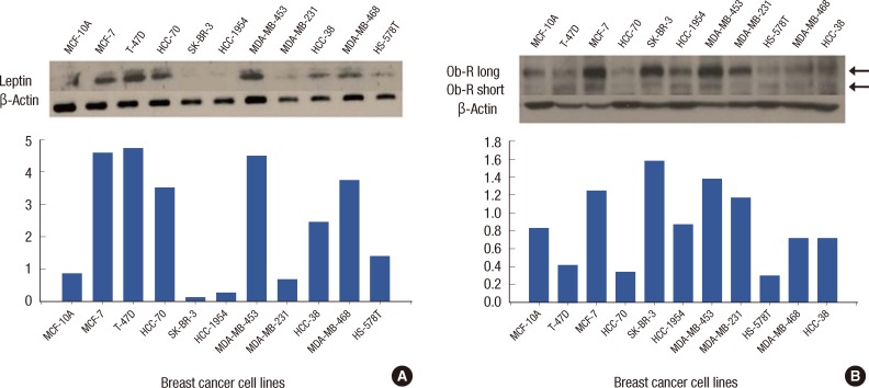
To study the effect of leptin on tamoxifen action, we first determined the expression level of leptin and Ob-R in various breast cancer cell lines. Leptin expression was found in MCF-7, T-47D, MDA-MB453, HCC-70, and MDA-MB468 cells. These results were confirmed by quantitative analysis using ImageJ densitometry (Figure 1A). Notably, leptin expression was increased in ER-positive breast cancer cell lines compared to other types of breast cancer cells. Ob-Rb was detected in the MCF-7, SK-BR3, MDA-MB453, and MDA-MB231 cell lines (Figure 1B). There was increased expression of Ob-Rb in breast cancer cells compared to normal breast cells. In contrast, there was no difference in the expression of Ob-Ra between breast cancer cells and normal breast cells.
Figure 1.
Leptin and leptin receptor (Ob-R) expression in various breast cancer cell lines. (A) Notably, there is increased leptin expression in estrogen receptor (ER)-positive cell lines (MCF-7, T-47D, and HCC-70) compared to other types of breast cancer cells. β-Actin was used for loading control in both cell lines. (B) In breast cancer cell lines, the Ob-R long (Ob-Rb) is the predominat receptor expressed compared to normal breast tissue, whereas there is no difference in Ob-R short (Ob-Ra) expression between breast cancer cell lines and normal breast tissue. Top, expression by Western blot assay; Bottom, quantitative analysis of Western blot results using densitometry.
Leptin enhances the proliferation of MCF-7 cells
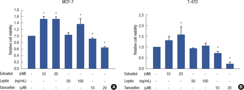
For cellular proliferation experiments, we chose to use MCF-7 and T-47D cells because they had differential expression patterns. MCF-7 cells have an ER-positive, leptin-positive, and Ob-Rb-positive expression profile, whereas T-47D cells have an ER-positive, leptin-positive, and Ob-Rb-negative expression profile. In the cell viability assay, E2 at 10 and 20 nM had a significant proliferative effect on MCF-7 cells compared to the respective E2 10 and 20 nM control treatments (p<0.001). Leptin at 50 ng/mL had no proliferative effect on MCF-7 cells (p=0.211), however, there was a significant proliferative effect of leptin at 100 ng/mL (p<0.001) (Figure 2A). In the T-47D cell line (Figure 2B), E2 at 10 nM increased cell proliferation but was not statistically significant (p=0.123). However, E2 at 20 nM had a significant proliferative effect compared to the control (p=0.048). Leptin at 50 and 100 ng/mL had no proliferative effect on T-47D cells (p=0.565 and p=0.641, respectively). Tamoxifen at 10 and 20 µM had a significant inhibitory effect on proliferation of both MCF-7 and T-47D cells in a dose dependent manner. However, leptin at 50 and 100 ng/mL had no additional proliferative effect on MCF-7 cells compared to E2-only treated control MCF-7 cells under both E2 10 and 20 nM treatments. Leptin did not affect the activity of tamoxifen treatment compared to tamoxifen-only treated control MCF-7 cells. Similar results were obtained in T-47D cells (data not shown). Thus, the results conclusively establish that leptin does not interfere with the effect of E2 or tamoxifen in either MCF-7 or T-47D cells.
Figure 2.
Cell viability after single treatment of estrogen, leptin, and tamoxifen in MCF-7 and T-47D cells. MCF-7 cells at 70% confluence were synchronized in serum-free medium for 24 hours and treated. Experiments were performed at least 3 times. Cell viability assays are shown as histograms. The bars represent relative cell number (±standard deviation). Cell viability was assessed by CellTiter-Glo® Luminescent Cell Viability assay. (A) Estrogen and leptin had proliferative effects on MCF-7 cells compared to no treatment. Tamoxifen had a significant inhibitory effect on MCF-7 cell proliferation in a dose-dependent manner. (B) Estrogen had proliferative effects at 20 nM, and leptin had no significant effect on T-47D cell proliferation.
*Statistical significance compared to no treatment (p<0.05).
Leptin inhibited the antiestrogenic effect of tamoxifen under E2 treated conditions
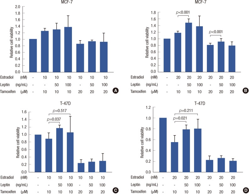
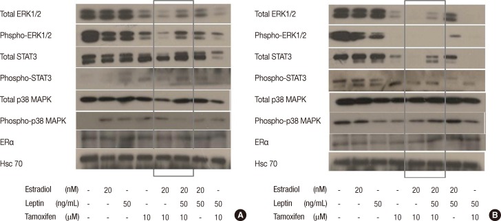
In MCF-7 cells, leptin at 50 and 100 ng/mL had no significant effect on tamoxifen response in E2 10 nM treated cells (Figure 3A). Interestingly, in E2 20 nM treated MCF-7 cells (Figure 3B), leptin at 50 ng/mL had a significant inhibitory effect on the response to both 10 and 20 µM tamoxifen (p<0.001 and p<0.001, respectively). In T-47D cells, when leptin 50 ng/mL was added to the combination of E2 10 nM and tamoxifen 10 µM, leptin significantly inhibited the antiestrogenic effect of tamoxifen (p=0.037) (Figure 3C). Leptin at 50 ng/mL also had a significant inhibitory effect on tamoxifen response with E2 at 20 nM (p=0.021) (Figure 3D). In cell signaling, when leptin was added to the combination treatment of E2 and tamoxifen, ERK1/2 and STAT3 were activated compared to the E2 and tamoxifen only combination at 1 hour in MCF-7 cells (Figure 4A). In contrast, in T-47D cells, when leptin was added to the combination of E2 and tamoxifen, leptin did not have any effect on ERK1/2 and STAT3 signaling (Figure 4B). The p38 MAPK and ERα signal transduction pathways were not activated or altered by treatment with leptin in either MCF-7 or T-47D cells.
Figure 3.
Leptin inhibition of the antiestrogenic effect of tamoxifen under estradiol stimulated conditions in MCF-7 and T-47D cells. In MCF-7 cells with estradiol 10 nM stimulation, leptin had no effect on the activity of tamoxifen for each combination treatment (A). At estradiol 20 nM stimulation, when leptin 50 ng/mL was added to tamoxifen treatment, leptin significantly inhibited the 10 and 20 µM tamoxifen responses (B). In T-47D cells, leptin 50 ng/mL had an inhibitory effect on tamoxifen 10 µM with estradiol 10 and 20 nM (C and D, respectively). There was no significant difference between leptin 100 ng/mL treatment and no leptin treatment. With tamoxifen 20 µM, cell viability was notably decreased compared to tamoxifen 10 µM treatment, and there was no difference between each combination. All cell viability results are expressed by the ratio of comparison to control (no treatment) group.
Figure 4.
Leptin activates multiple signal transduction pathways in MCF-7 cells compared to T-47D cells. MCF-7 and T-47D cells were synchronized in serum-free medium and then stimulated with estrogen (20 nM), leptin (50 ng/mL), and tamoxifen (10 µM). Stimulation of Ob-Rb was assessed at different time points from 5 minutes to 6 hours. Hsc70 was used as a loading control at each time point for both cell lines. Activation (phospho) and levels of ERK1/2, STAT3, p38 MAPK, and estrogen receptor α (ERα) were assessed by Western blotting in 25 µg of protein using specific antibodies. Figure 4 shows multiple signal transduction pathways being activated at 1 hour. Tamoxifen 10 µM effectively inhibited the activity of total- and phospho-ERK1/2 and STAT3 in both cell lines. (A) In MCF-7 cells, when leptin was added to the combination of estrogen and tamoxifen, leptin induced activation of total- and phospho-ERK1/2, total- and phospho-STAT3 signaling (grey box). There were no significant differences in total- and phospho-p38 MAPK, and ERα signaling when leptin was added to the combination of estrogen and tamoxifen. (B) In contrast to the results obtained in MCF-7 cells, in T-47D cells, there was no activation of the phospho-ERK1/2 and phospho-STAT3 signal transduction pathways. Also, total- and phospho-p38 MAPK, ERα signaling were not activated by addition of leptin (grey box).
DISCUSSION
The goal of hormone therapy is to block estrogen-induced proliferation of breast cancer cells. The ER is expressed in approximately 70% of patients with breast cancer. In spite of high levels of ER expression, many ER-positive patients demonstrate resistance to hormone therapy. The Ob-R and ERα have been found to be coexpressed in malignant mammary tissue and in breast cancer cell lines [20]. Therefore, it is possible that the Ob-R and ERα signal transduction pathways are involved in functional cross-talk that contributes to tumor development and progression. Leptin is also known to cross-talk with and transactivate several signal transduction pathways, including estrogen, the EGF receptor and HER2 signal transduction pathways, that are targets of breast cancer therapy [21]. Therefore, we anticipated that leptin would play some role in resistance to antiestrogenic treatment, and our results showed that leptin inhibited the antiestrogenic effect of tamoxifen in MCF-7 and T-47D ER-positive breast cancer cells under E2 stimulation. This finding was supported by a recent study by Chen et al. [22].
Leptin is known to activate the STAT3 signal transduction pathway in ERα-positive breast cancer cells which are already resistant to antihormones. Thus, high leptin levels in obese breast cancer patients may contribute to the development of antiestrogen resistance [23]. These results were confirmed by leptin-induced activation of multiple signal transduction pathways in the current study. When leptin was added to E2 and tamoxifen combination treatment, remarkably, leptin activated total- and phospho-ERK1/2, total- and phospho-STAT3 compared to E2 and tamoxifen combination alone in MCF-7 cells (Figure 4A). These results suggest that leptin inhibits the antiestrogenic effect of tamoxifen under E2 stimulation through activation of the ERK1/2 and STAT3 signal transduction pathways. In contrast to the results obtained in MCF-7 cells, in Ob-Rb negative T-47D cells in which leptin also had an inhibitory effect on tamoxifen acitivity (Figure 3C and 3D), the phospho-ERK1/2 and phospho-STAT3 signal transduction pathways were not activated but total ERK1/2 and total STAT3 were found to be activated (Figure 4B). These results suggest that leptin exerts its activity not only through the Ob-Rb, but also through cross-talk with other signal transduction pathways implicated in tumorigenesis. A recent discovery revealed that ERα turnover is differentially regulated depending on whether the receptor is free (not bound by ligand), agonist bound, or antagonist bound and whether signal transduction pathways (e.g., MAP kinases) are induced by other cell surface receptors [24]. Thus, it is possible that leptin can exert its action only on antiestrogen ICI 182,780-dependent estrogen receptor processing. Indeed, in different assays, no effects of leptin were observed in the basal or E2-induced activity of ERα [18]. This phenomenon was also shown in our study; leptin had no additional proliferative effect on E2-only treatment in both MCF-7 and T-47D cells. However, in E2 and tamoxifen combination treatment, leptin showed an inhibitory effect on tamoxifen in both MCF-7 (Ob-Rb positive) and T-47D (Ob-Rb negative) cells. That is, leptin can exert its action only on tamoixifen-dependent ER processing, displaying its maximal activity at a concentration of 50 ng/mL.
Previous studies have demonstrated in vitro that leptin stimulates cancer cell proliferation [25] as well as inhibits apoptosis [26] and induces angiogenesis [27]. Leptin was recently reported to stimulate the proliferation of various tumor cell types (prostate, colorectal, lung) [28]. Similar results have been described in breast cancer in which leptin was shown to stimulate the proliferation of various breast cancer cell lines [20]. Chen et al. [29] found no significant effect of surgical tumor removal on high serum leptin concentrations in their breast cancer patients. This finding is consistent with the hypothesis that production of leptin by tumor cells is a minor source of the adipokines, while unexcised adipose tissue remains the major contributor to circulating levels of leptin [3]. Therefore, serum leptin levels are considered an important factor which affects the tumor environment in breast cancer [30]. In the current study, leptin significantly inhibited the antiestrogenic effect of tamoxifen in the presence of E2 in ER-positive breast cancer cells. That is, increased serum leptin levels in breast cancer patients could be a cause of endocrine-resistance, especially for those with high serum E2 levels. Therefore, inhibition of leptin, including lowering of circulating leptin levels, could be an effective measure to overcome hormone resistance as well as antitumor activity. Although large-scale clinical studies are required to draw clinically meaningful conclusions, we found that leptin may be a promising target for the treatment of ER-positive breast cancer.
Footnotes
This experimental study was presented as a poster at the San Antonio Breast Cancer Symposium, December 6-10, 2011, San Antonio, TX, USA.
The authors declare that they have no competing interests.
References
- 1.Reinier KS, Vacek PM, Geller BM. Risk factors for breast carcinoma in situ versus invasive breast cancer in a prospective study of pre- and post-menopausal women. Breast Cancer Res Treat. 2007;103:343–348. doi: 10.1007/s10549-006-9375-9. [DOI] [PubMed] [Google Scholar]
- 2.Key TJ, Appleby PN, Reeves GK, Roddam A, Dorgan JF, Longcope C, et al. Body mass index, serum sex hormones, and breast cancer risk in postmenopausal women. J Natl Cancer Inst. 2003;95:1218–1226. doi: 10.1093/jnci/djg022. [DOI] [PubMed] [Google Scholar]
- 3.Vona-Davis L, Rose DP. Adipokines as endocrine, paracrine, and autocrine factors in breast cancer risk and progression. Endocr Relat Cancer. 2007;14:189–206. doi: 10.1677/ERC-06-0068. [DOI] [PubMed] [Google Scholar]
- 4.Pischon T, Nöthlings U, Boeing H. Obesity and cancer. Proc Nutr Soc. 2008;67:128–145. doi: 10.1017/S0029665108006976. [DOI] [PubMed] [Google Scholar]
- 5.Saxena NK, Sharma D, Ding X, Lin S, Marra F, Merlin D, et al. Concomitant activation of the JAK/STAT, PI3K/AKT, and ERK signaling is involved in leptin-mediated promotion of invasion and migration of hepatocellular carcinoma cells. Cancer Res. 2007;67:2497–2507. doi: 10.1158/0008-5472.CAN-06-3075. [DOI] [PMC free article] [PubMed] [Google Scholar]
- 6.Tartaglia LA, Dembski M, Weng X, Deng N, Culpepper J, Devos R, et al. Identification and expression cloning of a leptin receptor, OB-R. Cell. 1995;83:1263–1271. doi: 10.1016/0092-8674(95)90151-5. [DOI] [PubMed] [Google Scholar]
- 7.Bjørbaek C, Uotani S, da Silva B, Flier JS. Divergent signaling capacities of the long and short isoforms of the leptin receptor. J Biol Chem. 1997;272:32686–32695. doi: 10.1074/jbc.272.51.32686. [DOI] [PubMed] [Google Scholar]
- 8.Yamashita T, Murakami T, Otani S, Kuwajima M, Shima K. Leptin receptor signal transduction: OBRa and OBRb of fa type. Biochem Biophys Res Commun. 1998;246:752–759. doi: 10.1006/bbrc.1998.8689. [DOI] [PubMed] [Google Scholar]
- 9.Garofalo C, Koda M, Cascio S, Sulkowska M, Kanczuga-Koda L, Golaszewska J, et al. Increased expression of leptin and the leptin receptor as a marker of breast cancer progression: possible role of obesity-related stimuli. Clin Cancer Res. 2006;12:1447–1453. doi: 10.1158/1078-0432.CCR-05-1913. [DOI] [PubMed] [Google Scholar]
- 10.Rose DP, Gilhooly EM, Nixon DW. Adverse effects of obesity on breast cancer prognosis, and the biological actions of leptin (review) Int J Oncol. 2002;21:1285–1292. [PubMed] [Google Scholar]
- 11.Catalano S, Marsico S, Giordano C, Mauro L, Rizza P, Panno ML, et al. Leptin enhances, via AP-1, expression of aromatase in the MCF-7 cell line. J Biol Chem. 2003;278:28668–28676. doi: 10.1074/jbc.M301695200. [DOI] [PubMed] [Google Scholar]
- 12.Kitawaki J, Kusuki I, Koshiba H, Tsukamoto K, Honjo H. Leptin directly stimulates aromatase activity in human luteinized granulosa cells. Mol Hum Reprod. 1999;5:708–713. doi: 10.1093/molehr/5.8.708. [DOI] [PubMed] [Google Scholar]
- 13.Spicer LJ, Francisco CC. The adipose obese gene product, leptin: evidence of a direct inhibitory role in ovarian function. Endocrinology. 1997;138:3374–3379. doi: 10.1210/endo.138.8.5311. [DOI] [PubMed] [Google Scholar]
- 14.Ghizzoni L, Barreca A, Mastorakos G, Furlini M, Vottero A, Ferrari B, et al. Leptin inhibits steroid biosynthesis by human granulosa-lutein cells. Horm Metab Res. 2001;33:323–328. doi: 10.1055/s-2001-15419. [DOI] [PubMed] [Google Scholar]
- 15.Machinal-Quélin F, Dieudonné MN, Pecquery R, Leneveu MC, Giudicelli Y. Direct in vitro effects of androgens and estrogens on ob gene expression and leptin secretion in human adipose tissue. Endocrine. 2002;18:179–184. doi: 10.1385/ENDO:18:2:179. [DOI] [PubMed] [Google Scholar]
- 16.Bennett PA, Lindell K, Karlsson C, Robinson IC, Carlsson LM, Carlsson B. Differential expression and regulation of leptin receptor isoforms in the rat brain: effects of fasting and oestrogen. Neuroendocrinology. 1998;67:29–36. doi: 10.1159/000054295. [DOI] [PubMed] [Google Scholar]
- 17.Lindell K, Bennett PA, Itoh Y, Robinson IC, Carlsson LM, Carlsson B. Leptin receptor 5'untranslated regions in the rat: relative abundance, genomic organization and relation to putative response elements. Mol Cell Endocrinol. 2001;172:37–45. doi: 10.1016/s0303-7207(00)00382-8. [DOI] [PubMed] [Google Scholar]
- 18.Garofalo C, Sisci D, Surmacz E. Leptin interferes with the effects of the antiestrogen ICI 182,780 in MCF-7 breast cancer cells. Clin Cancer Res. 2004;10:6466–6475. doi: 10.1158/1078-0432.CCR-04-0203. [DOI] [PubMed] [Google Scholar]
- 19.Catalano S, Mauro L, Marsico S, Giordano C, Rizza P, Rago V, et al. Leptin induces, via ERK1/ERK2 signal, functional activation of estrogen receptor alpha in MCF-7 cells. J Biol Chem. 2004;279:19908–19915. doi: 10.1074/jbc.M313191200. [DOI] [PubMed] [Google Scholar]
- 20.Hu X, Juneja SC, Maihle NJ, Cleary MP. Leptin: a growth factor in normal and malignant breast cells and for normal mammary gland development. J Natl Cancer Inst. 2002;94:1704–1711. doi: 10.1093/jnci/94.22.1704. [DOI] [PubMed] [Google Scholar]
- 21.Surmacz E. Obesity hormone leptin: a new target in breast cancer? Breast Cancer Res. 2007;9:301. doi: 10.1186/bcr1638. [DOI] [PMC free article] [PubMed] [Google Scholar]
- 22.Chen X, Zha X, Chen W, Zhu T, Qiu J, Røe OD, et al. Leptin attenuates the anti-estrogen effect of tamoxifen in breast cancer. Biomed Pharmacother. 2013;67:22–30. doi: 10.1016/j.biopha.2012.10.001. [DOI] [PubMed] [Google Scholar]
- 23.Binai NA, Damert A, Carra G, Steckelbroeck S, Löwer J, Löwer R, et al. Expression of estrogen receptor alpha increases leptin-induced STAT3 activity in breast cancer cells. Int J Cancer. 2010;127:55–66. doi: 10.1002/ijc.25010. [DOI] [PubMed] [Google Scholar]
- 24.Marsaud V, Gougelet A, Maillard S, Renoir JM. Various phosphorylation pathways, depending on agonist and antagonist binding to endogenous estrogen receptor alpha (ERalpha), differentially affect ERalpha extractability, proteasome-mediated stability, and transcriptional activity in human breast cancer cells. Mol Endocrinol. 2003;17:2013–2027. doi: 10.1210/me.2002-0269. [DOI] [PubMed] [Google Scholar]
- 25.Onuma M, Bub JD, Rummel TL, Iwamoto Y. Prostate cancer cell-adipocyte interaction: leptin mediates androgen-independent prostate cancer cell proliferation through c-Jun NH2-terminal kinase. J Biol Chem. 2003;278:42660–42667. doi: 10.1074/jbc.M304984200. [DOI] [PubMed] [Google Scholar]
- 26.Rouet-Benzineb P, Aparicio T, Guilmeau S, Pouzet C, Descatoire V, Buyse M, et al. Leptin counteracts sodium butyrate-induced apoptosis in human colon cancer HT-29 cells via NF-kappaB signaling. J Biol Chem. 2004;279:16495–16502. doi: 10.1074/jbc.M312999200. [DOI] [PubMed] [Google Scholar]
- 27.Bouloumié A, Drexler HC, Lafontan M, Busse R. Leptin, the product of Ob gene, promotes angiogenesis. Circ Res. 1998;83:1059–1066. doi: 10.1161/01.res.83.10.1059. [DOI] [PubMed] [Google Scholar]
- 28.Tessitore L, Vizio B, Jenkins O, De Stefano I, Ritossa C, Argiles JM, et al. Leptin expression in colorectal and breast cancer patients. Int J Mol Med. 2000;5:421–426. doi: 10.3892/ijmm.5.4.421. [DOI] [PubMed] [Google Scholar]
- 29.Chen DC, Chung YF, Yeh YT, Chaung HC, Kuo FC, Fu OY, et al. Serum adiponectin and leptin levels in Taiwanese breast cancer patients. Cancer Lett. 2006;237:109–114. doi: 10.1016/j.canlet.2005.05.047. [DOI] [PubMed] [Google Scholar]
- 30.Madaio RA, Spalletta G, Cravello L, Ceci M, Repetto L, Naso G. Overcoming endocrine resistance in breast cancer. Curr Cancer Drug Targets. 2010;10:519–528. doi: 10.2174/156800910791517226. [DOI] [PubMed] [Google Scholar]


