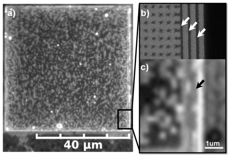
Fig. 1.

a) Pixilation pattern of coexisting dark Lo and bright Ld phase lipid multibilayers supported by a square lattice pattern of PMMA features (bumps) on silica surrounded by 3 PMMA walls; imaging by fluorescence microscopy. b) SEM image demonstrating that the underlying square lattice of bumps has a spacing of 200 nm and the 3 walls/fences (arrows) are 180 nm in thickness spaced 250 nm apart as produced by e-beam lithography. c) Alignment and elongation of dark Lo phase domains (e.g. arrow), supported by this wall pattern, along the long axis of the walls.
