Sir,
We present a case of juvenile choroidal xanthogranuloma (JXG) in a child, mimicking non-accidental injury.
Case report
A 3-year-old boy presented with a 2-week history of drowsiness and left eye redness. His father reported that the child fell from a height of about 20 cm. The fact that a doubt had arisen for the cause of the eye problem raised the suspicion of non-accidental injury. No abnormality was detected on physical examination and magnetic resonance imaging of the brain. His past medical history was unremarkable. On ophthalmological examination, visual acuity was 0.2 LogMAR in the right eye and perception of light in the left eye. Slit-lamp examination revealed a total hyphaema in the left eye, while the right eye showed no ocular abnormality. The intraocular pressure was 63 mm Hg in the left eye and dropped to 10 mm Hg after washing-out of the blood from the anterior chamber. No iris lesions were evident. Additionally, there was no fundal view of the left eye due to a dense vitreous haemorrhage. Ocular ultrasound examination showed a thicker choroid temporally, but no evidence of retinal detachment or calcification.
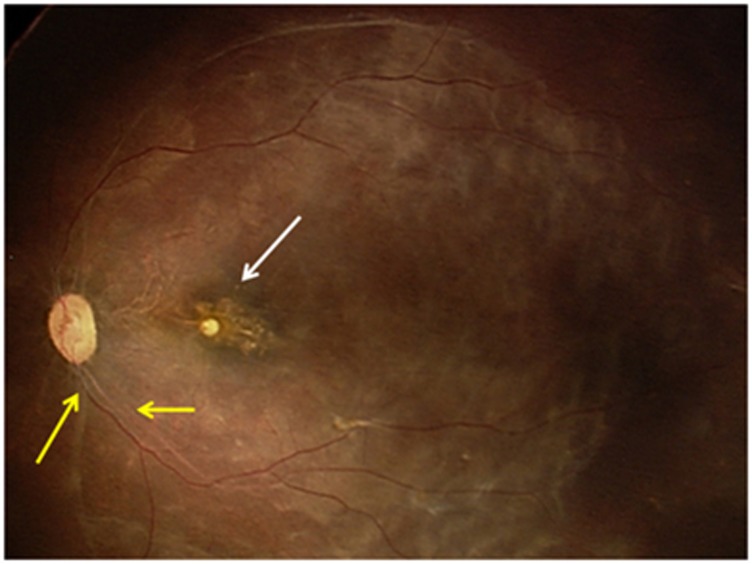
Following a left eye vitrectomy and lens aspiration, the retina demonstrated retinal infiltrates and a pale optic disc (Figure 1). A vitreous biopsy was taken, and immunocytochemistry analysis showed the cells to stain strongly with CD68 and weakly with S-100. PAS was found to be positive within the cytoplasm. A diagnosis of choroidal JXG was made.
Figure 1.
Fundus photograph of the left eye showing posterior segment involvement with sclerosed blood vessels and a pale optic disc (yellow arrows) and retinal infiltrates (white arrow).
The family had been assessed by social services and no concerns had been raised. Fourteen months after the initial presentation, there is no involvement of the fellow eye.
Comment
Our case is the first to use a vitreous biopsy to diagnose this condition and the fourth in the literature of JXG with ocular involvement and absence of cutaneous manifestations.1, 2, 3 If spontaneous hyphaema and vitreous haemorrhage are present, one should include in the differential diagnosis the possibility of JXG, along with non-accidental injury and malignancy, even in the absence of skin or systemic lesions. Consideration of this condition may prevent needless investigations for child protection.
Acknowledgments
We would like to acknowledge Mr K K Nischal (Director, Paediatric Ophthalmology, Strabismus and Adult Motility, UPMC Children's Hospital, Pittsburgh) for his involvement with this case.
The authors declare no conflict of interest.
References
- Wertz FD, Zimmerman LE, McKeown CA, Croxatto JO, Whitmore PV, LaPiana FG. Juvenile xanthogranuloma of the optic nerve, disc, retina, and choroid. Ophthalmology. 1982;89:1331–1335. doi: 10.1016/s0161-6420(82)34637-0. [DOI] [PubMed] [Google Scholar]
- DeBarge LR, Chan CC, Greenberg SC, McLean IW, Yannuzzi LA, Nussenblatt RB. Chorioretinal, iris, and ciliary body infiltration by juvenile xanthogranuloma masquerading as uveitis. Surv Ophthalmol. 1994;39:65–71. doi: 10.1016/s0039-6257(05)80046-3. [DOI] [PubMed] [Google Scholar]
- Zamir E, Wang RC, Krishnakumar S, Aiello Leverant A, Dugel PU, Rao NA. Juvenile xanthogranuloma masquerading as paediatric chronic uveitis: a clinicopathologic study. Surv Ophthalmol. 2001;46:164–171. doi: 10.1016/s0039-6257(01)00253-3. [DOI] [PubMed] [Google Scholar]


