Abstract
Purpose
To study the predisposing factors for late in-the-bag intraocular lens (IOL) dislocation and to analyze the outcomes after explantation surgery.
Methods
In this retrospective multicentre study, 61 eyes were enrolled. The main inclusion criterion was in-the-bag spontaneous IOL dislocation after uneventful phacoemulsification cataract extraction. Only eyes with serious dislocation that required IOL explantation were eligible. Follow-up after explantation surgery of at least 3 months was required. Exclusion criteria were complicated cataract surgery, out-of-the-bag IOL dislocation, and dislocations that occurred in the first year after the cataract surgery. The main outcome measures were predisposing factors for dislocation, interval between cataract surgery and dislocation, surgical treatment at the time of explantation, preoperative and postoperative corrected distance visual acuity (CDVA), and postoperative complications.
Results
High myopia was detected in 12 cases (19.7%) and it was the main predisposing factor. Mean time interval from cataract surgery to dislocation was 7.5 (SD 5.2) years. The dislocated in-the-bag IOL was replaced with a scleral fixated IOL (36.1%), angle-supported anterior chamber IOL (31.1%), sulcus repositioning (18%), or posterior chamber iris sutured IOL (4.9%). Finally, 9.8% of the patients were left aphakic. Mean CDVA improved significantly after surgery (P=0.005). Final CDVA of 20/40 or better was achieved in 29 cases (47.5%).
Conclusions
High myopia was the main risk factor for late in-the-bag IOL dislocation. Surgical treatment significantly improved the CDVA in our sample and was associated with a low complication rate.
Keywords: IOL dislocation, pseudoexfoliation, high myopia, cataract surgery and explantation
Introduction
Late in-the-bag dislocation of intraocular lenses (IOLs) is a rare but potentially serious complication after cataract surgery. The incidence of surgery specifically due to late dislocated IOL is 0.032–0.28%.1, 2 In a recent observational study, it was shown that the cumulative risk of IOL dislocation at 5, 10, 15, 20, and 25 years after cataract extraction was 0.1%, 0.1%, 0.2%, 0.7%, and 1.7% respectively.3 Nevertheless, the pseudophakic population has been growing very quickly in recent years as a result of the longer lifespan, the new phacorefractive procedures, and the improvement in the quality and safety of phacoemulsification surgery. As a result of this, late in-the-bag dislocation may become a more common issue in the future.
Bag dislocation is due to a progressive zonular dehiscence many years after uneventful surgery. The risks factors for this condition include pseudoexfoliation (PEX), connective tissue disorders, uveitis, retinitis pigmentosa, high myopia, and patients who underwent vitreoretinal surgery.4, 5, 6 Bag dislocation in these situations usually occurs a long time after cataract surgery, with a mean interval of 6.9–8.5 years.5, 6, 7, 8
Surgical management of this condition is usually complex and may be associated to new complications. Explantation of the whole capsular bag with implantation of a sulcus sutured IOL or an anterior chamber IOL is often required.
The optimum approach for each case remains unclear because each surgical option may result in new complications. Therefore, more scientific evidence is needed in order to support a surgical decision in these cases.
The aim of this paper is to study the weight of the different predisposing factors for late in-the-bag IOL dislocation and to analyze the outcomes and complications following the explantation surgery for a better understanding of the management of these cases.
Materials and methods
Observational retrospective multicentre study. Three different centers participated in this study, two of them are private settings (Vissum Corporation Alicante, Spain, and Centro de Oftalmología Barraquer, Barcelona, Spain) and the other one is a public hospital (Hospital Universitario Ramón y Cajal, Madrid, Spain). The three centers have different geographic locations in the country.
A total of 61 eyes of 56 patients fulfilled the criteria of the study.
Inclusion and exclusion criteria
Consecutive patients with in-the-bag spontaneous IOL dislocation after uneventful phacoemulsification cataract extraction were included. Only eyes with serious dislocation that required IOL explantation were eligible. Follow-up after explantation surgery of at least 3 months was required. Only patients with axial length ≥27 mm were labeled in the high-myopia group.
The exclusion criteria were: complicated cataract surgery including posterior capsule tear (with or without vitreous loss), any zonular deficiencies noticed during the surgery, and problems related to the capsulorrhexis step. The remaining exclusion criteria were out-of-the-bag IOL dislocation, and dislocations that occurred in the first year after the cataract surgery.
Following these inclusion and exclusion criteria, we evaluated the consecutive cases that were explanted due to in-the-bag dislocation in these three centers from 2004 to 2011. The explantation surgeries were performed by different experienced surgeons, and no specific protocol was followed during the surgery. Implanting the IOL with scleral sutures was performed using an ab externo scleral fixation technique.
Main outcome measures
The main outcome measures of the study were predisposing factors for dislocation, interval between cataract surgery and dislocation, surgical treatment at the time of explantation, preoperative and postoperative corrected distance visual acuity (CDVA), and complications associated to the explantation surgery.
Statistical analysis
All data were analyzed using SPSS for windows software (version 15.0; SPSS, Inc., Chicago, IL, USA).
When comparing CDVA preoperatively and postoperatively, Student's t-test for related sample was used. Kruskal–Wallis test was used to analyze if there were differences in the increase of visual acuity among groups depending on the final IOL position (scleral fixated IOL, angle-supported anterior chamber IOL, sulcus repositioning, posterior chamber iris sutured IOL, and aphakia).
Mann–Whitney U-test was performed to compare the time interval from cataract surgery to in-the-bag IOL dislocation between the high-myopia and the PEX groups. The same test was also used to compare age at cataract surgery and age at explantation surgery in these two groups.
Results
Table 1 shows the patients' demographic data. Some predisposing factors were detected; the most prevalent ones were high myopia in 12 cases (19.7%), PEX in 10 eyes (16.4%), vitrectomy in 8 cases (13.1%), previous traumatism in 4 cases (6.6%), retinitis pigmentosa in 2 eyes (3.3%), uveitis in 1 case (1.6%), and both high myopia and PEX together in 1 more case (1.6%). There was no identifiable cause in 23 eyes (37.7%). The interval between the uneventful cataract surgery and the dislocation was 7.5 (SD 5.2) years. There was no statistically significant difference in the time interval from the cataract surgery to the dislocation between the eyes with PEX and those with high myopia (P=0.819).
Table 1. Patients' demographic data.
| Parameter | All patients, mean (SD; (range)) | High-myopia group, median (p25–p75) | Pseudoexfoliation group, median (p25–p75) |
|---|---|---|---|
| Age at cataract surgery | 63.5 (SD 12.8; (33–88)) years old | 52.9 (50.7–65) years olda | 78 (68–85) years olda |
| Age at explantation surgery | 71.2 (SD12.5; (41–97)) years old | 65 (61.2–74.7) years oldb | 87 (76.5–95) years oldb |
| Sex (%) | |||
| ♂: Male | ♂:68.9% | ♂: 58.3% | ♂: 50% |
| ♀: Female | ♀:31.1% | ♀: 41.7% | ♀: 50% |
Statistically significant differences were found between both groups, Mann–Whitney U-test (P=0.002).
Statistically significant differences were found between both groups, Mann–Whitney U-test (P=0.001).
However, high-myopic eyes had cataract surgery and explantation surgery at a younger age than the PEX eyes (P=0.002 and 0.001, respectively).
The dislocated in-the-bag IOL was replaced with a scleral fixated IOL in 22 cases (36.1%), angle-supported anterior chamber IOL in 19 cases (31.1%), sulcus repositioning in 11 cases (18%), or posterior chamber iris sutured IOL in 3 cases (4.9%). Finally, six eyes (9.8%) were left aphakic. There was no statistically significant difference in the increase of CDVA after the surgery among these groups (P=0.693).
Preoperative mean LogMAR CDVA was 0.64 (SD 0.6 (0–2.8)), whereas mean postoperative LogMAR CDVA was 0.45 (SD 0.4 (0–2)). The improvement in the CDVA after surgery was statistically significant (P=0.005). Final LogMAR CDVA of 0.3 (Snellen 20/40) or better was achieved in 29 cases (47.5%) and 0.18 (Snellen 20/30) or better in 17 eyes (27.9%).
In a subanalysis comparing the two main risk-factor groups (high myopia and PEX), we found that the median (p25–p75) preoperative LogMAR CDVA was 0.49 (1–0.15) and 0.45 (0.99–0.33), respectively, with no significant differences (P=0.76). Postoperative median LogMAR CDVA was 0.26 (0.89–0.11) in the high-myopia group and 0.39 (0.84–0.19) in the PEX group. No statistically significant difference was detected (P=0.49).
We did not detect statistically significant differences in the gain of CDVA after the explantation surgery between both groups (P=0.176).
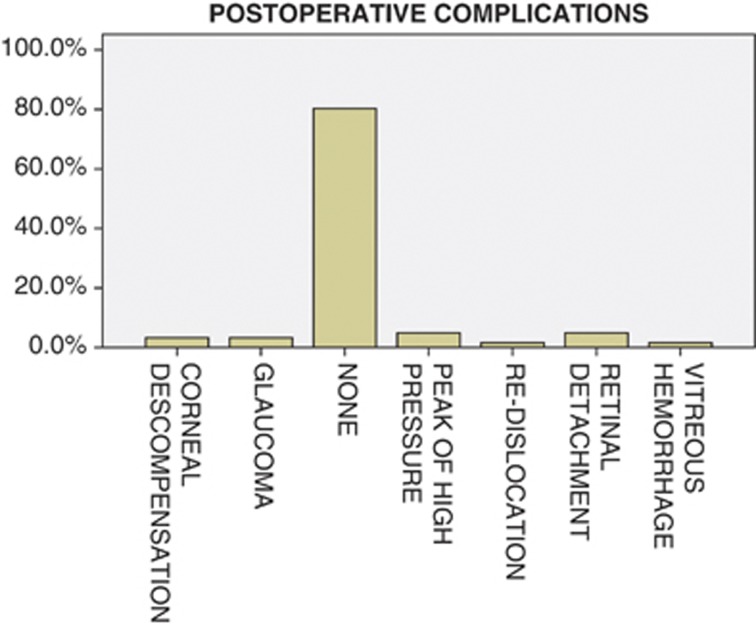
The overall complication rate after the explantation surgery was 19.7%. These complications associated to the surgical procedure are shown in Figure 1. Peak of high pressure the day after the surgery (three eyes) and retinal detachment in the early postoperative period (three eyes) were the two types of complications most frequently found after the explantation surgery.
Figure 1.
Complications after the explantation surgery.
Discussion
In recent years, there has been an increase in the number of papers reporting late IOL dislocations due to zonular failure after uneventful cataract surgery. In these studies, some features are detected as predisposing factors. Zonular weakness has been associated to PEX,4, 5, 6, 7, 9 uveitis,6 trauma,6, 10 high myopia,11 vitreoretinal surgery,12 retinitis pigmentosa,13 and connective tissue disorders, such as Marfan's syndrome, homocystinuria, hyperlysinemia, Ehler-Danlos, scleroderma, and Weil–Marchesani.14
In our study, the most prevalent risk factor was high myopia in 12 cases (19.7%). To our knowledge, this is the first long-term case series of late in-the-bag IOL dislocation showing high myopia as the main risk factor. High myopia has always been mentioned as a risk factor for late IOL dislocation in some single case reports,11, 15 in studies analyzing capsular bags with scanning electron microscopy,16 and in the long-term case-series reports that are discussed later. However, high myopia has never been detected before as the main risk factor for late spontaneous in-the-bag IOL dislocation. High-myopic eyes show some typical alterations due to thinning and degeneration of several eye layers as lacker cracks, chorioretinal atrophy, or posterior staphyloma.17 We hypothesize that as well as the previously mentioned alterations, these eyes may be also more prone to zonular failure due to excessive elongation of the zonular fibers that have to support greater stress than in normal axial length eyes. This theory is supported by the outcomes of a study using high-resolution magnetic resonance imaging, which demonstrated that myopic eyes are larger in all three dimensions (ie, equatorial, antero-posterior, and vertical axes).18 Moreover, this fact is also evident in the lens–iris diaphragm retropulsion syndrome that usually occurs when performing cataract surgery in high-myopic eyes.19
PEX has always been the most recognized predisposing factor for late dislocation as described above. PEX is likely to produce zonular insufficiency by two mechanisms. First, PEX accumulations mechanically weaken the zonular lamella and impair zonular anchoring to the epithelial basement membrane at both its origin and insertion.20 Furthermore, patients with PEX also exhibit an increase in elastinolysis that weakens the zonula. Second, PEX has shown to facilitate the anterior capsule contraction syndrome that if left untreated usually leads to zonular failure.21, 22
PEX was the second most important risk factor in our sample. It was described in 10 eyes (16.4%). On the contrary, in the other case series published to date, PEX was the most important risk factor.1, 6, 8, 23 Lorente et al.,7 in a recent study also performed in Spain, showed that PEX was found in 66.6% of the cases (30 eyes), whereas high myopia was only detected in 1 case (2.22%). This emphasizes that PEX incidence depends highly on the geographical location, even within a small country such as Spain. In Madrid, PEX incidence is 0.5%, whereas in some places in the north of Spain, the incidence is 25–30%.24 This geographical variability is represented in our study because the patients were from three different hospitals, located in three different Spanish regions.
In our study, most of the patients who underwent explantation due to late IOL dislocation were males (68.9%). This surprising result has also been found by other authors.6, 23, 25 This is difficult to explain because more women than men undergo cataract surgery26 and have PEX.27 Thus, some investigators suggest that there may be a gender-related difference that results in weaker zonulae in men with PEX. Another explanation may be that males are more prone to ocular trauma that might have happened a long time ago and was forgotten at the time of cataract surgery.25
The mean age of our patients at explantation surgery was 71.2 (SD:12.5 (41–97). Patients in the high-myopia group were younger at time of explantation than patients from the PEX group (P=0.001). Other authors also had the same finding.8
The mean time interval from cataract surgery to explantation due to late dislocation was 7.5±5.2 years. This coincides with previous reports that also showed a mean interval of around 8 years between both surgeries.1, 5, 6, 7, 8
The high-myopia group had cataract surgery and explantation surgery at a younger age than the PEX group (P=0.001). However, we did not detect any differences when comparing the time interval from the cataract surgery to the explantation surgery between both groups (P=0.819), or differences among the other risk factors regarding this time interval. Other authors had the same findings.8
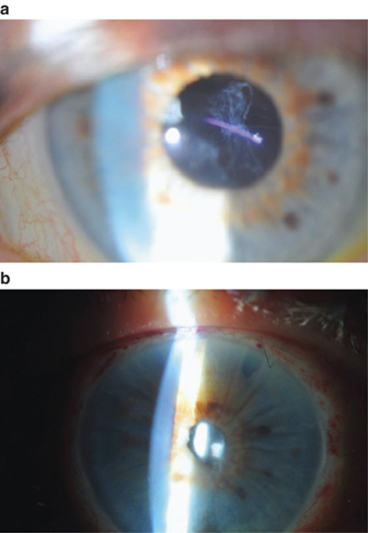
Different surgical techniques can be used to reposition a dislocated IOL. In our series, all the patients had IOL explantation because this was one of the inclusion criteria. A new IOL was placed after the explantation during the same surgery. A scleral fixated IOL was placed in most of the cases (36.1%) (Figure 2).
Figure 2.
(a) High-myopic eye with late in-the-bag IOL dislocation. (b) The same high-myopic eye after the explantation surgery. A scleral fixated IOL was implanted in this case.
At present, there is no consensus on what technique to use and several surgical procedures have good results.4, 6 Most authors agree that it is desirable to preserve and reposition the existing IOL if possible in order to avoid a large corneal wound, thus minimizing surgical trauma and major complications such as choroidal expulsive hemorrhage.1, 28, 29, 30 However, in some advanced dislocations, explantation is the only valid alternative.
We found a statistically significant improvement in CDVA after the explantation surgery (P=0.005). A final LogMAR CDVA of 0.3 (Snellen 20/40) or better was achieved in 29 cases (47.5%) and 0.18 (Snellen 20/30) or better in 17 eyes (27.9%). These results are similar to other reports.3 However, other authors have recently found better results; Lorente et al7 showed that 62.22% of patients had a final CDVA of 20/40 or better. We think that this difference is explained because in our sample all the patients had IOL exchange, and this is a more aggressive surgical technique performed in the worst cases when isolated IOL repositioning is not possible. Furthermore, in contrast to our results, other authors reporting outcomes from an IOL-exchange case series did not achieve a statistically significant improvement in CDVA after the surgery.23
Complications after surgery are shown in Figure 1. We had a complications incidence after surgery of 19.7%. This incidence is slightly higher than that reported by Lorente et al.7 In our opinion, it may be explained because this author reported a case series that included both IOL repositioning and IOL-exchange cases, while in our series the only possible treatment was IOL exchange, and as it has been previously mentioned, it is a more invasive surgery, thus associated with more complications. Indeed, we had lower complications than other IOL-explantation series that reported a complications incidence of 42.1%.23 In our series, the most common complications were intraocular pressure rise in three cases and retinal detachment in three more eyes. Vitrectomy was successfully performed in these retinal detachment cases. In previous IOL-explantation reports, intraocular pressure rise was also the most frequently found complication, whereas more severe complications like retinal detachment, corneal descompensation, or redislocation were rarely detected.23
Our study has some limitations: first, those related to its retrospective and multicenter nature. Another limitation is that the follow-up after the explantation surgery was at least 3 months but not much longer. However, we think that this is enough time to detect most of the postsurgical complications. Obviously, late-onset complications like pseudophakic bullous keratopathy in the group of patients with AC IOL or suture degrading in the group of patients with scleral fixated IOLs cannot be detected. Most of the series published1, 6, 7, 23 have a follow-up of approximately 1 year, which is not enough to detect these late-onset complications.
In conclusion, our study highlights that high myopia is an important risk factor for the late in-the-bag serious IOL dislocation after uneventful cataract surgery. This is the first long-term case-series study that shows that high myopia may be even more prevalent than PEX in eyes which developed spontaneous late in-the-bag dislocation. Furthermore, dislocation in high myopia occurred at a younger age than in PEX, hence affecting patients with bigger visual demands.
Lens exchange with scleral suture-fixated IOL was the most frequently chosen treatment. CDVA improved significantly after surgery with a low rate of complications.

Acknowledgments
Two of the three centers that participated in this study (Vissum Corporation and Centro de Oftalmología Barraquer) belong to the ‘RETICS' (Red Temática de Investigación Oftalmológica), ‘Patología ocular del envejecimiento, calidad visual y calidad de vida', Subproyecto de Calidad Visual (RD07/0062), which is a cooperative network for ophthalmic research sponsored by the Spanish Ministry of Health. Thus, this study has been supported in part by a grant from the Spanish Ministry of Health, Instituto Carlos III. The study also adhered to the Tenets of the Declaration of Helsinki.
The authors declare no conflict of interest.
References
- Jakobsson G, Zetterberg M, Lundstrom M, Stenevi U, Grenmark R, Sundelin K. Late dislocation of in-the-bag and out-of-the bag intraocular lenses: ocular and surgical characteristics and time to lens repositioning. J Cataract Refract Surg. 2010;36:1637–1644. doi: 10.1016/j.jcrs.2010.04.042. [DOI] [PubMed] [Google Scholar]
- Clark A, Morlet N, Ng JQ, Preen DB, Semmens JB. Whole population trends in complications of cataract surgery over 22 years in Western Australia. Ophthalmology. 2011;118 (6:1055–1061. doi: 10.1016/j.ophtha.2010.11.001. [DOI] [PubMed] [Google Scholar]
- Pueringer SL, Hodge DO, Erie JC. Risk of late intraocular lens dislocation after cataract surgery, 1980-2009: a population-based study. Am J Ophthalmol. 2011;152:618–623. doi: 10.1016/j.ajo.2011.03.009. [DOI] [PMC free article] [PubMed] [Google Scholar]
- Gimbel HV, Condon GP, Kohnen T, Olson RJ, Halkiadakis I. Late in-the-bag intraocular lens dislocation: incidence, prevention, and management. J Cataract Refract Surg. 2005;31:2193–2204. doi: 10.1016/j.jcrs.2005.06.053. [DOI] [PubMed] [Google Scholar]
- Jehan FS, Mamalis N, Crandall AS. Spontaneous late dislocation of intraocular lens within the capsular bag in pseudoexfoliation patients. Ophthalmology. 2001;108:1727–1731. doi: 10.1016/s0161-6420(01)00710-2. [DOI] [PubMed] [Google Scholar]
- Gross JG, Kokame GT, Weinberg DV. In-the-bag intraocular lens dislocation. Am J Ophthalmol. 2004;137:630–635. doi: 10.1016/j.ajo.2003.10.037. [DOI] [PubMed] [Google Scholar]
- Lorente R, de Rojas V, Vazquez de Parga P, Moreno C, Landaluce ML, Domínguez R, et al. Management of late spontaneous in-the-bag intraocular lens dislocation: Retrospective analysis of 45 cases. J Cataract Refract Surg. 2010;36:1270–1282. doi: 10.1016/j.jcrs.2010.01.035. [DOI] [PubMed] [Google Scholar]
- Davis D, Brubaker J, Espandar L, Stringham J, Crandall A, Werner L, et al. Late in-the-bag spontaneous intraocular lens dislocation: evaluation of 86 consecutive cases. Ophthalmology. 2009;116:664–670. doi: 10.1016/j.ophtha.2008.11.018. [DOI] [PubMed] [Google Scholar]
- Naumann GO, Schlotzer-Schrehardt U, Kuchle M. Pseudoexfoliation syndrome for the comprehensive ophthalmologist. Intraocular and systemic manifestations. Ophthalmology. 1998;105:951–968. doi: 10.1016/S0161-6420(98)96020-1. [DOI] [PubMed] [Google Scholar]
- Marin MI, Tejero TR, Dominguez FM, Gutierrez ME. Ocular injuries in midfacial fractures. Orbit. 1998;17:41–46. doi: 10.1076/orbi.17.1.41.7950. [DOI] [PubMed] [Google Scholar]
- Zech JC, Tanniere P, Denis P, Trepsat C. Posterior chamber intraocular lens dislocation with the bag. J Cataract Refract Surg. 1999;25:1168–1169. doi: 10.1016/s0886-3350(99)00124-8. [DOI] [PubMed] [Google Scholar]
- Yasuda A, Ohkoshi K, Orihara Y, Kusano Y, Sakuma A, Yamaguchi T. Spontaneous luxation of encapsulated intraocular lens onto the retina after a triple procedure of vitrectomy, phacoemulsification, and intraocular lens implantation. Am J Ophthalmol. 2000;130:836–837. doi: 10.1016/s0002-9394(00)00630-9. [DOI] [PubMed] [Google Scholar]
- Hayashi K, Hayashi H, Matsuo K, Nakao F, Hayashi F. Anterior capsule contraction and intraocular lens dislocation after implant surgery in eyes with retinitis pigmentosa. Ophthalmology. 1998;105:1239–1243. doi: 10.1016/S0161-6420(98)97028-2. [DOI] [PubMed] [Google Scholar]
- Cionni RJ.Surgical management of the congenitally subluxated crystalline lens using the modified capsular tension ringIn: Steinert RF (ed)Cataract Surgery: Technique, Complications, and Management2nd ed.Saunders: Philadelphia; 2004305–313. [Google Scholar]
- Laude A, Agrawal A. Spontaneous partial dislocation of an aphakic capsular bag in high myopia. J Cataract Refract Surg. 2011;37:427–428. doi: 10.1016/j.jcrs.2010.11.018. [DOI] [PubMed] [Google Scholar]
- Hirata A, Okinami S, Hayashi K. Occurrence of capsular delamination in the dislocated in-the-bag intraocular lens. Graefes Arch Clin Exp Ophthalmol. 2011;249:1409–1415. doi: 10.1007/s00417-010-1605-5. [DOI] [PubMed] [Google Scholar]
- Kanski Jack J.Clinical Ophthalmology. A Systematic Approach6th ed.Elsevier: Madrid, Spain; 2009 [Google Scholar]
- Cheng HM, Singh OS, Kwong KK, Xiong J, Woods BT, Brady TJ. Shape of the myopic eye as seen with high-resolution magnetic resonance imaging. Optom Vis Sci. 1992;69:698–701. doi: 10.1097/00006324-199209000-00005. [DOI] [PubMed] [Google Scholar]
- Wilbrandt HR, Wilbrandt TH. Pathogenesis and management of the lens-iris diaphragm retropulsion syndrome during phacoemulsification. J Cataract Refract Surg. 1994;20:48–53. doi: 10.1016/s0886-3350(13)80043-0. [DOI] [PubMed] [Google Scholar]
- Conway RM, Schlotzer-Schrehardt U, Kuchle M, Naumann GO. Pseudoexfoliation syndrome: pathological manifestations of relevance to intraocular surgery. Clin Experiment Ophthalmol. 2004;32:199–210. doi: 10.1111/j.1442-9071.2004.00806.x. [DOI] [PubMed] [Google Scholar]
- Davison JA. Capsule contraction syndrome. J Cataract Refract Surg. 1993;19:582–589. doi: 10.1016/s0886-3350(13)80004-1. [DOI] [PubMed] [Google Scholar]
- Auffarth GU, Tsao K, Wesendahl TA, Sugita A, Apple DJ. Centration and fixation of posterior chamber intraocular lenses in eyes with pseudoexfoliation syndrome. An analysis of explanted autopsy eyes. Acta Ophthalmol Scand. 1996;74:463–467. doi: 10.1111/j.1600-0420.1996.tb00600.x. [DOI] [PubMed] [Google Scholar]
- Hayashi K, Hirata A, Hayashi H. Possible predisposing factors for in-the-bag and out-of-the-bag intraocular lens dislocation and outcomes of intraocular lens exchange surgery. Ophthalmology. 2007;114:969–975. doi: 10.1016/j.ophtha.2006.09.017. [DOI] [PubMed] [Google Scholar]
- Lorente R, de Rojas V.Luxación tardía del complejo saco capsular y lente intraocularIn: Lorente R, Mendicute J (eds)Cirugía Del Cristalino Sociedad Española de Oftalmología: Madrid, Spain; 20081751–1767. [Google Scholar]
- Monestam EI. Incidence of dislocation of intraocular lenses and pseudophakodonesis 10 years after cataract surgery. Ophthalmology. 2009;116:2315–2320. doi: 10.1016/j.ophtha.2009.05.015. [DOI] [PubMed] [Google Scholar]
- Klein BE, Klein R, Moss SE. Incident cataract surgery: the Beaver Dam eye study. Ophthalmology. 1997;104:573–580. doi: 10.1016/s0161-6420(97)30267-x. [DOI] [PubMed] [Google Scholar]
- Astrom S, Linden C. Incidence and prevalence of pseudoexfoliation and open-angle glaucoma in northern Sweden: I. Baseline report. Acta Ophthalmol Scand. 2007;85:828–831. doi: 10.1111/j.1600-0420.2007.00992.x. [DOI] [PubMed] [Google Scholar]
- Chan CC, Crandall AS, Ahmed II. Ab externo scleral suture loop fixation for posterior chamber intraocular lens decentration: clinical results. J Cataract Refract Surg. 2006;32:121–128. doi: 10.1016/j.jcrs.2005.06.050. [DOI] [PubMed] [Google Scholar]
- Hoffman RS, Fine IH, Packer M. Scleral fixation without conjunctival dissection. J Cataract Refract Surg. 2006;32:1907–1912. doi: 10.1016/j.jcrs.2006.05.029. [DOI] [PubMed] [Google Scholar]
- Mello MO, Jr., Scott IU, Smiddy WE, Flynn HW, Feuer W. Surgical management and outcomes of dislocated intraocular lenses. Ophthalmology. 2000;107:62–67. doi: 10.1016/s0161-6420(99)00017-2. [DOI] [PubMed] [Google Scholar]


