SUMMARY
Septoplasty is performed to resolve breathing problems, but it often becomes pivotal to correct external nasal deviation, representing a central step in rhinoplasty surgery. Even in patients with no functional problems, septal surgery may represent the best solution for obtaining a proper realignment of the external nasal pyramid. One-stage septorhinoplasty has become the standard of treatment for a deviated nose, hence septoplasty cannot be considered as a separate procedure to perform before or after rhinoplasty or as a partial operation subject to later revision. The aim of this article is to discuss the close relationship between the nasal septum and the aesthetics of the nose, and how a graduated surgical approach for the correction of septal deviations could affect the external deviated nose.
KEY WORDS: Septorhinoplasty, Deviated septum, Septoplasty, Cosmetic rhinoplasty
RIASSUNTO
La rinosettoplastica one-stage rappresenta ormai il trattamento standard per le deviazioni nasali e la settoplastica non può essere considerata né come una procedura indipendente da effettuare prima o dopo la rinoplastica, né come un intervento parziale soggetto a successiva revisione. L'intervento di settoplastica, normalmente finalizzato al recupero funzionale nei quadri di ostruzione respiratoria nasale, ha spesso un ruolo centrale nella correzione dei difetti estetici della piramide nasale ed è uno step chirurgico chiave negli interventi di rinosettoplastica. La chirurgia del setto può rappresentare anche l'unica procedura per ottenere un corretto riallineamento della piramide nasale. Il presente lavoro ha il fine di discutere sia la stretta relazione fra la chirurgia del setto nasale e l'estetica del naso, sia il ruolo ricoperto da un approccio chirurgico graduale volto alla correzione del setto nasale nel trattamento delle deviazioni della piramide nasale.
Introduction
Successful correction of the deviated nose is accomplished when all anatomic constituents involved in the defect are adequately identified and surgically straightened. However, in the rhinoplasty algorithm, septum abnormalities and septal surgery are often underestimated, and inaccurately labelled as "complementary" to tip surgery or nasal vault surgery.
As a rule, septoplasty is performed to correct breathing problems, but often it becomes pivotal to correct external nasal deviation, representing a central step in rhinoplasty surgery. Even in patients with no functional problems, septal surgery may represent one solution for obtaining a proper realignment of the external nasal pyramid. Therefore, the septum deserves attention not only for functional, but also for aesthetic surgery.
About 30 years ago, an historical landmark in the literature outlined the intimate relationship between the septum and plastic nasal operations 1. Even before, Frank Lloyd Wright said "form and function should be one, joined in a spiritual union" 2. For this reason, one-stage septorhinoplasty has become the standard of treatment for a deviated nose and cannot be considered as a separate procedure to perform before or after the rhinoplasty or as a partial operation subject to later revision.
The multidisciplinary improvement obtained from the knowledge of the anatomy, physiology and surgical techniques about internal and external nasal surgery provide the conclusion that they are inseparable. In fact, surgeons from several specialties (head and neck surgeons, oral and maxillofacial surgeons, plastic and reconstructive surgeons) have actively concurred to create a more extensive scientific background, providing functional and cosmetic improvements in the literature of their respective fields. Hence, each rhinoplasty surgeon, regardless of specific training, needs a firm knowledge of both the functional and cosmetic aspects of this surgery.
The aim of this article is to discuss the close relationship between the nasal septum and the aesthetic of the nose, and how a graduated surgical approach for the correction of septal deviations can affect the external deviated nose.
Nasal septal surgical anatomy and preoperative septum analysis in the decision-making process
The nasal septum consists of posterior bony and anterior cartilaginous parts. The anatomic relationship between cartilage and bony segments of the septum play a key role both in incision selection and approach to the septum.
The cartilaginous septum represents the central pillar of the nose, supporting the skeleton for the outer appearance as well as for the inner functional corridors. Fibrous connections and ligaments attach the septum to adjacent anatomical landmarks, providing flexibility in the most anterior (and visible) part and the stiffness toward the inner part. The silhouette, shape and position of the septum, together with the internal and external nasal valve area and the turbinates, play an essential role in nasal function.
The anterior cartilaginous part, consisting of a quadrangular cartilage and two upper lateral cartilages, is a very important supporting structure of the nose. The cartilaginous septum contributes to the contours of the external cartilaginous nose and an efficient airway. Therefore, anatomic malformations of the cartilaginous septum can cause functional and aesthetic criticisms 3.
Cartilaginous septal deviations in the anterior nose are often the main reason for functional problems. This area is the narrowest part of the nose, and therefore small abnormalities can cause significant nasal obstruction.
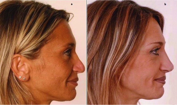
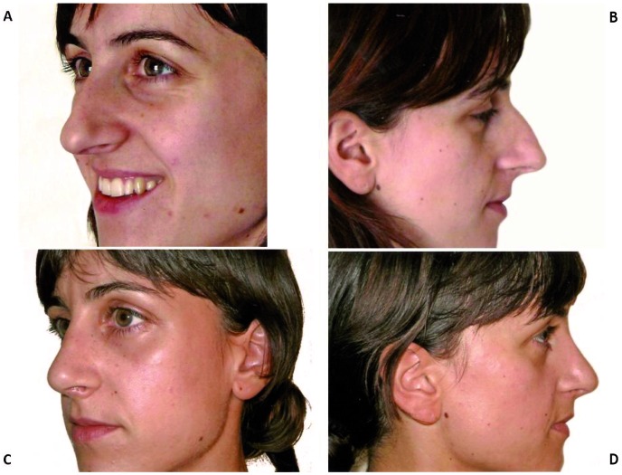
Cosmetic complaints result from high and anterior cartilaginous septal deviations, leading to a "twisted" cartilaginous nasal dorsum (Fig. 1), an asymmetric nasal tip or columella. A septal dorsal defect produces a depression of the nasal dorsum (Fig. 2). On the other hand, an overdeveloped dorsal or columellar septal cartilage determines, respectively, a dorsal hump, a rounded nasolabial angle or an "excessive columellar show"(Fig. 3) 4.
Fig. 1.
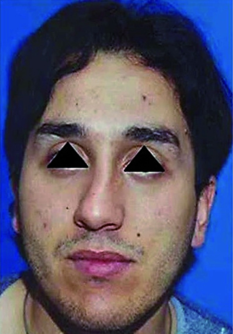
Patient with a twisted cartilaginous nasal dorsum as a result of high cartilaginous septal deviation.
Fig. 2.
a. Patient with a small defect of the dorsum of the cartilaginous septum after primary rhinoplasty; b. Postoperative view after filling the defect with patient's own cartilage.
Fig. 3.
a, b. Preoperative view of a patient with a bony-cartilaginous dorsal hump ("tension nose"); c, d. Postoperative view after resection of the hump and refinement of the anterior septal angle. Indirect change of the ala, nostril and nasal tip can be observed.
The bony part of the septum is easier to handle. It does not have the same aesthetic involvement (less supporting function on "visible nose") and is related to less functional problems, because of the much wider section of the nose at this level.
Rudimentary understanding of these anatomic nasal landmarks is of paramount importance in the decision-making process for safe and efficient septal surgery. The preservation of the supporting dorsal and caudal areas avoid a "saddle nose" deformity or a collapse and ptosis of the nasal tip, respectively, which can determine poor aesthetic results and airway obstruction.
A very critical functional area is the internal nasal valve, anatomically related to the relationship between the dorsal septum and the upper lateral cartilage. The patency of this area has a remarkable impact on the airflow passage, and the extramucosal cartilage resection represents a key point to the maintenance of an adequate section, and once again reinforces the conclusion that septoplasty and rhinoplasty affect each other considerably. The surgeon should be able to identify and treat a hypertrophied inferior turbinate that obviously, contributes to the obstruction of the internal nasal valve area.
Proper pre-operative analysis of septal deformity and evaluation of the nasal airway should be performed in all patients presenting for rhinoplasty because, in addition to functional and aesthetic expectation, they are crucial in the decision-making process.
Visual examination of the outer shape of the nose at rest, during smiling and with forced inspiration are the starting points in clinical examination; therefore, a C-shaped or Sshaped deflection should be recognized. The second step is the palpation of the nose to evaluate the cartilaginous and osseous framework, nasal tip stability and caudal deflections. The patient should be examined for collapse of the external nasal valves on deep inspiration. A Cottle manoeuvre is helpful to recognize inner valve stenosis 5. Internal nasal examination should be performed with nasal endoscopy before and after decongestion, and narrowing or collapse of the internal valves with inspiration should be noted 6-10 as well as inferior turbinate hypertrophy, septal deformities 11 12 (including deviation, tilt, maxillary crest, spurs and perforations). In secondary procedures, the availability of septal cartilage is assessed, as this is the primary source of autogenous graft material 13 14. The presence of nasal polyps or tumours should be excluded.
Rhinomanometry should be performed to assess nasal flow. CT scan can also be useful, especially in patients with concomitant sinusitis, even if it is not obligatory 15.
Septal surgery: resection or preservation?
In the past, the septum was exposed to wide resections of bone and cartilage with potentially negative functional and aesthetic consequences. The most remarkable (and famous) advancements in the surgical approach came from Killian (19th century), advocating the preservation of an "L" strut of at least 1.5 cm on the dorsal and caudal areas of the quadrangular cartilaginous septum 16. This technical aspect is "required" for good long-term cosmetic and functional results.
A few decades later, Metzenbaum, Peer, Huffmann and Lierle emphasized the principle of a more conservative septoplasty with the swinging door technique and septal columellar graft 17. In 1958, Cottle proposed the maxillapremaxilla approach, with elevation of mucoperichondrium on only one side and more conservative septal surgery. The extracorporeal septum reconstruction, proposed by King, Ashley and Perret, and refined by Gubish 18, has become increasingly popular and represents a good alternative (for some authors the standard of care) in the case of severely deviated septum or revision surgery. In this technique, the cartilaginous and bony septum is removed in one piece. The removed septum is then addressed accordingly to abnormalities. It can be straightened by scoring unilaterally the septum to reduce tension on cartilage; redundant bone or cartilage can be resected or even drilled. Cartilaginous or bony fragments can be used to reconstitute the stability and shape of the L-strut with suturing techniques or as batten grafts.
At present, because of the supporting role of the septum and its possible use as graft material, surgeons try to operate as conservatively as possible. This may be interpreted in many ways, from not performing the submucous resection at all during the course of rhinoplasty or possibly performing a minimal procedure from the standpoint of resecting skeletal septal structures.
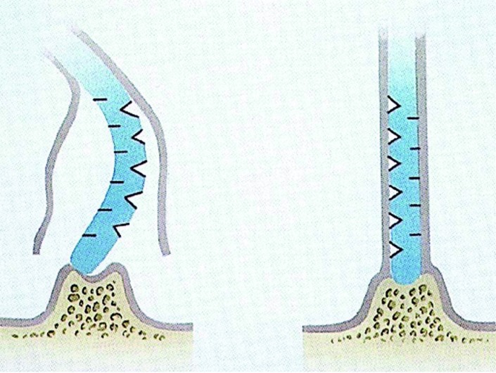
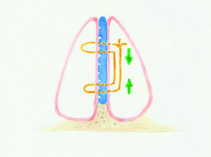
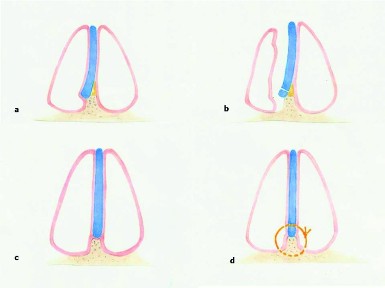
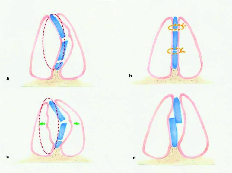
In this way, the most advisable technique is to straighten the cartilage by scoring it on the concave side. Concurrently, the attached contralateral mucoperichondrium on the convex side allows a realignment of the septum and contributes to the stability of the scored cartilage, even though a cut through of the entire cartilage has been performed. Multiple scores are made when the tension of the curvature is high (Fig. 4). In particular, in some cases these manoeuvres are not sufficient to straighten the septum, and hence cartilage carving on the convex side has to be combined with a multiple cutting on the concave one (Fig. 5). Traditionally, the scalpel is used for scoring cartilage, but some authors proposed other tools such as the holmium:YAG laser and radiofrequency energy 19 20. Further straightening of the weakened cartilage can be obtained by a through-andthrough suture above and beneath the deviation, securing the knots on the convex side (Fig. 6).
Fig. 4.
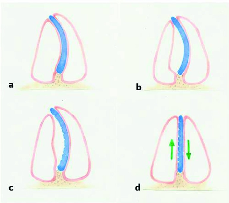
a. Deviated anterior septal cartilage; b. Right anterior septum tunnel; c. Multiple scores are made on the concave side; d. Straightening of the cartilage: the attached contralateral mucoperichondrium flap helps in the straightening process.
Fig. 5.
a. A cartilage carving on the convex side is combined with a multiple cutting on the concave one; b. A weakening and straightening effect on the cartilage is obtained.
Fig. 6.
Through-and-through suture above and beneath the deviation, securing the knots on the convex side. This procedure, combined with weakening process, stabilizes the new position of the septum.
If scoring and weakening of the cartilage is inadequate, resection of the convex septal portion is often obligatory. Therefore, spurs, replications and bows of the septum, if not properly resected, can lead to the septum returning to its original deviated position. A cartilaginous redundancy is most common at the inferior aspect of the septum with its connection to adjacent maxillary crest and at the posterior cartilaginous septum with its connection to bony septum (Fig. 7). The goal of resection is to remove a proper amount of deviated septum preserving the supporting functions of the maxillary crest to avoid a deprojection of the nose as a result of loss of septum support. For reducing the risk of septal perforation in a submucosal fashion, preserving the overlying mucoperichondral flap, is required. Some authors suggest to leave in place (if possible) the mucoperichondrium of the contralateral side of the septum resection, in order to preserve its supporting function (Fig. 8).
Fig. 7.
a. Deviated septal cartilage on the maxillary crest; b. Anterior superior and anterior inferior tunnel; c. Midline repositioning of the septum; d. Fixed cartilaginous septum by sutures to the nasal spine.
Fig. 8.
a. Cartilaginous resection on the fractured site; b. The preservation of the contralateral mucoperichondral flap, together with the fixation using through-and-through sutures, stabilizes the mobile pieces of the cartilage. c, d. Unstable fractured cartilaginous fragments resulting from the bilateral mucoperichondral flap and the absence of fixating sutures.
Surgical approach
The approach to septoplasty is usually established on grade and location of deformity, goals and the surgeon's preference and experience. If technically achievable, the endonasal approach should be preferred, because it preserves fibrous connections and ligaments as much as possible, such as around the anterior nasal spine or the K-area and the internal nasal valve. Therefore, it should be the first choice in this surgery, reserving extracorporeal reconstruction to severe septal pathologies 1. An endoscopic approach is also an option, but this is beyond the scope of this article.
Routinely, a hemitransfixion incision, located at the end of the caudal septum, 2 mm behind the septal border, parallel to its contour, preserves membranous septum and represents the preferred incision. It is related to a better healing and less scar contracture if compared to Killian's more aggressive resection techniques. This incision allows correction of deflections at the caudal border and access to nasal spine when required. Moreover, bilateral flap elevation permits wide access to the entire septum (bony and cartilaginous framework). Due to all the aforementionated reasons, hemitransfixion incision is very suitable for the correction of any kind of deflection, bony spurs, cartilage scoring, suture techniques and grafts to straighten the septum. Combined with inter- cartilaginous incisions, hemitransfixion incision can be efficiently used for most rhinoplasty procedures. The full transfixion incision is an extension of the hemitransfixion, with the complete release of the fibrous connection to the medial crura, advisable when a de-projection of the tip is desired. However, if this is not the case an excessive de-projection and/or a columellar retraction could become an unexpected result.
Finally, an open rhinoplasty approach provides direct and extensive access to the entire septum, especially after separation of the upper lateral cartilage from the dorsal septal cartilage. With this approach, the surgeon can accomplish all septal corrections and straightening techniques including all harvesting procedures and graft insertion 21. Most surgeons reserve this approach to severe septal deviations or revision surgery because of the potential damage of the internal nasal valve in the scarring process. On the other hand, some authors employ this approach in all patients, regardless of the severity of the case 22-26. Granted that the ancestral debate on open vs. endonasal rhinoplasty is out of the scope of this article, our policy is to perform an endonasal approach as much as possible. Nonetheless, an external approach is mandatory in the armamentarium of the rhinoplasty surgeon because some complicated cases (tip surgery, crooked nose, revision cases) cannot be managed through an endonasal approach. Consequently, the driving factor in the choice of the approach is not the confidence with the technique, but rather the goal of the surgery together with correct identification of deformities.
Indications for aesthetic corrections of the septum
As described, a thorough and detailed analysis of the septum can indicate the surgical corrections needed to achieve cosmetic results with septoplasty. There are several indications for combining septoplasty with cosmetic rhinoplasty: columellar strut for tip support, spreader graft to improve internal nasal valve narrowing, batten graft to avoid collapse of the external nasal valve; all these procedures involve the nasal septum and are both aesthetic and functional in nature 2. Nevertheless, all these functional and aesthetic grafts are primarily harvested from the septum, and so require "septal" surgery.
While the aim of this paper is to emphasize the relevant aesthetic goals that can be obtained through corrections of the nasal septum, regardless of combined corrections (osteotomies, tip surgery) of the other frameworks of the nose. The indications for cosmetic correction of the septum include: septal deflection with a twisted cartilaginous nasal dorsum, overdeveloped hanging columella, deviation of the caudal edge of the septum, cartilaginous nasal hump, C-shaped deformity, S-shaped deformity and crooked nose.
Crooked cartilaginous nasal dorsum and high dorsal septal deviation
High septal deviations are very difficult to correct, and especially when located in the K-area disruption of this supporting zone can lead to a de-projection of the nose.
Generally speaking, to prevent cartilaginous saddling, the vertical incision next to the osseocartilaginous junction should terminate 1-1.5 cm below the nasal dorsum. If the K-area is involved, some authors suggest to crush the cartilage to weaken and straighten the cartilage, while preserving its supporting function 3 27.
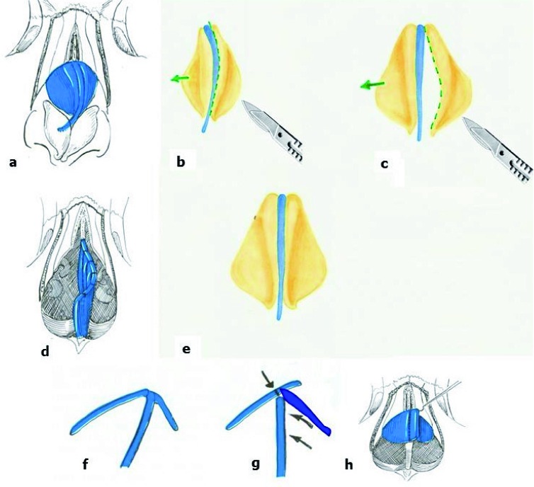
In some cases, high cartilaginous septal deviation can explain solely as a twisted nasal dorsum on the outside. In these circumstances, the upper lateral cartilage is often asymmetric, therefore, routine septal straightening may not be sufficient and separation of the upper lateral cartilage from one side or two is demanded (starting with the convex side; Fig. 9). In order to reduce tension on the dorsum, the cartilaginous dorsal septum can be detached from the skin, by a hemitransfixion incision 28. If these manoeuvres are not sufficient, a spreader graft between the septum and the upper lateral cartilage, is indicated. In patients with high septal deviations associated with a bony crooked nose, osteotomies are required 29.
Fig. 9.
a. Twisted dorsal septum with asymmetric upper lateral cartilage; b, c. Separating the upper lateral cartilage on the convex side; d. weakening the cartilage by scores and carving on the cartilage; e. Realignment of the septum; f, g, h. A redundant upper lateral cartilage is removed (not always required).
Deflection of the caudal septum
A deflection of the caudal septum is combined with a nostril obstruction and a distorted columella (Fig. 10). If the tip is well supported and an overdeveloped columella is present, the solution is a simple resection of the caudal cartilage together with removal of the overlying mucosa and skin. When the columella has a normal length, the septal cartilage must be scratched on the concave side and positioned on the midline (after the creation of a columellar pocket). Alternatively, a vertical strip resection on the fractured line may be indicated. In this latter case, some authors suggest inserting a thin and perforated part of the perpendicular plate acting as a batten graft upon the resected area 17 28 to obtain an effective long-term result. All these procedures can be performed by a hemitransfixion incision.
Fig. 10.
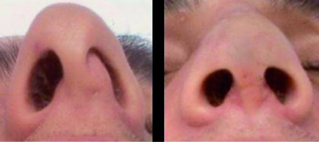
a. Preoperative view of a patient with a deviation of the caudal septum and almost complete obstruction of the left nostril. b. Postoperative view of the same patient after correction of the caudal septum.
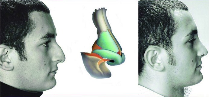
An anterior septal angle and caudal border of the cartilaginous septum that is too prominent frequently results in a downward rotation of the tip. A simple resection of the cartilage of the anterior septal angle and the caudal edge, in combination with resection of the overlying mucosa, can correct this deformity. An upward rotation of the nasal tip will result from elasticity of the nasal dorsum 3 30 (Fig. 11).
Fig. 11.
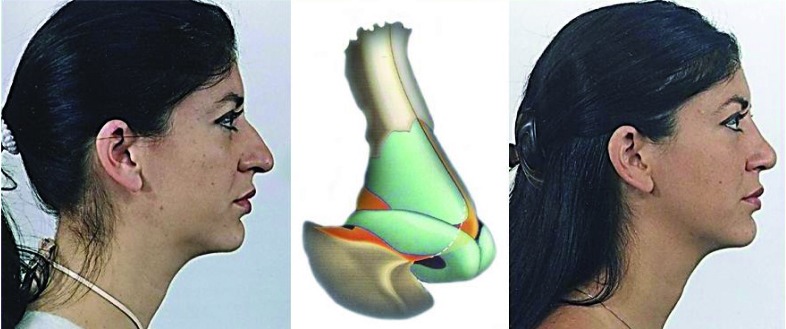
a. Patient with a prominent anterior septal angle and adjacent caudal edge of the cartilaginous septum; b. Dotted line: resection area of anterior septal angle; c. Postoperative view after resection of the anterior septal angle, an upward rotation of the nasal tip can be observed.
In severe conditions, where the nasal tip is also deviated because of severe caudal septum deviation, extracorporeal septoplasty may be indicated. In this case, an open approach provides better access for the mattress-sutures and for placement/fixation of a spreader graft (extended spreader grafts in cases that required lateralization of both the upper lateral cartilage and the nasal bone) 21 31.
Columellar show
This anatomical feature describes an excessive columellar show on profile view (normal alar-columellar relationship is 2-4 mm) so called hanging columella. This can be managed by a transfixion incision that allows resection of the whole caudal edge of the septum together with the overlying mucosa and skin. Closure of the defect, by suturing it primarily, is sufficient to retract the columella posteriorly.
A caudal septum that is too long can be associated with a blunt naso-labial angle, often in an asymmetrical way; therefore, a combined nasal spine resection is needed 4 32 (Fig. 12).
Fig. 12.
a. Patient with a blunt naso-labial angle and downward rotation of the tip. b. Dotted line: resection area of the nasal spine together with adjacent caudal edge of the septum; c. Postoperative view after resection of the caudal edge of the septum and the nasal spine, as well as resection of the anterior septal angle.
Overprojected cartilaginous nasal dorsum
An overdeveloped cartilaginous dorsal septum leads to a "tension" nose, by creating a supratip prominence of the lower dorsum. This can be corrected through dorsal resection of the quadrangular cartilage and can be accomplished easily by either an external or endonasal approach. The latter is accomplished through a transfixion incision combined with an intercartilaginous one, on both sides. To prevent "polly beak" deformity, the anterior septal angle has to be resected according to the cartilage dorsal lowering.
Cartilaginous nasal hump resection is not such a simple procedure, in fact, excess removal results in loss of tip support, a "scooped" look or even "saddle" nose deformity. Therefore, even experienced surgeons consider it challenging to draw the exact amount and place of dorsal reduction 33 34.
C-shaped deviation
A C-shaped deformity, which describes a convexity in vertical or horizontal dimension of the cartilaginous nasal dorsum can be addressed through endonasal weakening of the cartilage on the concave side, by a scratching procedure or full-thickness incisions. Even in this case, some authors suggest the placement of a cartilaginous or bony batten graft fixed by mattress-sutures to the convex side (cross-bar spreader graft) 35. Some severe C-shaped deviations need extracorporeal septal correction and could be combined with spreader graft and osteotomies, if required 36.
Crooked nose and S-shaped deviation
A crooked nose can become from a congenital or traumatic deformity, almost always associated with a septal deformity (Fig. 13). In such conditions mobilization and straightening of the "cosmetic" part of the septum (caudal and dorsal aspect of the cartilaginous septum) can be technically difficult. Therefore, a complete radical excision and reinsertion after corrections in an extracorporeal way, by an open approach, is indicated. The stabilization and realignment of the nasal dorsum can be challenging. The open technique provides an excellent exposure to perform complex manoeuvres (mattress suturing, scoring, drilling, spreader grafts, batten grafts) under direct view. Some authors suggest removing the cartilaginous septum together with the adjacent perpendicular plate because this part is long and straight enough to rebuild the new dorsum after a 90° anterior rotation of the whole septum. This way, the osseocartilaginous junction becomes the new dorsum and the previous dorsum becomes the new caudal-anterior border 18 37 38. After septal correction, other procedures are needed to straighten the crooked nose (medial and or lateral osteotomies, splints), but this is beyond the scope of this article.
Fig. 13.
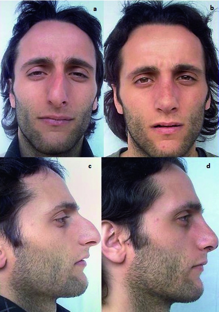
a, c. Preoperative view of a patient with post-traumatic crooked nose. Note the dorsal and caudal involvement of the septum and the "S-shaped" deviation; b, d. Postoperative view after correction through radical excision and reinsertion in an extracorporeal fashion using an open approach.
Conclusions
To achieve successful correction of the deviated nose all anatomic components involved in the deformity have to be recognized and treated accordingly. Septal surgery plays a crucial role in the management of the deviated nose. Even in the absence of functional problems, correction of minor and major septal deviations can determine realignment of the external nose. Hence, one-stage rhinoplasty becomes the standard of care in the management of a deviated nose. The endonasal or open approach should be determined by detailed preoperative evaluation. As a rough guide, an endonasal approach can overcome almost all deviations and allow placement of simple grafts (septal batten grafts, caudal grafts). Alternatively, an open approach is indicated for treating a twisted nose, heavy deformities and S-shaped deviations, malformations such as the cleft nose, complicated revision cases or when a difficult grafting procedure is demanded. A partial or complete extracorporeal reconstruction should be combined when a crooked nose is present.
References
- 1.Bernstein D. Septal surgery in rhinoplasty. Bull N Y Acad Med. 1981;57:293–303. [PMC free article] [PubMed] [Google Scholar]
- 2.Nease CJ, Deal RC. Septoplasty in conjunction with cosmetic rhinoplasty. Oral Maxillofac Surg Clin North Am. 2012;24:49–58. doi: 10.1016/j.coms.2011.10.006. [DOI] [PubMed] [Google Scholar]
- 3.Otten FWA. Septoplasty-Basic technique. The nasal septum in rhinoplasty. In: Trenitè N, editor. The rhinoplasty. The Hague, the Netherlands: Kugler Publications; 2005. [Google Scholar]
- 4.Park SS, Holt GR. Rhinoplasty and septoplasty. Part 1. Otolaryngol Clin North am. 1999;32:IX–IX. doi: 10.1016/s0030-6665(05)70164-x. [DOI] [PubMed] [Google Scholar]
- 5.Cottle MH, Loring RM, Fisher GG, et al. The maxilla-premaxilla approach to extensive nasal septum surgery. AMA Arch Otolaryngol. 1958;68:301–313. doi: 10.1001/archotol.1958.00730020311003. [DOI] [PubMed] [Google Scholar]
- 6.Constantian MB, Clardy RB. The relative importance of septal and nasal valvular surgery in correcting airway obstruction in primary and secondary rhinoplasty. Plast Reconstr Surg. 1996;98:38–54. doi: 10.1097/00006534-199607000-00007. [DOI] [PubMed] [Google Scholar]
- 7.Wittkopf M, Wittkopf J, Ries WR. The diagnosis and treatment of nasal valve collapse. Curr Opin Otolaryngol Head Neck Surg. 2008;16:10–13. doi: 10.1097/MOO.0b013e3282f396ef. [DOI] [PubMed] [Google Scholar]
- 8.Rhee JS, Arganbright JM, McMullin BT, et al. Evidence supporting functional rhinoplasty or nasal valve repair: A 25-year systematic review. Otolaryngol Head Neck Surg. 2008;139:10–20. doi: 10.1016/j.otohns.2008.02.007. [DOI] [PubMed] [Google Scholar]
- 9.Spielmann PM, White PS, Hussain SS. Surgical techniques for the treatment of nasal valve collapse: a systematic review. Laryngoscope. 2009;119:1281–1290. doi: 10.1002/lary.20495. [DOI] [PubMed] [Google Scholar]
- 10.Rhee JS, Weaver EM, Park SS, et al. Clinical consensus statement: Diagnosis and management of nasal valve compromise. Otolaryngol Head Neck Surg. 2010;143:48–59. doi: 10.1016/j.otohns.2010.04.019. [DOI] [PMC free article] [PubMed] [Google Scholar]
- 11.Gunter JP, Rohrich RJ. Management of the deviated nose: The importance of septal reconstruction. Clin Plast Surg. 1988;15:43–55. [PubMed] [Google Scholar]
- 12.Guyuron B, Uzzo CD, Scull H. A practical classification of septonasal deviation and an effective guide to septal surgery. Plast Reconstr Surg. 1999;104:2202–2209. doi: 10.1097/00006534-199912000-00039. [DOI] [PubMed] [Google Scholar]
- 13.Guyuron B, Behmand RA. Caudal nasal deviation. Plast Reconstr Sur. 2003;111:2449–2457. doi: 10.1097/01.PRS.0000060802.70218.FE. [DOI] [PubMed] [Google Scholar]
- 14.Mowlavi A, Masouem S, Kalkanis J, et al. Septal cartilage defined: Implications for nasal dynamics and rhinoplasty. Plast Reconstr Surg. 2006;117:2171–2174. doi: 10.1097/01.prs.0000218182.73780.d2. [DOI] [PubMed] [Google Scholar]
- 15.Tombu S, Daele J, Lefebvre P. Rhinomanometry and acoustic rhinometry in rhinoplasty. B-ENT. 2010;6(Suppl. 15):3–11. [PubMed] [Google Scholar]
- 16.Killian G. Die submukose Fensterresektion der Nasenscheidewand. Arch Laryng Rhin (Berlin) 1904;16:326–326. [Google Scholar]
- 17.Heppt W, Gubisch W. Septal surgery in rhnoplasty. Facial Plast Surg. 2011;27:167–178. doi: 10.1055/s-0030-1271297. [DOI] [PubMed] [Google Scholar]
- 18.Gubisch W. The extracorporeal septum plasty: a technique to correct difficult nasal deformities. Plast Reconstr Surg. 1995;95:672–682. doi: 10.1097/00006534-199504000-00008. [DOI] [PubMed] [Google Scholar]
- 19.Ovchinnikov Y, Sobol E, Svistushkin V, et al. Laser septochondrocorrection. Arch Facial Plast Surg. 2002;4:180–185. doi: 10.1001/archfaci.4.3.180. [DOI] [PubMed] [Google Scholar]
- 20.Keefe MW, Rasouli A, Telenkov SA, et al. Radiofrequency cartilage reshaping: efficacy, biophysical measurements, and tissue viability. Arch Facial Plast Surg. 2003;5:46–52. doi: 10.1001/archfaci.5.1.46. [DOI] [PubMed] [Google Scholar]
- 21.Gunter JP. The merits of the open approach in rhinoplasty. Plast Reconstr Surg. 1997;99:863–867. doi: 10.1097/00006534-199703000-00040. [DOI] [PubMed] [Google Scholar]
- 22.Constantian MB. Differing characteristics in 100 consecutive secondary rhinoplasty patients following closed versus open surgical approaches. Plast Reconstr Surg. 2002;109:2097–2111. doi: 10.1097/00006534-200205000-00048. [DOI] [PubMed] [Google Scholar]
- 23.Perkins SW. The evolution of the combined use of endonasal and external columellar approaches to rhinoplasty. Facial Plast Surg Clin North Am. 2004;12:35–50. doi: 10.1016/S1064-7406(03)00112-3. [DOI] [PubMed] [Google Scholar]
- 24.Simons RL. A personal report: Emphasizing the endonasal approach. Facial Plast Surg Clin North Am. 2004;12:15–34. doi: 10.1016/S1064-7406(03)00122-6. [DOI] [PubMed] [Google Scholar]
- 25.Simons RL, Greene RM. Rhinoplasty pearls: Value of the endonasal approach and vertical dome division. Clin Plast Surg. 2010;37:265–283. doi: 10.1016/j.cps.2009.11.005. [DOI] [PubMed] [Google Scholar]
- 26.Tebbetts JB. Open and closed rhinoplasty (minus the "versus"): analyzing processes. Aesthet Surg J. 2006;26:456–459. doi: 10.1016/j.asj.2006.06.003. [DOI] [PubMed] [Google Scholar]
- 27.Rohrich RJ, Muzaffar AR, Janis JE. Component dorsal hump reduction: The importance of maintaining dorsal aesthetic lines in rhinoplasty. Plast Reconstr Surg. 2004;114:1298–1308. doi: 10.1097/01.prs.0000135861.45986.cf. [DOI] [PubMed] [Google Scholar]
- 28.Foda HM. The role of septal surgery in management of the deviated nose. Plast Reconstr Surg. 2005;115:406–415. doi: 10.1097/01.prs.0000149421.14281.fd. [DOI] [PubMed] [Google Scholar]
- 29.Rohrich RJ, Gunter JP, Deuber MA, et al. The deviated nose: Optimizing results using a simplified classification and algorithmic approach. Plast Reconstr Surg. 2002;110:1509–1523. doi: 10.1097/01.PRS.0000029975.08760.25. [DOI] [PubMed] [Google Scholar]
- 30.Dobratz EJ, Tran V, Hilger PA. Comparison of techniques used to support the nasal tip and their long-term effects on tip position. Arch Facial Plast Surg. 2010;12:172–179. doi: 10.1001/archfacial.2010.33. [DOI] [PubMed] [Google Scholar]
- 31.Gruber RP. Open rhinoplasty. Clin Plast Surg. 1988;15:95–114. [PubMed] [Google Scholar]
- 32.Matarasso A, Greer SE, Longaker MT. The true hanging columella: simplified diagnosis and treatment using a modified direct approach. Plast Reconstr Surg. 2000;106:469–474. doi: 10.1097/00006534-200008000-00038. [DOI] [PubMed] [Google Scholar]
- 33.Rohrich RJ, Janis JE, Muzaffar AR, et al. Evaluation and surgical approach to the nasal dorsum: Component dorsal hump reduction. In: Gunter JP, Rohrich RJ, Adams WP Jr, editors. Dallas Rhinoplasty: Nasal Surgery by the Masters. 2nd ed. St. Louis: Quality Medical; 2007. pp. 221–244. [Google Scholar]
- 34.Dresner HS, Hilger PA. An overview of nasal dorsal augmentation. Semin Plast Surg. 2008;22:65–73. doi: 10.1055/s-2008-1063566. [DOI] [PMC free article] [PubMed] [Google Scholar]
- 35.Boccieri A, Pascali M. Septal crossbar graft for the correction of the crooked nose. Plast Reconstr Surg. 2003;111:629–638. doi: 10.1097/01.PRS.0000042205.27330.E4. [DOI] [PubMed] [Google Scholar]
- 36.Hsiao YC, Kao CH, Wang HW, et al. A surgical algorithm using open rhinoplasty for correction of traumatic twisted nose. Aesthetic Plast Surg. 2007;31:250–258. doi: 10.1007/s00266-006-0185-6. [DOI] [PubMed] [Google Scholar]
- 37.Byrd HS, Salomon J, Flood J. Correction of the crooked nose. Plast Reconstr Surg. 1998;102:2148–2157. doi: 10.1097/00006534-199811000-00055. [DOI] [PubMed] [Google Scholar]
- 38.Kim DW, Toriumi DM. Management of posttraumatic nasal deformities: The crooked nose and the saddle nose. Facial Plast Surg Clin North Am. 2004;12:111–132. doi: 10.1016/S1064-7406(03)00124-X. [DOI] [PubMed] [Google Scholar]


