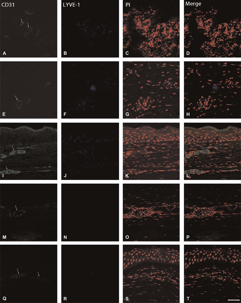
FIGURE 2.
Immunohistochemical visualization of the CD31+/LYVE-1− phenotype. Specimens exhibiting vascular structures were immunostained for CD31 (green), LYVE-1 (blue), and propidium iodide (PI, red) to differentiate between HA and LA. Arrows indicate neovascular formations. Representative specimens showing (A–D) fungal keratitis (subject 16), (E–H) HSV keratitis (subject 24), (I–L) inflammatory pannus (subject 27), (M–P) inflammatory pannus (subject 25), and (Q–T) chronic keratitis (subject 8); scale bar, 47.62 µm.

