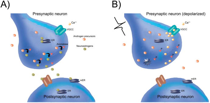
Figure 2.
Synaptocrine Signalling:Testosterone acts as a precursor (orange hexagons) diffusing from the extracellular space to be available as a substrate for the enzyme aromatase (red triangles) located within the presynaptic bouton. Estrogens such as 17-beta-estradiol (green hexagons) synthesized within the presynaptic bouton are then available to bind presynaptic estrogen receptors (ERs; grey ovals) and modulate presynaptic physiology. Synaptocrine estrogens may also diffuse across the synaptic cleft to interact with postsynaptic ion channel receptors (brown channel), and/or postsynaptic ERs (grey ovals, cytosolic) or membrane ERs (mER; grey ovals, membrane-bound). Synaptocrine estrogens could therefore modulate postsynaptic neurotransmitter receptors, cell signaling pathways, and/or have genomic effects on the postsynaptic cell. Because the activity of the aromatase enzyme is highest in a dephosphorylated state, the synaptocrine actions of estradiol are most likely to occur when the cell membrane is at rest (i.e., not depolarized) and when voltage-gated calcium channels (VGCCs) are closed. In contrast, (right panel), depolarization of the presynaptic neuron leads to calcium influx into the presynaptic bouton. This can activate a calcium-dependent kinase (kin) to phosphorylate presynaptic aromatase (P) and thereby cause a rapid reduction in the synthesis of synaptocrine estrogens, while simultaneously potentially increasing the local concentration of androgens. Adapted from Remage-Healey et al., 2011, Frontiers in Endocrinology, (ref 14).

