Summary
Muscular architecture involves the organization of fibres in the muscle and is one of the most important factors of muscular function. Studies have demonstrated an association with muscular architecture and contraction, with an increase of the pennation angle in muscles.
The aim of the study was to evaluate the change of muscular pennation angle after therapy with warm thermal water (crenotherapy).
Participants:
45 amateur athletes undertaking different sporting activities;
Group A: 30 runners;
Group B: 15 swimmers.
All the athletes underwent muscular ultrasound and clinical examination before and after the 10 sessions of the thermal protocol. At baseline the groups showed different values of pennation angle (group A = 19.1° ± 3.8° vs group B = 16.7° ± 2.4°; p=0.05). Following the thermal therapy protocol, significant variation of pennation angle was detected at rest in Group A which had significantly lower values than before the treatment (17.5° ± 2.9°; p=0.01). No differences were detected in group B.
Conclusions:
thermal therapy induced the greatest effect on runners (Group A) as pennation angle at rest was significantly lower after the period of crenotherapy and this variation may be as a result of a smaller muscular contracture.
Keywords: pennation angle, crenotherapy, muscular architecture
Introduction
Muscular architecture involves the organization of the fibers in the muscle and is the most important factor of muscular function1,2.
A direct association between muscular architecture and muscle volume has been identified, suggesting that muscular hypertrophy induces a higher pennation angle3. However a direct association between pennation angle and muscular strength has yet to be found. In 1993 Kawakami et al. found an association between the grade of hypertrophy and pennation angle in sedentary and body builders4.
The relationship between muscular architecture and contraction has been identified in participants whose muscle has been subjected to endurance training with an increase in the pennation angle being identified5. Strength training affects the muscular architecture both immediately after effort6, and also after long period of training, demonstrating hypertrophy of fibres and varying angles of pennation angle7,8. The effect of water immersion on recovery after exercise has been studied9 alongside the positive effects of immersion in warm water to reduce muscular stress after exercise10. However, there are no studies identifying the effects of thermal therapy on muscle architecture.
The aim of the study was to evaluate the change of muscular pennation angle after therapy with warm thermal water
Methods
Participants
45 males amateur athletes undertaking two different sporting activities, 2 or 3 times a week. The participants were divided into two groups:
Group A: 30 amateur runners;
Group B: 15 amateur swimmers.
The local Ethics Committee approved the study, and all participants gave written informed consent. Participants suspended their usual training during the study period (three weeks).
Protocols
All participants underwent muscular ultrasound and clinical examination before and after the 10 sessions of the thermal protocol performed in the “Terme Stufe di Nerone” di Bacoli, Napoli, (Italy) (Tab. 1), with the following protocol:
5 minutes in thermal cave at 30°C;
10 minutes in thermal cave at 32°C;
10 minutes in a warm swimming pool at 40°C;
Sauna (54°C) for 5 minutes;
Warm-cold shower;
5 minutes in thermal cave at 32°C;
10 minutes in thermal cave at 30°C;
Thermal adjustment for 15 minutes;
15 minutes in a warm swimming pool at 35°C.
Table 1.
Analysis of “Terme Stufe di Nerone” water hypertermal Salso-Bromo-Iodica-Solfato-Alcalina-Terrosa water temperature to the source: 79.60°C, hardness: 95.7°F, fixed residue at 180°: 16742 mg/L.
| ION | RESULTS mg/L |
|---|---|
| Silicon | 127,70 |
| Bicarbonates | 373,80 |
| Chlorides | 10932,16 |
| Sulfates | 526,10 |
| Sodium | 6997,45 |
| Potassium | 257,11 |
| Calcium | 338,23 |
| Magnesium | 128,57 |
| Iron | 0,03 |
| Phosphorus | 0,30 |
| Strontium | 1,39 |
| Litio | 1,68 |
| Bromine | 1,95 |
| Iodine | 3,00 |
| Fluorine | 3,20 |
Measurements were taken with the patient in a prone position, the knees extended and the ankle of the end of the table, to allow the physician to effect the maximum bending of the ankle. A 12 MHz probe was used and the transducer was placed in a sagittal plane and moved medially to the Achilles tendon to identify the pennation angle of the soleus muscle. Measurements were taken on the left leg in two positions of the ankle:
At rest in a natural position;
After passive maximal bending performed by the physician.
The same operator performed all the measurements, and the point from where the measurements were taken was marked with a marker pen and identified by metric coordinates in comparison to the apex of the heel.
Statistics
Statistical analysis was performed using the SPSS statistical package for windows 16.0. Comparison between the values of pennation angle obtained in each group was performed using a paired t-test. Comparison between the values of pennation angle obtained in the two groups was performed using the Student t-test for different samples. Statistical significance was defined as P< 0.05.
To calculate the intra-observer variation the main author measured pennation angle on the soleus of a single subject, repeating the measurements 20 times for each position (natural position and hyperflexion of the ankle). The coefficient of variation (CV) was calculated using the formula: CV= SD/mean × 100, where SD is the square root of the variance and the mean is the combined mean of the measurements. The CV for the pennation angle in the natural position was 4.5% and in hyperflexion was 3.1%.
Results
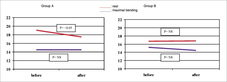
24 athletes completed the protocol in Group A (mean age 45.04 ± 14.29) and 14 in Group B (mean age 53.30 ± 7.65). 7 athletes were excluded because did not completed the protocol so as stated. In group A pennation angle was 19.1° ± 3.8 at rest, in the natural position and 14.5° ± 4.2° after maximal bending, with a mean variation of 24.2 ± 14.3%. After the cycle of thermal therapy the pennation angle was found to be 17.5° ± 2.9° at rest which was significantly lower (p=0.01) and 14.5° ± 2.8° in maximal bending with a mean variation of 16.28 ± 12.18%, significantly lower than the one detected at rest (p= 0.03).
In Group B the pennation angle before the treatment was 16.7° ± 2.4° in the natural position and 15.2° ± 2.3° in the maximal bending position. Following the thermal therapy the pennation angle was almost the same with mean values of 17.3° ± 2.4° in the natural position and 14.5° ± 2.7° in the maximal bending (p= NS). There were significant differences of the pennation angle in the natural position prior to the thermal therapy between the two groups, whereas, after the treatment, the angles were almost the same. The measurements detected after passive stretching showed no differences before and after thermal therapy.
Discussion
The study demonstrated the variation in muscle architecture induced by a cycle of crenotherapy. The two groups were evaluated before and after a cycle of thermal therapy, and have shown differences in values of pennation angle.
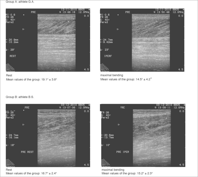
Before the treatment the groups showed different values of pennation angle (group A= 19.1° ± 3.8° vs group B= 16.7° ± 2.4°; p= 0.05 - Fig. 1), which confirms what has been found previously that pennation angle can vary in athletes involved in different sporting activities. Fast runners have higher pennation angle than other athletes, because of the big muscular mass and the high speed of contraction11,12. However, these differences disappear after stretching the ankle with maximal bending, and following stretching both groups reached smaller values of pennation angle (Group A= 14.5° ± 4.2° vs Group B= 15.2° ± 2.3°; p= NS). Abellaneda et al., have shown increasing the bend of the ankle leads to a decrease in pennation angle with minimal differences between individuals13. This study confirms that individuals involved in different sporting activities can give different pennation angles at rest. However, when the muscle has been subjected to passive stretching the pennation angle becomes almost the same. Genetic factors and the age of the athlete may have more of a role to play in the changes in muscular pennation angles than the particular sport practised as confirmed by the work of14,15 who identify that pennation angle is related to the stiffness of muscle-tendon unit determined by genetic features and age14,15.
Figure 1.
Images of pennation angle detected in one athlete of each group at rest and after maximal bending.
After thermal therapy there was a significant variation of pennation angle at rest in Group. A which had significantly lower values than before the treatment, however, no differences were detected in group B (Fig. 2). Similarly, no differences were detected after passive stretching in maximal bending and both groups had similar values of pennation angle after the cycle of thermal therapy. Examining the two groups we see that thermal therapy has the greatest effect on Group A where the pennation angle at rest was significantly lower after the period of crenotherapy. This variation may be due to a smaller muscular contracture at rest, as a greater muscle contraction corresponds with a greater pennation angle16,17.
Figure 2.
Variation of pennation angle detected in both groups before and after thermal therapy.
Group B, showed no meaningful variation in muscular architecture, which is probably because of the nature of the sport (swimming) which requires a smaller involvement of muscle soleus during sporting activity.
Conclusions
This study demonstrates that thermal therapy induces a lowering of pennation angle in runners, probably decreasing the state of basal contraction. Crenotherapy can be a useful way to guarantee a state of muscular relaxation in runners in future studies. However, it would be ideal to also identify the role of other factors such as age and genetics when evaluating the role of thermal therapy on recovery following sporting activities.
References
- 1.Savelberg HH, Schamhardt HC. The influence of inhomogeneity in architecture on the modelled force-length relationship of muscles. J Biomech. 1995;28:187–197. doi: 10.1016/0021-9290(94)00050-e. [DOI] [PubMed] [Google Scholar]
- 2.Morse CI, Thom JM, Birch KM, Narici MV. Changes in triceps surae muscle architecture with sarcopenia. Acta Physiol Scand. 2005;183:291–298. doi: 10.1111/j.1365-201X.2004.01404.x. [DOI] [PubMed] [Google Scholar]
- 3.Rutherford OM, Jones DA. Measurement of fibre pennation using ultrasound in the human quadriceps in vivo. Eur J Appl Physiol Occup Physiol. 1992;65:433–437. doi: 10.1007/BF00243510. [DOI] [PubMed] [Google Scholar]
- 4.Kawakami Y, Abe T, Fukunaga T. “Muscle fiber pennation angles are greater in hypertrophied than in normal muscles”. J Appl Physiol. 1993;74(6):2740–2744. doi: 10.1152/jappl.1993.74.6.2740. [DOI] [PubMed] [Google Scholar]
- 5.Aagaard P, Andersen JL, Dyhre-Poulsen P, et al. “A mechanism for increased contractile strength of human pennate muscle in response to strength training: changes in muscle architecture”. J Physiol. 2001;534(Pt.2):613–623. doi: 10.1111/j.1469-7793.2001.t01-1-00613.x. [DOI] [PMC free article] [PubMed] [Google Scholar]
- 6.Brancaccio P, Limongelli FM, D'Aponte A, Narici M, Maffulli N, et al. Changes in skeletal muscle architecture following a cycloergometer test to exhaustion in athletes. Journal of Science and Medicine in Sport. 2008:538–541. doi: 10.1016/j.jsams.2007.05.011. [DOI] [PubMed] [Google Scholar]
- 7.Matta T, Simao R, de Salles BF, Spineti J, Oliveira LF. Strenght training's chronic effects on muscle architectures parameters of different arm sites. J strenght Cond res. 2011;25:1711–1717. doi: 10.1519/JSC.0b013e3181dba162. [DOI] [PubMed] [Google Scholar]
- 8.Duclay J, Martin A, Duclay A, Cometti G, Pousson M. Behaviour of fascicles and the myotendinous junction of human medial gastrocnemius following eccentric strenght training. Muscle Nerve. 2009;39:819–827. doi: 10.1002/mus.21297. [DOI] [PubMed] [Google Scholar]
- 9.Wilcock IM, Cronin JB, Hing WA. Physiological response to water immersion: a method for sport recovery? Sports Med. 2006;36:747–765. doi: 10.2165/00007256-200636090-00003. [DOI] [PubMed] [Google Scholar]
- 10.Skurvydas A, Kamandulis S, Stanislovaitis A, Streckis V, Mamkus G, Drazdauskas A. Leg Immersion in Warm Water, Stretch-Shortening Exercise, and Exercise-Induced Muscle Damage. J Athl Train. 2008;43:592–599. doi: 10.4085/1062-6050-43.6.592. [DOI] [PMC free article] [PubMed] [Google Scholar]
- 11.Abe T, Kumagai K, Brechue WF. Fascicle lenght of leg muscles is greater in sprinters than in distance runners. Med Sci Sports Exerc. 2000;32:1125–1129. doi: 10.1097/00005768-200006000-00014. [DOI] [PubMed] [Google Scholar]
- 12.Kanehisa H, Muraoka Y, Kawakami Y, Fukunaga T. Fascicle arrangements of vastus lateralis and gastrocnemius muscles in highly trained soccer players and swimmers of both genders. Int J Sports Med. 2003;24:90–95. doi: 10.1055/s-2003-38197. [DOI] [PubMed] [Google Scholar]
- 13.Abellaneda S, Guissard N, Duchateau J. The relative lenghtening of the myotendinous structures in the medial gastrocnemius during passive stretching differs among individuals. J Appl Physiol. 2009;106:169–177. doi: 10.1152/japplphysiol.90577.2008. [DOI] [PubMed] [Google Scholar]
- 14.Baudry S, Lecoeuvre G, Duchateau J. Age-related changes in the behavior of the muscle-tendon unit of the gastrocnemius medialis during upright stance. J Appl Physiol. 2011 Oct 27; doi: 10.1152/japplphysiol.00913.2011. [DOI] [PubMed] [Google Scholar]
- 15.O'Brien TD, Reeves ND, Baltzopoulos V, Jones DA, Maganaris CN. Muscle-tendon structure and dimensions in adults and children. J Anat. 2010;216(5):631–642. doi: 10.1111/j.1469-7580.2010.01218.x. [DOI] [PMC free article] [PubMed] [Google Scholar]
- 16.Stark JH, Schilling N. A novel method of studying fascicle architecture in relaxed and contracted muscles. Biomech. 2010;43:2897–2903. doi: 10.1016/j.jbiomech.2010.07.031. 16. [DOI] [PubMed] [Google Scholar]
- 17.Shi J, Zheng Y, Yan Z. The relationship between SEMG and change in pennation angle of brachialis. Conf Proc IEEE Eng Med Biol Soc. 2007;2007:4802–4805. doi: 10.1109/IEMBS.2007.4353414. [DOI] [PubMed] [Google Scholar]


