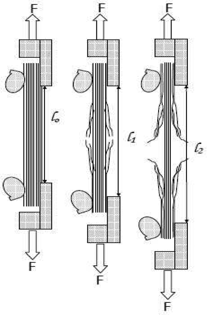
Figure 1.
Side view of anticipated degradation progression as a function of time. In the dynamic enzyme-induced creep experiment, the tissue will degrade from the external surfaces transferring load to the interior fibrils. Left frame (pre-exposure), the corneal tissue is intact and holding the load securely in the cam grips, the gage length is l0. Center frame (short exposure time). The tissue, just having been exposed to enzyme, has begun to degrade, since the load is fixed, the loss of load bearing area results in extension of the strip to a new gage length, l1. Right frame (long exposure), the tissue has been exposed to the enzyme for a significant amount of time and has strained considerably. The combination of strain and diffusion limitations should begin to slow the degradation rate.

