
Fig. 2.
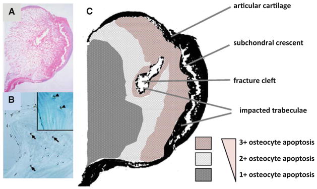
Glucocorticoid-induced osteonecrosis is osteocyte apoptosis. Abundant apoptotic osteocytes are present in sections of whole femoral heads obtained during total hip replacement for glucocorticoid- induced osteonecrosis (a, b). In c is a prevalence map of osteocyte apoptosis made from the section of the femoral head shown in (a). Osteocyte apoptosis was most prevalent (3?) adjacent to the subchondral crescent and fracture cleft and decreased (1?) as the examination progressed more distally. Osteocyte apoptosis was anatomically juxtaposed to the osteonecrotic fracture (adapted from Weinstein et al. [40]. Copyright 2000. The Endocrine Society)