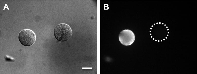
Figure 1.
The detection of AQP1 proteins in oocytes by immunofluoresence. Mouse oocytes at the germinal vesicle stage were injected with cRNA of human AQP1, and cultured for 12-14 h. The expression of AQP1 was detected by immunofluoresent-staining using anti-rat AQP1 rabbit antibody and FITC-conjugated anti-rabbit immunoglobin goat antibody. A, an uninjected oocyte (right) and a cRNA-injected oocyte (left); B, the same two cells under fluorescence microscope. The bar indicates 50 μm. Edashige et al. (2003, 2007) have obtained analogous immunofluorescence data for AQP3 proteins in mouse oocytes.

