

Madam,
A successful endodontic treatment very much depends on the endodontist's ability to recognize unusual root canal anatomy. Most teeth have shown accessory canals, multiple foramina, fins and deltas.
In 1984, Vertucci[1] reported that the clinician must treat the tooth by assuming that there is presence of accessory root canals unless proven otherwise. In 1979, Cooke and Cox[2] first described the term C-shaped root canal. They reported three cases where the root canals were like the English capital letter ‘C’, in which canals were connected by a continuous slit. C-shaped canals are commonly found in permanent mandibular second molars. A great deal of variations can present, especially in the canal configuration of the mandibular second molar. Mandibular second molars usually have two roots and three root canals but variations in the number of roots as well as canal morphology are not uncommon. The mandibular second molars with C-shaped root canals vary in their configurations and many methods have been used to classify such canals.[1]
C-shaped root canal was classified into three categories by Melton (1991) [Table 1].[3]
Table 1.
Types of C-shaped root canal[3]
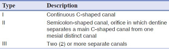
A C-shaped canal appears when fusion of either the buccal or lingual aspect of the mesial and distal roots occurs. This fusion remains irregular and the two roots stay connected by an interradicular ribbon.[4] Two or three canals may be found in the C-shaped groove, or the C-shape may be continuous throughout the length of the root. The floor of the pulp chamber is deep and has an unusual anatomical appearance.[4] The report of teeth with C-shaped canal has drawn the attention of dental practitioners, especially endodontists due to the lower success rate of root canal therapy. Clearly, the root canal anatomy cannot always be predicted, and this can be the source of failure. A solid understanding of the tooth anatomy including the number and course of various root canals represents the basis for successful endodontics’ therapy. Zheng, et al.,[5] showed almost equal occurrence of C-shaped canal among different age groups which implies that age is not a strong determinant of the occurrence of a C-shaped canal. Melton, et al.,[3] found that Type III C-shaped canal has the highest frequency followed by Type II and lastly Type I. Melton also commented on the large amount of debris in instrumented canal space found in histological sections of these teeth, for which many authors agree on using ultrasonic files to facilitate their removal.
C-shaped canal appears when fusions of either the buccal or lingual aspect of the mesial and distal roots occur. These fusions remain irregular, and two roots stay connected by an interradicular ribbon.[4] C-shaped root canals may occur among Malaysian people especially Chinese racial. There is a very high occurrence in Chinese racial (14.6%) compared to that of non-Chinese racial (1.04%). Among the C-shaped canal that were found, there are 7 out of 9 cases or 77.8% in Chinese racial, and this finding is consistent with what was reported in a previous study by Yang, et al.,[6] where a high prevalence of C-shaped canal was found in the Chinese population in a study conducted in China. Besides, all the teeth that possess C-shaped root canal configurations are lower molars. Hence, such an unusual configuration of the root canal should be recognized earlier and precaution should be taken during management or referral to endodontist should be made. It is important to be familiar with variations in tooth anatomy and characteristic features in various racial groups since such knowledge can aid location and negotiation of canals, as well as their subsequent management.
REFERENCES
- 1.Vertucci FJ. Root canal anatomy of the human permanent teeth. Oral Surg Oral Med Oral Pathol. 1984;58:589–99. doi: 10.1016/0030-4220(84)90085-9. [DOI] [PubMed] [Google Scholar]
- 2.Cooke HG, Cox FL. C-shaped canal configurations in mandibular molars. J Am Dent Assoc. 1979;99:836–9. doi: 10.14219/jada.archive.1979.0402. [DOI] [PubMed] [Google Scholar]
- 3.Melton DC, Krell KV, Fuller MW. Anatomical and histological features Of C-shaped canals in mandibular second molars. J Endod. 1991;17:384–8. doi: 10.1016/S0099-2399(06)81990-4. [DOI] [PubMed] [Google Scholar]
- 4.Bolger WL, Schindler WG. A mandibular first molar with a C-shaped root configuration. J Endod. 1988;14:515–9. doi: 10.1016/S0099-2399(88)80110-9. [DOI] [PubMed] [Google Scholar]
- 5.Zheng Q, Zhang L, Zhou X, Wang Q, Wang Y, Tang L, et al. C-shaped root canal system in mandibular second molars in a Chinese population evaluated by cone-beam computed tomography. Int Endod J. 2011;44:857–62. doi: 10.1111/j.1365-2591.2011.01896.x. [DOI] [PubMed] [Google Scholar]
- 6.Yang ZP, Yang SF, Lin YC, Shay JC, Chi CY. C-shaped root canals in mandibular second molars in a Chinese population. Endod Dent Traumatol. 1988;4:160–3. doi: 10.1111/j.1600-9657.1988.tb00315.x. [DOI] [PubMed] [Google Scholar]