Introduction
The characterization of native-like structures of small helical transmembrane (TM) proteins is particularly challenging. The high fraction of hydrophobic amino acid residues in the TM domain leads to weak interactions between helices and an increased significance for the interactions of the protein with its lipid environment1. Consequently, the characterization of these proteins in membrane mimetic environments that do not provide interactions with the protein similar to those of the native environment can lead to non native-like structures2. The detergent based environments used in X-ray crystallography have resulted in very few crystal structures of proteins with less than four TM helices due, in part, to the lack of a stabilizing membrane mimetic environment. While there are more solution NMR structures of such proteins the validity of detergent micelles as an adequate environment for stabilizing native-like structures has been questioned2,3. Solid-state NMR (ssNMR) spectroscopy has a unique capability to characterize these structures in a native-like lipid environment, even a liquid crystalline lipid bilayer environment. While such promise has been extant for more than a decade4,5, there have been significant challenges to overcome before this potential could be routinely achieved. Today, these challenges have been addressed and ssNMR’s potential for achieving native-like structures validated.
In the past year it has become clearer what properties of the native membrane protein environment need to be adequately modeled in the membrane mimetic environment for structural characterization2. While the hydrophobic thickness of the membrane mimetic can be modulated by the protein, it is also clear that the membrane can influence the tilt of the TM helices6. Dual hydrophilic surfaces constraining the TM helices to span a bilayer environment can be important in contrast to the single surface of a detergent micelle that permits hydrophilic sidechains from the center of a TM helix to interact with the polar surface without disrupting the interaction of the terminal regions with the aqueous interface. Similarly, it is necessary for the membrane mimetic to have a dramatic dielectric gradient and water concentration gradient, such that a span of at least 20Å is very hydrophobic and so the interfacial region is well defined2. It may also be important for the membrane mimetic to accurately model the lateral pressure profile of the native membrane.
Two ssNMR approaches have been used for achieving atomic resolution structural restraints of membrane proteins. Proteoliposome preparations for Magic Angle Spinning (MAS) spectroscopy have been used for torsional and distance restraints resulting in numerous studies of membrane proteins7–12. Recently, MAS spectroscopy has been used to obtain orientational restraints13. Indeed, the structure of the G-protein coupled receptor, CXCR1 has recently been characterized using orientational restraints from MAS spectroscopy of a proteoliposome preparation14. Uniformly oriented samples through magnetically aligned bicelles or mechanically oriented bilayers on glass surfaces have more frequently been used to obtain orientational restraints from Oriented Sample (OS) NMR15–19. Each of these approaches for structural characterization has advantages and significant challenges, but by combining the two approaches we can take advantage of both techniques to minimize the challenges and maximize the quality of the structural results20,21.
Numerous publications on the expression, isotopic labeling, purification and reconstitution of membrane proteins have been published in recent years demonstrating that the production of enough protein for solid state NMR spectroscopy is routinely possible22. Detailed protocols for the preparation of high q (ratio of lipid to detergent) bicelle samples necessary for achieving uniform orientation have been published23. Uniform orientation of bilayers on glass slides had been more of an art than a science, but recently with a greater understanding of how to minimize detergents from the purification and reconstitution steps for the final samples this art form has been transformed into a science (Murray et al., unpublished). Currently, we are working on 6 full length membrane proteins that have been uniformly oriented, one in bicelles and five using glass slides. As a result, the preparation of such oriented samples does not appear to be a significant limitation for the structural characterization of small helical membrane proteins. Furthermore, in the preparation of the mechanically oriented samples, proteoliposomes are prepared that can be used directly as a MAS sample. As a result it is not necessary to develop two different sample preparation protocols in order to take advantage of the structural restraints obtained from both techniques.
The proteins used as examples here include the M2 protein from Influenza A. This is a proven drug target that has multiple functions including a proton channel formed by a tetramer of the single TM of this protein24–26. The conductance domain (residues 22–62) has the same proton conductance properties as the full length protein27. In addition, we discuss two membrane proteins from Mycobacterium tuberculosis, CrgA that has two TM helices and is involved in cell division28 and Rv1861 that has three TM helices and binds nucleotide triphosphates29.
Structural Restraints
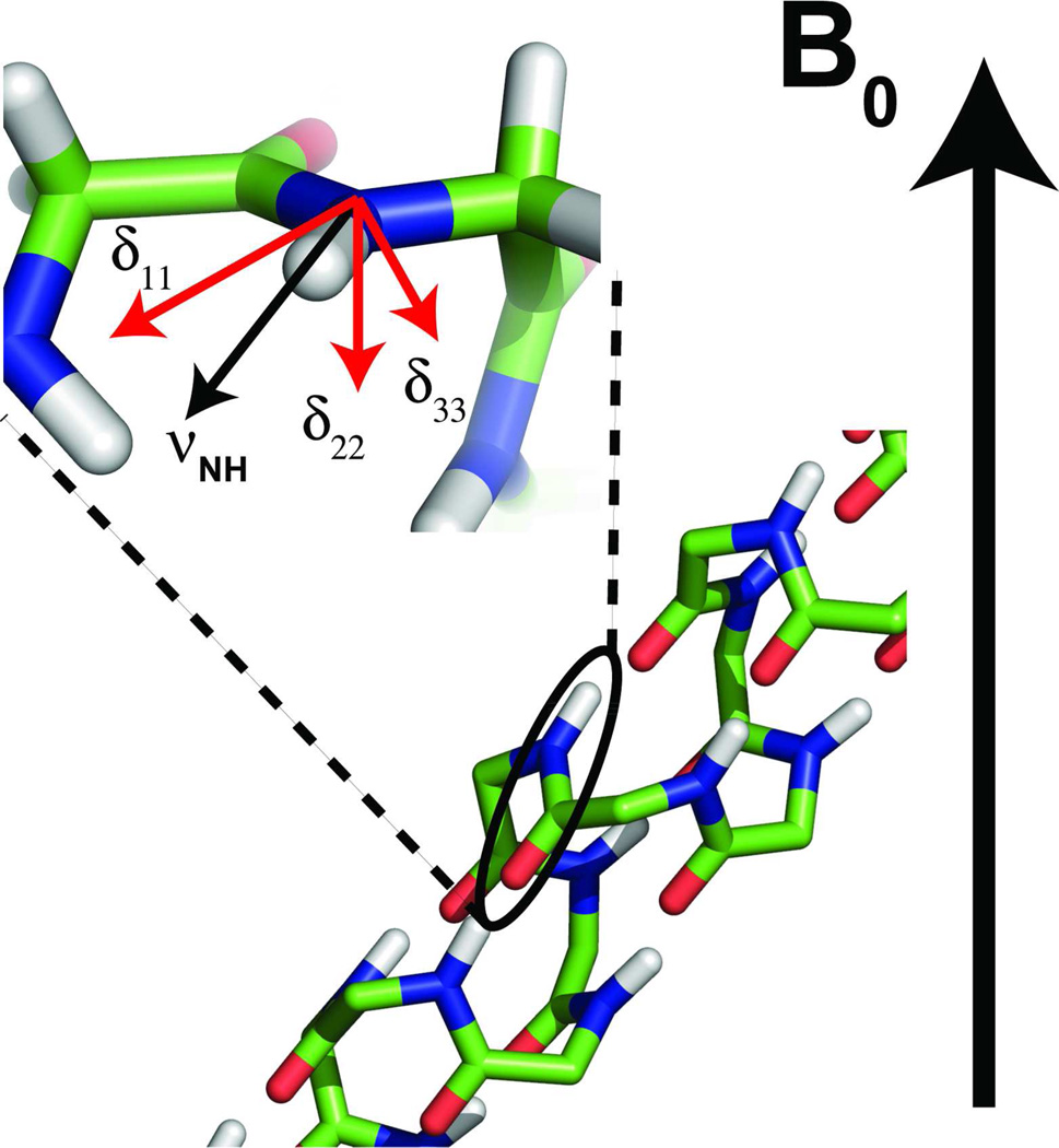
Orientational restraints are typically obtained from Separated Local Field (SLF) spectroscopy when the anisotropic chemical shift and dipolar interactions are correlated. More specifically the PISEMA and SAMPI4 experiments30,31 (and recent enhancements on these experiments32,33) are routinely used to obtain 15N-1H dipolar interactions and the anisotropic 15N chemical shifts. These experiments have focused on the 15N spins in proteins because the homonuclear 15N-15N spin interactions are small, whereas 13C-13C homonuclear interactions in uniformly labeled protein are substantial, thereby further complicating the spectra. Considerable amino acid specific labeling is often required for resolving resonances from multiple helices and for achieving the residue specific resonance assignments. While this approach has been criticized for being laborious, the need for native-like structures warrants such an effort. Furthermore, the expression and sample preparation for this spectroscopy is no longer such a time consuming process and therefore the preparation of multiple samples is not a major impediment for structural characterization. The orientational restraints result from observations of the anisotropic spin interaction component parallel to the magnetic field axis (Fig. 1). The orientational dependence of the spin interaction ((3cos2θ-1)/2- where θ is the angle of the spin interaction tensor element with respect to the magnetic field) leads to a ready interpretation of the data as a structural restraint for the atomic site. The analysis of multiple sites leads to accurate characterizations of individual TM helices. Typically, the helix tilt and rotation angles are determined first with the data also supplying precise backbone torsion angles for atomic resolution structure determination. However, these restraints do not provide tertiary structural restraints between helices.
Figure 1.
From an 15N labeled site, such as in the inset image of a peptide plane from a TM helix, the component of the chemical shift tensor (δii) and dipolar interaction (νNH) parallel to the magnetic field (Bo) can be assessed as structural restraints.
Torsional restraints are often obtained from extensively 13C and 15N labeled samples with MAS spectroscopy34. The isotropic chemical shifts of the carbonyl, Cα and Cβ carbons provide significant ϕ,ψ torsion angle restraints for the polypeptide backbone. However, MAS resonance assignments are particularly challenging in the TM helices of membrane proteins because of broad lineshapes and the uniformity of both the helical structures and their environment, all of which results in very little frequency dispersion for each amino acid type. The dominance of non-polar amino acid residues contributes to the uniformity of the resonance frequencies. Typically the best linewidths for membrane proteins have been achieved from crystallized samples in detergent environments as opposed to proteoliposome preparations. However, Ladizhansky has demonstrated excellent linewidths from proteoliposome preparations of sensory rhodopsin following extensive lipid screening efforts35. Whether this spectral resolution is unique to the rhodopsin family of membrane proteins or not has yet to be determined.
Distance restraints have also been obtained from extensively 13C and 15N labeled samples by MAS spectroscopy and like the torsional restraints they are dependent of resonance assignments. Here, the assignments are even more challenging as the interhelical distances are typically obtained between sidechain resonances. The dispersion in the TM helices for the sidechains is even less than in the polypeptide backbone confounding the challenges for spectral interpretation. Once again this appears to be the result of the relatively uniform apolar environment. As a result it is difficult to collect the large number of distance restraints necessary for characterizing a tertiary structure of a helical membrane protein by MAS ssNMR alone.
The precision and accuracy of the orientational restraints are dictated by knowledge of the various spin interaction tensor element magnitudes and orientations relative to the molecular frame, the local and global dynamics present for a given set of sample conditions, and the quality or mosaic spread of the sample orientation. While the 15N-1H dipolar interaction magnitude and orientation is highly uniform from site to site, the magnitude of the 15N chemical shift tensor elements varies significantly, especially that for glycine compared to the other amino acids36. The dynamics of the helices in TM proteins includes the global rotational dynamics about the bilayer normal which has no impact on the orientational restraints from mechanically oriented samples and is a requirement for bicelle oriented samples. Local motions in the helical backbone are minimal as reflected in an order parameter of >0.95. The quality of the sample alignment can influence the spectral linewidths, however, typical linewidths in both the dipolar and chemical shift dimensions suggest a mosaic spread of bilayer normal orientations of less than 1° for both mechanically aligned samples and for magnetic alignment of bicelles37.
As a result of the uniform alignment of the samples and the high sensitivity of the observed resonances to the orientation of the tensors with respect to the magnetic field, the orientational restraints have high structural precision. Even with the 10 ppm variation in the chemical shift tensor element magnitudes, typical 15N chemical shift orientational restraints have an error bar of less than 3°38. In comparison the torsional restraints from the isotropic chemical shifts have a larger error bar and the distance restraints obtained from proton driven spin diffusion experiments, while very important are qualitative.
Orientational restraints are absolute restraints38, meaning that the error from one site does not add to the error of other sites along the helix. For orientational restraints, the structure at each site is restrained independently to the laboratory frame of reference defined by the axis of the NMR magnet field, which is also the protein alignment axis. However, for distance restraints the error accumulates across the molecular structure, in other words the error between points A&B and between B&C are added when discussing the error between points A&C, and so these restraints are known as relative restraints. Similarly, torsional restraints based on the isotropic chemical shifts from MAS spectroscopy are relative restraints. The number of restraints needed to adequately define a structure is significantly greater when using relative restraints alone. By using absolute restraints the total number of restraints required for a well defined structure can be substantially reduced.
Obtaining Orientational Restraints
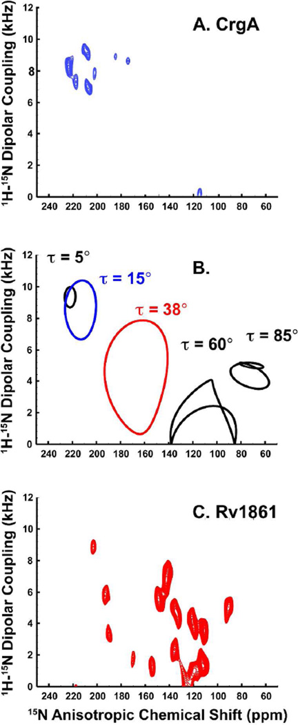
The interpretation of orientational restraints is a two stage process. The spectra of TM helices provide images of helical wheels in the SLF spectra with 3.6 resonances per turn around a wheel-like pattern of resonances known as a PISA (Polar Index Slant Angles) wheel (Fig. 2)39,40. The spectral dispersion is dependent on the helical tilt angle with respect to the bilayer normal: 0–10° provide very little dispersion, while 10–20° provide more, 30–60° provide maximal dispersion. Amphipathic helices on the bilayer surface are further complicated, because the plane orthogonal to the magnetic field is also a symmetry plane for the resonance frequencies. Therefore, a helix at exactly 90° to the bilayer normal displays a pattern of resonances in which a full circuit of the PISA wheel is achieved in a 180° arc of the helical wheel. Wheels associated with helical tilts either less than or more than 90° morph this 180° wheel into a 360° wheel associated with helical tilt angles of less than 70° or more than 110° (e.g. Fig. 2B, 85° helical tilt). Also note that spectra are symmetric about 0 kHz and that the PISA wheels retain their asymmetry when they span this symmetry axis for the data.
Figure 2.
Initial analysis of PISEMA spectra. A) PISEMA spectrum of CrgA 15N Phe labeled sample (residues: 33, 37, 51, 79 & 81). B) Calculated PISA wheels for different helical tilt angles. T=15° is consistent with the CrgA data in A and 38° is consistent with the data in C. C) PISEMA spectrum of Rv1861 15N Val labeled protein.
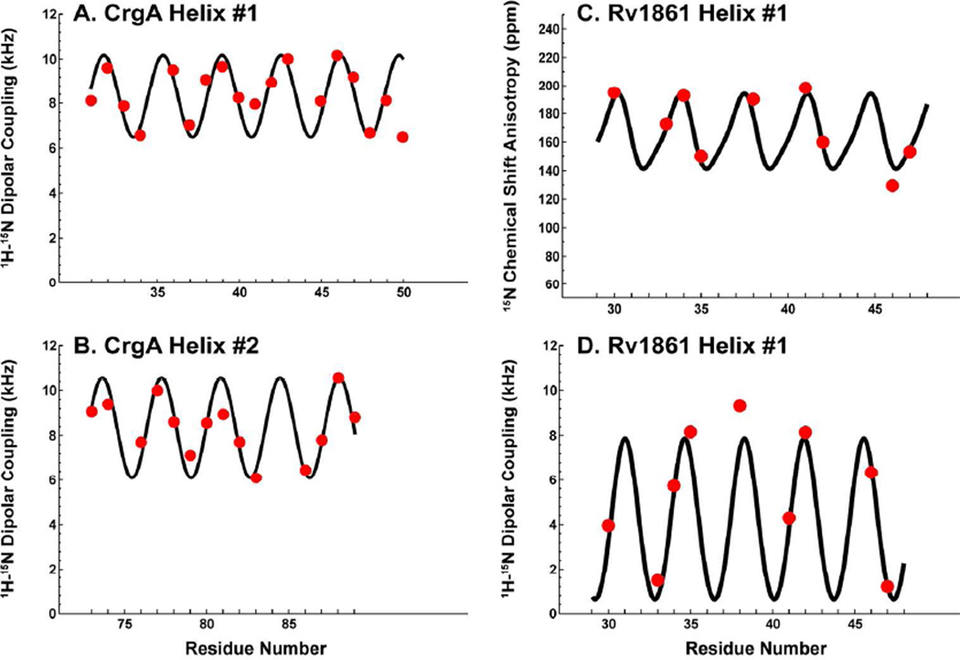
The uniformity of the helical structure is seen more clearly in the dipolar and chemical shift waves in Fig. 3. The influence of varying tensor element magnitudes, tensor orientations, and torsion angles have all been studied41. Indeed, the helical torsion angles in a lipid environment (ϕ, ψ = −60, −45°) are significantly different from helices in an aqueous environment (ϕ, ψ = −65, −40°), where there is competition for the amide hydrogen bonding by water that is absent in the lipid environment. The uniformity of the structure in helical segments has been confirmed by high resolution crystal structures, as has the shift in helical torsion angles41,42.
Figure 3.
The uniform oscillation of the anisotropic chemical shift (C) and dipolar interactions (A,B & D) is displayed more clearly with these wave patterns with exactly 3.6 residues per cycle. CrgA has two helices (A&B). Rv1861 has three helices, but only the data from helix #1 is displayed here (C&D).
The dependence on helical tilt for the pattern of resonances causes not only a change in the dispersion (the size of the wheel), but also in the center of mass of the resonances (Fig. 2B). A kink in the helix would typically result in a change in the helical tilt and a change in the center of mass for the helical segment with a different tilt angle. As a result such deformations are readily identified in the spectra43. While, it is not necessary to have the resonance assignments to determine the helical tilt, it is necessary to have minimal assignments for characterizing the rotational orientation of the helix39. Because of the resonance pattern it is only necessary to assign a single sequence specific resonance to determine the rotational orientation of a uniform helical segment. Typically, a single amino acid specific labeled sample is adequate to accomplish this goal. Likewise, a change in rotational orientation induced by a kink or bend within a helix can typically be assessed with a second amino acid specifically labeled protein.
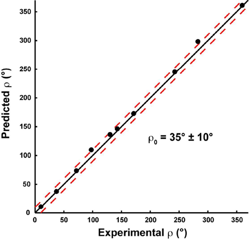
The complete spectral assignments can be achieved through multiple amino acid specific labels and through some reverse labeling (growing bacteria on uniform 15N labeling media with unlabeled amino acids added to avoid labeling these residues).44,45 Often resonances in the first turn or the last turn of a TM helix display less helical uniformity, potentially the result of a single hydrogen bond from within the helix per peptide plane and a more polar environment resulting in secondary hydrogen bonds to the amide backbone sites. The result is to induce more scatter in the resonance frequencies about the helical wheel. Consequently, the resonance assignments are best defined from the core of the helical segment toward the ends of the helical segments. Assignments are facilitated by plotting theoretical r values (100° per residue) versus the experimentally characterized values from the PISA wheel analysis (Fig. 4). Occasionally, two resonances that are very close to each other (such as two Ala residues in i and i+7 positions in the sequence) may be difficult to assign with this approach. Interestingly, a mistaken assignment for two such resonances in a PISA wheelhas very little impact on the structure, because resonances that are so close to each other give rise to nearly identical orientational restraints and hence only a marginal difference (less than 1–2°) in the orientations for these sites.
Figure 4.
Experimental ρ values from PISA wheel analysis are plotted against predicted values (100°/residue) for the same helix #1 residues of Rv1861 shown in Fig. 3 C&D.
Of course the resonance pattern(s) become more complex when there is more than one helix often causing the resonance patterns to overlap. Judicious choice for amino acid labels can lead to identification of the PISA wheels even when they are severely overlapped. Taking advantage of i to i+4 or i+7 patterns within a specific amino acid label or focusing on the distribution of the resonances at the core of the helix can be useful aids in identifying a PISA wheel within a TM helical sequence. We have recently assigned the resonances for Rv1861 having three TM helices, as well as the resonances from the two helices of CrgA (Murray et al., unpublished and Das et al., unpublished). These both represented challenging problems; the two helices of CrgA have small helical tilts (15° and 16°; Fig. 2A and 3A&B). The fact that the tilt angles are small leads to only a modest dispersion in the resonances and both PISA wheels are severely overlapped. For CrgA the presence of unique amino acids in both TM helices (valine in TM1 and alanine in TM2) were helpful for determining the tilts and rotation angles for both helices. For Rv1861 the helical tilt angles are much greater, but once again the PISA wheels are overlapped due to the similarity of the tilt angles, 38°, 40° and 46°. Virtually all hydrophobic amino acid labels were needed to clinch the resonance assignments. There are two resonance correlation techniques that have recently been demonstrated46,47 suggesting that more sophisticated and less labor intensive approaches for resonance assignments or their validation may become routine with further enhancements in sensitivity.
It has also been possible to characterize a helix that includes a bend or kink. The M2TM domain has a bent helix when the antiflu drug, amantadine, is bound. Kinks may be required for the protein function, but exposure of polar atoms in the low dielectric environment will always be minimized for the sake of tertiary structural stability that is often marginal. As a result the torsion angle space available for the bend or kink is limited. Mathematically this has been approached for the situation in which there is only two non-helical torsion angles to show that unique solutions can be obtained if the tilt and rotational angles are known for the helical segments on either side of a kink48.
The resonance frequencies for amide 15N sites of a TM helix can be used as high resolution orientational restraints, but there are multiple degeneracies both in the restraints defining the orientation of a peptide plane and in the de novo determination of torsion angles37. However, with knowledge that the data is from an α-helix having torsion angles with a variation of ±5° eliminates all of the degenerate solutions except for the trivial case in which the peptide plane is nearly parallel to Bo (within ±4°). Consequently, the interpretation of orientational restraints within a helix has a unique solution.
The result is a set of TM helices with accurately defined short range structure, i.e. torsion angles and precise orientational order with respect to the membrane environment. Recall, that these restraints are absolute restraints and consequently the solution for a set of torsion angles results in a helix whose tilt and rotational angles cannot be changed during refinement. The remaining flexibility in packing a set of helices is limited to rotations about the bilayer normal and translations that are limited to the X,Y axes with a potential of a few Å of flexibility in the Z axis. Because the sidechain conformations have not been determined there is some additional complexity in packing the helices. However, because of the scarcity of interhelical hydrogen bonds, TM helices are typically packed so as to maximize the van der Waals interactions and interhelical backbone-backbone electrostatic interactions. In other words the packing takes advantage of the small residues such as glycine, alanine and serine to pack the backbones of adjacent helices closely together49. In fact, conserved glycines appear to be rarely, if ever, exposed to the fatty acyl environment of the lipid bilayer, presumably because glycine residues would expose the polar atoms of the backbone to the apolar environment of the membrane2. Consequently, it may be possible at this stage to generate models for helix packing based on these restraints. Such models can be used to predict distances (i.e. crosspeaks in MAS spectra), thereby solving the docking problem with sparse distance restraints between the helices.
Orientational restraints can also be obtained for 15N histidine and tryptophan sidechains50. Because both χ1 and χ2 are variables it is rare that unique solutions can be obtained with just the anisotropic 15N-1H dipolar and 15N anisotropic chemical shift restraints. However, these sidechains are substantially restrained by such data.
Obtaining Distance and additional Torsional Restraints
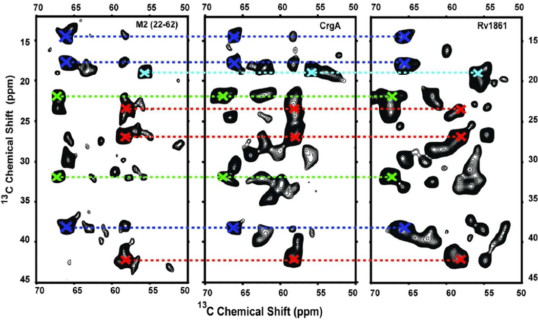
The same liposome preparation used for preparing the oriented samples on glass slides can be used for MAS spectroscopy samples, although it is often possible to increase the protein/lipid ratio and hence the sensitivity of these samples. For sparse distance restraints we focus on the unique or rare amino acid residues in the TM helices for both assignments and restraints. As mentioned previously, the limited chemical shift dispersion within a residue type, especially for hydrophobic amino acid residues, coupled with many residues of the same type and linewidths that are relatively broad generates ambiguities for resonance assignments. Fig. 5 shows numerous resonance envelopes for Leu, Val, Ile, and Ala that occur at essentially the same frequencies in the spectra of all three proteins. For these three proteins the LVIA residues account for 50–70% of all the residues in the TM helices. In Rv1861 the three TM helices also include 16 glycine residues. However, with only sparse distance restraints needed to complete the structure assignments can be achieved for the rare residues. Obviously, for those amino acids that are unique in the TM domain, a unique assignment can be achieved even if there are multiple residues of this type in the water soluble domain. Chemical shifts, reflecting an α-helix coupled to neighboring amino acids can confirm this assignment. In addition, observation of residue pairs through NCOCX or through CAN(CO)CX experiments with sparse labeling can lead to additional sequence specific assignments. Such labeling can be achieved with 2-13C or 1,3-13C glycerol or by reverse labeling as described above. From these assigned resonances it is possible to search for a few specific interhelical distance restraints.
Figure 5.
DARR (Dipolar Assisted Rotational Resonance) MAS ssNMR spectra (243K, 50ms mixing & 10kHz spinning) from three proteins and their mixing times: A) M2 protein, B) CrgA, and C) Rv1861. The aliphatic resonance envelopes for Leu (red), Val (green), Ile (blue) and Ala (cyan) are highlighted with dotted lines showing nearly identical positions for their resonance envelopes in the three spectra.
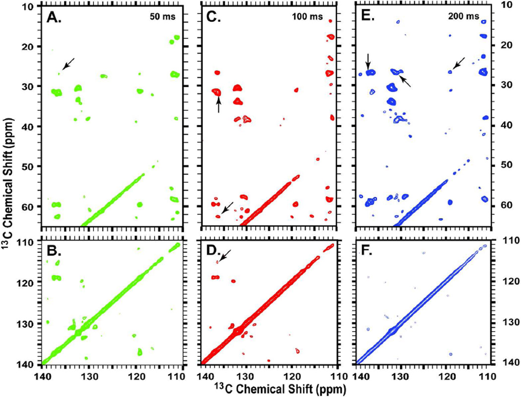
In studies of the M2 conductance domain that includes the single TM helix and the C-terminal amphipathic helix we have been able to sequence specifically assign all of the TM helix backbone except for the numerous leucine residues. These assignments were achieved using uniform 13C and 15N labeled samples. However, even with these assignments, the sidechain resonance overlap is so severe between the sidechain resonances of the hydrophobic residues that crosspeaks cannot be unambiguously assigned and hence they lead to degenerate structural restraints. Unique residues in the TM helix, Ser31, Gly34 and His37 were then used to search for interhelical distances to restrain the tetrameric structure. None of these residues are exposed to the lipid environment and therefore, they have the potential to generate interhelical restraints. In particular, the His37 sidechain forms a dimer of dimers structure through imidazole-imidazolium hydrogen bonds that were originally characterized by Nδ1 and Nε2 sidechain labeling51 and more recently through sparse labeling52. The unique chemical shifts of His37 displayed numerous DARR crosspeaks with residues in the vicinity20 (Fig.6). While some of these distances were ambiguous in that the crosspeaks could be assigned to multiple sidechains, multiple uniquely assigned restraints were obtained to adequately restrain the tetrameric structure.
Figure 6.
DARR MAS ssNMR spectra from M2 protein (residues 22–62) showing a number of crosspeaks correlated with interhelical distance restraints (arrows). A&B) 50 ms mixing time; C&D) 100 ms mixing time; E&F) 200 ms mixing time.
Structural Refinement
Importantly, it is necessary to refine the structure in the same environment in which the structural restraints were obtained. The environment provides many of the interactions that stabilize the tertiary structure and therefore this should become a standard protocol for membrane protein structural refinement. All of the experimental restraints are used in a restrained molecular dynamics protocol. While only a some of the sidechains are experimentally restrained the molecular dynamics force field can be anticipated to achieve a very realistic model of the remaining sidechain orientations. Of course many of these sidechain conformations are of little consequence since approximately half of them face the lipid environment and can be anticipated to have significant dynamics.
Outlook
Especially for small helical membrane proteins the structural characterization in a native-like environment can be very important. Interhelical hydrogen bonds or Coulombic interactions between charged sidechains are rare in the TM domain of these proteins. Van der Waals interactions are non-specific and consequently lead to relatively little stability for a given tertiary or quaternary structure. The result is that the interaction with the protein environment can often significantly influence the structure. OS and MAS ssNMR methods have been rapidly developing as a methodology for characterizing membrane protein structure in a lipid bilayer environment. By combining restraints from these two approaches the limitations of each can be overcome to achieve a robust technique for the characterization of important membrane protein structures in environments that accurately reproduce the biophysical properties of the membrane environment and thereby lead to a native-like membrane protein structure.
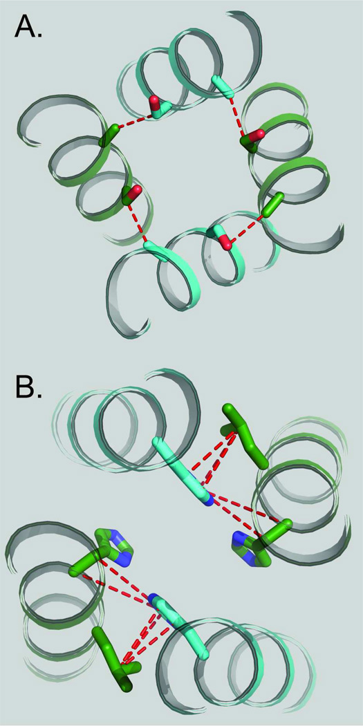
Figure 7.
Images of the M2 (22–62) protein structure showing the sparse interhelical distance restraints that uniquely constrain the quaternary structure.
Unlike water soluble proteins, the structures of helical transmembrane proteins depend on a very complex environment. These proteins sit in the midst of dramatic electrical and chemical gradients and are often subject to variations in the lateral pressure profile, order parameters, dielectric constant, and other properties. Solid state NMR is a collection of tools that can characterize high resolution membrane protein structure in this environment. Indeed, prior work has shown that this complex environment significantly influences transmembrane protein structure. Therefore, it is important to characterize such structures under conditions that closely resemble its native environment.
Researchers have used two approaches to gain protein structural restraints via solid state NMR spectroscopy. The more traditional approach uses magic angle sample spinning to generate isotropic chemical shifts, much like solution NMR. As with solution NMR, researchers can analyze the backbone chemical shifts to obtain torsional restraints. They can also examine nuclear spin interactions between nearby atoms to obtain distances between atomic sites. Unfortunately, for membrane proteins in lipid preparations, the spectral resolution is not adequate to obtain complete resonance assignments.
Researchers have developed another approach for gaining structural restraints from membrane proteins, the use of uniformly oriented lipid bilayers, provides a method for obtaining high resolution orientational restraints. When the bilayers are aligned with respect to the magnetic field of the NMR spectrometer, researchers can obtain orientational restraints in which atomic sites in the protein are restrained relative to the alignment axis. However, this approach does not allow researchers to determine the relative packing between helices.
By combining the two approaches we can take advantage of the information acquired from each technique to minimize the challenges and maximize the quality of the structural results. By combining the distance, torsional and orientational restraints we can characterize high resolution membrane protein structure in native-like lipid bilayer environments.
Acknowledgments
This work was supported by the National Institutes of Health (AI-074805, AI-023007, AI-073891) and the National Science Foundation through Cooperative Agreement 0654118 between the Division of Material Research and the State of Florida.
Biographies
Dylan T. Murray received a B.S. in Physics from The State University of New York at Plattsburgh in 2004. From 2004 to 2007 he worked at the Center for X-Ray Crystallography at the University of Vermont. Since 2007 he has been in the Molecular Biophysics Ph.D. program at Florida State University working in the laboratory of Prof. Cross. His research focuses on the development of solid state NMR techniques aimed at determining the structures for membrane proteins in native-like environments. He earned a University Fellowship in 2008 and received the Michael Kasha Student Publication Award in 2011.
Nabanita Das received her B.Sc and M.Sc Biotechnology degree from Bangalore University, India (2001–2006). Afterward, she worked as Scientist in Jubilant Biosys and BioBase International, Bangalore, India (2006–2008). She entered the PhD Molecular Biophysics program at Florida State University in Fall 2008. Since 2009 till date she is pursuing her thesis research under the supervision of Prof. Cross on the M. tuberculosis membrane protein CrgA and CwsA drug targets structure and functional studies by solid state NMR spectroscopy.
Timothy A. Cross received his Ph.D. from the University of Pennsylvania in 1981 studying membrane protein structure by solution NMR spectroscopy. Since 1985 he has been on the faculty of Florida State University in the Department of Chemistry and Biochemistry where his laboratory has been developing strategies for the characterization of membrane protein structure and dynamics in native like environments using solid state NMR spectroscopy. He also heads up the NMR and MRI User Program at the National High Magnetic Field Laboratory in Tallahassee.
References
- 1.Page RC, Li C, Hu J, Gao FP, Cross TA. Lipid bilayers: an essential environment for the understanding of membrane proteins. Magn Reson Chem. 2007;45:S2–S11. doi: 10.1002/mrc.2077. [DOI] [PubMed] [Google Scholar]
- 2.Dong H, Sharma M, Zhou HX, Cross TA. Glycines: Role in alpha-helical membrane protein structures and a potential indicator of native conformation. Biochemistry. 2012;51:4779–4789. doi: 10.1021/bi300090x. [DOI] [PMC free article] [PubMed] [Google Scholar]
- 3.Cross TA, Sharma M, Yi M, Zhou HX. Influence of solubilizing environments on membrane protein structures. Trends Biochem Sci. 2011;36:117–125. doi: 10.1016/j.tibs.2010.07.005. [DOI] [PMC free article] [PubMed] [Google Scholar]
- 4.Ketchem RR, Roux B, Cross TA. High-Resolution Polypeptide Structure in a Lamellar Phase Lipid Environment from Solid-State NMR Derived Orientational Constraints. Structure. 1997;5:1655–1669. doi: 10.1016/s0969-2126(97)00312-2. [DOI] [PubMed] [Google Scholar]
- 5.Opella SJ, Marassi FM, Gesell JJ, Valente AP, Kim Y, Oblatt-Montal M, Montal M. Structures of the M2 channel-lining segments from nicotinic acetylcholine and NMDA receptors by NMR spectroscopy. Nat Struct Biol. 1999;6:374–379. doi: 10.1038/7610. [DOI] [PMC free article] [PubMed] [Google Scholar]
- 6.Duong-Ly KC, Nanda V, DeGrado WF, Howard KP. The conformation of the pore region of the M2 prooton channel depends on lipid bilayer environment. Protein Sci. 2005;14:856–861. doi: 10.1110/ps.041185805. [DOI] [PMC free article] [PubMed] [Google Scholar]
- 7.McDermott A. Structure and dynamics of membrane proteins by magic angle spinning solid-state NMR. Annual review of biophysics. 2009;38:385–403. doi: 10.1146/annurev.biophys.050708.133719. [DOI] [PubMed] [Google Scholar]
- 8.Renault M, Cukkemane A, Baldus M. Solid-state NMR spectroscopy on complex biomolecules. Angew Chem Int Ed Engl. 2010;49:8346–8357. doi: 10.1002/anie.201002823. [DOI] [PubMed] [Google Scholar]
- 9.Tang M, Sperling LJ, Berthold DA, Schwieters CD, Nesbitt AE, Nieuwkoop AJ, Gennis RB, Rienstra CM. High-resolution membrane protein structure by joint calculations with solid-state NMR and X-ray experimental data. Journal of biomolecular NMR. 2011;51:227–233. doi: 10.1007/s10858-011-9565-6. [DOI] [PMC free article] [PubMed] [Google Scholar]
- 10.Lange A, Giller K, Hornig S, Martin-Eauclaire MF, Pongs O, Becker S, Baldus M. Toxin-induced conformational changes in a potassium channel revealed by solid-state NMR. Nature. 2006;440:959–962. doi: 10.1038/nature04649. [DOI] [PubMed] [Google Scholar]
- 11.Andronesi OC, Becker S, Seidel K, Heise H, Young HS, Baldus M. Determination of membrane protein structure and dynamics by magic-angle-spinning solid-state NMR spectroscopy. Journal of the American Chemical Society. 2005;127:12965–12974. doi: 10.1021/ja0530164. [DOI] [PubMed] [Google Scholar]
- 12.Higman VA, Varga K, Aslimovska L, Judge PJ, Sperling LJ, Rienstra CM, Watts A. The Conformation of Bacteriorhodopsin Loops in Purple Membranes Resolved by Solid-State MAS NMR Spectroscopy. Angew Chem Int Ed Engl. 2011 doi: 10.1002/anie.201100730. [DOI] [PubMed] [Google Scholar]
- 13.Das BB, Nothnagel HJ, Lu GJ, Son WS, Tian Y, Marassi FM, Opella SJ. Structure determination of a membrane protein in proteoliposomes. Journal of the American Chemical Society. 2012;134:2047–2056. doi: 10.1021/ja209464f. [DOI] [PMC free article] [PubMed] [Google Scholar]
- 14.Park SH, Das BB, Casagrande F, Tian Y, Nothnagel HJ, Chu M, Kiefer H, Maier K, De Angelis AA, Marassi FM, Opella SJ. Structure of the chemokine receptor CXCR1 in phospholipid bilayers. Nature. 2012;491:779–783. doi: 10.1038/nature11580. [DOI] [PMC free article] [PubMed] [Google Scholar]
- 15.Cross TA, Opella SJ. Protein structure by solid state nuclear magnetic resonance. Residues 40 to 45 of bacteriophage fd coat protein. Journal of molecular biology. 1985;182:367–381. doi: 10.1016/0022-2836(85)90197-4. [DOI] [PubMed] [Google Scholar]
- 16.Moll F, 3rd, Cross TA. Optimizing and characterizing alignment of oriented lipid bilayers containing gramicidin D. Biophys J. 1990;57:351–362. doi: 10.1016/S0006-3495(90)82536-4. [DOI] [PMC free article] [PubMed] [Google Scholar]
- 17.Mahalakshmi R, Marassi FM. Orientation of the Escherichia coli Outer Membrane Protein OmpX in Phospholipid Bilayer Membranes Determined by Solid-State NMR. Biochemistry. 2008 doi: 10.1021/bi800362b. [DOI] [PMC free article] [PubMed] [Google Scholar]
- 18.Verardi R, Shi L, Traaseth NJ, Walsh N, Veglia G. Structural topology of phospholamban pentamer in lipid bilayers by a hybrid solution and solid-state NMR method. Proceedings of the National Academy of Sciences of the United States of America. 2011;108:9101–9106. doi: 10.1073/pnas.1016535108. [DOI] [PMC free article] [PubMed] [Google Scholar]
- 19.De Angelis AA, Howell SC, Nevzorov AA, Opella SJ. Structure determination of a membrane protein with two trans-membrane helices in aligned phospholipid bicelles by solid-state NMR spectroscopy. Journal of the American Chemical Society. 2006;128:12256–12267. doi: 10.1021/ja063640w. [DOI] [PMC free article] [PubMed] [Google Scholar]
- 20.Can TV, Sharma M, Hung I, Gor'kov PL, Brey WW, Cross TA. Magic angle spinning and oriented sample solid-state NMR structural restraints combine for influenza A M2 protein functional insights. Journal of the American Chemical Society. 2012 doi: 10.1021/ja3004039. [DOI] [PMC free article] [PubMed] [Google Scholar]
- 21.Cady SD, Schmidt-Rohr K, Wang J, Soto CS, Degrado WF, Hong M. Structure of the amantadine binding site of influenza M2 proton channels in lipid bilayers. Nature. 2010;463:689–692. doi: 10.1038/nature08722. [DOI] [PMC free article] [PubMed] [Google Scholar]
- 22.Hu J, Qin H, Gao FP, Cross TA. A systematic assessment of mature MBP in membrane protein production: overexpression, membrane targeting and purification. Protein expression and purification. 2011;80:34–40. doi: 10.1016/j.pep.2011.06.001. [DOI] [PMC free article] [PubMed] [Google Scholar]
- 23.De Angelis AA, Opella SJ. Bicelle samples for solid-state NMR of membrane proteins. Nat Protoc. 2007;2:2332–2338. doi: 10.1038/nprot.2007.329. [DOI] [PubMed] [Google Scholar]
- 24.Holsinger LJ, Lamb RA. Influenza virus M2 integral membrane protein is a homotetramer stabilized by formation of disulfide bonds. Virology. 1991;183:32–43. doi: 10.1016/0042-6822(91)90115-r. [DOI] [PubMed] [Google Scholar]
- 25.Wang C, Takeuchi K, Pinto LH, Lamb RA. Ion channel activity of influenza A virus M2 protein: characterizations of the amantadine block. J Virol. 1993;67:5585–5594. doi: 10.1128/jvi.67.9.5585-5594.1993. [DOI] [PMC free article] [PubMed] [Google Scholar]
- 26.Sugrue RJ, aH AJ. Structural Characteristics of the M2 Protein of Influenza A Viruses: Evidence that it forms a Tetrameric Channel. Virology. 1991;180:617–624. doi: 10.1016/0042-6822(91)90075-M. [DOI] [PMC free article] [PubMed] [Google Scholar]
- 27.Ma C, Polishchuk AL, Ohigashi Y, Stouffer AL, Schon A, Magavern E, Jing X, Lear JD, Freire E, Lamb RA, DeGrado WF, Pinto LH. Identification of the functional core of the influenza A virus A/M2 proton-selective ion channel. Proceedings of the National Academy of Sciences of the United States of America. 2009;106:12283–12288. doi: 10.1073/pnas.0905726106. [DOI] [PMC free article] [PubMed] [Google Scholar]
- 28.Plocinski P, Ziolkiewicz M, Kiran M, Vadrevu SI, Nguyen HB, Hugonnet J, Veckerle C, Arthur M, Dziadek J, Cross TA, Madiraju M, Rajagopalan M. Characterization of CrgA, a new partner of the Mycobacterium 13 tuberculosis peptidoglycan polymerization complexes. Journal of bacteriology. 2011;193:3246–3256. doi: 10.1128/JB.00188-11. [DOI] [PMC free article] [PubMed] [Google Scholar]
- 29.Li C, Gao P, Qin H, Chase R, Gor'kov PL, Brey WW, Cross TA. Uniformly aligned full-length membrane proteins in liquid crystalline bilayers for structural characterization. Journal of the American Chemical Society. 2007;129:5304–5305. doi: 10.1021/ja068402f. [DOI] [PMC free article] [PubMed] [Google Scholar]
- 30.Nevzorov AA, Opella SJ. Selective averaging for high-resolution solid-state NMR spectroscopy of aligned samples. J Magn Reson. 2007;185:59–70. doi: 10.1016/j.jmr.2006.09.006. [DOI] [PubMed] [Google Scholar]
- 31.Ramamoorthy A, Wu CH, Opella SJ. Three-dimensional solid-state NMR experiment that correlates the chemical shift and dipolar coupling frequencies of two heteronuclei. J Magn Reson B. 1995;107:88–90. doi: 10.1006/jmrb.1995.1063. [DOI] [PubMed] [Google Scholar]
- 32.Tang W, Knox RW, Nevzorov AA. A spectroscopic assignment technique for membrane proteins reconstituted in magnetically aligned bicelles. Journal of biomolecular NMR. 2012;54:307–316. doi: 10.1007/s10858-012-9673-y. [DOI] [PubMed] [Google Scholar]
- 33.Gopinath T, Verardi R, Traaseth NJ, Veglia G. Sensitivity enhancement of separated local field experiments: application to membrane proteins. The journal of physical chemistry. B. 2010;114:5089–5095. doi: 10.1021/jp909778a. [DOI] [PMC free article] [PubMed] [Google Scholar]
- 34.Shen Y, Delaglio F, Cornilescu G, Bax A. TALOS+: a hybrid method for predicting protein backbone torsion angles from NMR chemical shifts. Journal of biomolecular NMR. 2009;44:213–223. doi: 10.1007/s10858-009-9333-z. [DOI] [PMC free article] [PubMed] [Google Scholar]
- 35.Shi L, Ladizhansky V. Magic angle spinning solid-state NMR experiments for structural characterization of proteins. Methods Mol Biol. 2012;895:153–165. doi: 10.1007/978-1-61779-927-3_12. [DOI] [PubMed] [Google Scholar]
- 36.Poon A, Birn J, Ramamoorthy A. How Does an Amide-N Chemical Shift Tensor Vary in Peptides? The journal of physical chemistry. B. 2004;108:16577–16585. doi: 10.1021/jp0471913. [DOI] [PMC free article] [PubMed] [Google Scholar]
- 37.Cross TA, Quine JR. Protein Structure in Anisotropic Environments: Development of Orientational Constraints. Concepts in NMR. 2000;12:55–70. [Google Scholar]
- 38.Wang J, Kim S, Kovacs F, Cross TA. Structure of the transmembrane region of the M2 protein H(+) channel. Protein Sci. 2001;10:2241–2250. doi: 10.1110/ps.17901. [DOI] [PMC free article] [PubMed] [Google Scholar]
- 39.Wang J, Denny J, Tian C, Kim S, Mo Y, Kovacs F, Song Z, Nishimura K, Gan Z, Fu R, Quine JR, Cross TA. Imaging membrane protein helical wheels. J Magn Reson. 2000;144:162–167. doi: 10.1006/jmre.2000.2037. [DOI] [PubMed] [Google Scholar]
- 40.Marassi FM, Opella SJ. A solid-state NMR index of helical membrane protein structure and topology. J Magn Reson. 2000;144:150–155. doi: 10.1006/jmre.2000.2035. [DOI] [PMC free article] [PubMed] [Google Scholar]
- 41.Page RC, Kim S, Cross TA. Transmembrane helix uniformity examined by spectral mapping of torsion angles. Structure. 2008;16:787–797. doi: 10.1016/j.str.2008.02.018. [DOI] [PMC free article] [PubMed] [Google Scholar]
- 42.Kim S, Cross TA. Uniformity, ideality, and hydrogen bonds in transmembrane alpha-helices. Biophys J. 2002;83:2084–2095. doi: 10.1016/S0006-3495(02)73969-6. [DOI] [PMC free article] [PubMed] [Google Scholar]
- 43.Hu J, Asbury T, Achuthan S, Li C, Bertram R, Quine JR, Fu R, Cross TA. Backbone structure of the amantadine-blocked trans-membrane domain M2 proton channel from Influenza A virus. Biophys J. 2007;92:4335–4343. doi: 10.1529/biophysj.106.090183. [DOI] [PMC free article] [PubMed] [Google Scholar]
- 44.Shi L, Kawamura I, Jung KH, Brown LS, Ladizhansky V. Conformation of a seven-helical transmembrane photosensor in the lipid environment. Angew Chem Int Ed Engl. 2011;50:1302–1305. doi: 10.1002/anie.201004422. [DOI] [PubMed] [Google Scholar]
- 45.Andreas LB, Eddy MT, Pielak RM, Chou J, Griffin RG. Magic angle spinning NMR investigation of influenza A M2(18–60): support for an allosteric mechanism of inhibition. Journal of the American Chemical Society. 2010;132:10958–10960. doi: 10.1021/ja101537p. [DOI] [PMC free article] [PubMed] [Google Scholar]
- 46.Mote KR, Gopinath T, Traaseth NJ, Kitchen J, Gor'kov PL, Brey WW, Veglia G. Multidimensional oriented solid-state NMR experiments enable the sequential assignment of uniformly 15N labeled integral membrane proteins in magnetically aligned lipid bilayers. Journal of biomolecular NMR. 2011;51:339–346. doi: 10.1007/s10858-011-9571-8. [DOI] [PubMed] [Google Scholar]
- 47.Knox RW, Lu GJ, Opella SJ, Nevzorov AA. A resonance assignment method for oriented-sample solid-state NMR of proteins. Journal of the American Chemical Society. 2010;132:8255–8257. doi: 10.1021/ja102932n. [DOI] [PMC free article] [PubMed] [Google Scholar]
- 48.Murray DT, Lu Y, Cross TA, Quine JR. Geometry of kinked protein helices from NMR data. J Magn Reson. 2011;210:82–89. doi: 10.1016/j.jmr.2011.02.012. [DOI] [PubMed] [Google Scholar]
- 49.Javadpour MM, Eilers M, Groesbeek M, Smith SO. Helix packing in polytopic membrane proteins: Role of glycine in transmembrane helix association. Biophys. J. 1999;77:1609–1618. doi: 10.1016/S0006-3495(99)77009-8. [DOI] [PMC free article] [PubMed] [Google Scholar]
- 50.Nishimura K, Kim S, Zhang L, Cross TA. The Closed State of a H+ Channel Helical Bundle: Combining Precise orientational and Distance Restraints from Solid State NMR. Biochemistry. 2002;41:13170–13177. doi: 10.1021/bi0262799. [DOI] [PubMed] [Google Scholar]
- 51.Hu J, Fu R, Nishimura K, Zhang L, Zhou HX, Busath DD, Vijayvergiya V, Cross TA. Histidines, heart of the hydrogen ion channel from influenza A virus: toward an understanding of conductance and proton selectivity. Proceedings of the National Academy of Sciences of the United States of America. 2006;103:6865–6870. doi: 10.1073/pnas.0601944103. [DOI] [PMC free article] [PubMed] [Google Scholar]
- 52.Andreas LB, Eddy MT, Chou JJ, Griffin RG. Magic-angle-spinning NMR of the drug resistant S31N M2 proton transporter from influenza A. Journal of the American Chemical Society. 2012;134:7215–7218. doi: 10.1021/ja3003606. [DOI] [PMC free article] [PubMed] [Google Scholar]


