Abstract
Objectives To present a critical evaluation of our experience using an expanded endoscopic endonasal approach (EEEA) to clival lesions and evaluate, based on the location of residual tumor, what the anatomic limitations to the approach are.
Design A retrospective review of all endoscopic endonasal operations performed at our institution identified 19 patients with lesions involving the clivus. Extent of resection was determined by preoperative and postoperative tumor volumes.
Results Three patients underwent planned subtotal resections. Of the remaining patients, gross total resection was achieved in 8/16 (50%), > 95% in 5/16 (31%), and < 95% in 3/16 (19%). Residual tumor occurred, most commonly with extension posterior and lateral to the internal carotid artery, with inferior, lateral invasion of the occipital condyle and with deep inferior extension to the midportion of the dens.
Conclusions The EEEA represents a safe and effective technique for the resection of clival lesions. Despite excellent overall visualization of this region we found that adequate exposure of the most lateral and inferior portions of large tumors is often difficult. Knowledge of these limitations allows us to determine which tumors are best suited for an EEEA and which may be more appropriate for an open skull base or combined technique.
Keywords: clival lesions, endoscopy, minimally invasive, transnasal transsphenoidal
Introduction
Lesions involving the clivus present a surgical challenge to skull base surgeons because of their central and deep location.1 As a result, several approaches have traditionally been utilized for the removal of these lesions with varying rates of success. “Open” approaches (e.g., frontotemporal, orbitozygomatic,2 subfrontal transbasal,3 subtemporal-infratemporal,4 transpetrosal,5 and extreme lateral transcondylar6) offer lateral trajectories to access clival lesions but, depending upon the location of the tumor, may entail brain retraction and require the surgeon to traverse critical neurovascular structures to gain access to the tumor. Anterior skull base approaches including rhinoseptal,7 transoral,8 transfacial,9 and transcervical10 reduce the need for brain retraction by providing more direct access to the clivus but may be less desirable due to facial incisions, palatal dysfunction, high cerebrospinal fluid (CSF) leak rates, and/or technical difficulty.11
Laws12 in the 1980s and Maira et al13 in the early 1990s were the first to utilize a more natural corridor to the clivus with a microscopic extended transnasal, transsphenoidal approach. Although effective at reducing some of the morbidity associated with the removal of these lesions, access to the more paramedian, lateral, and inferior portions of a tumor is restricted in part due to the confines of the nasal speculum.14,15 The adoption of the endoscope in skull base surgery has resulted in improved visualization, increased lateral exposure and greater maneuverability when utilizing a transsphenoidal corridor. Despite these advantages, however, previous reports have demonstrated varying success rates in achieving gross total tumor resection (GTR) when utilizing an expanded endoscopic endonasal approach (EEEA) to remove lesions of the clivus.14,16,17,18,19,20
The panoramic view of the clivus and ventral brainstem that can be achieved with the endoscope has been demonstrated with anatomic studies in cadaveric specimens. Critical neurovascular structures, primarily the paraclival internal carotid artery (ICA) segments and the lower cranial nerves, are generally considered to represent the lateral limits of safe exposure.19,21,22 Clinically, clival lesions are often found to displace, invaginate around, and extend beyond these structures, thus restricting the success of an EEEA.23 Understanding the specific anatomic limitations associated with this approach is important to better predict preoperatively whether an expanded endoscopic, lateral, or anterior “open” procedure would be most effective in safely achieving the greatest amount of tumor removal.
In this study we present a critical evaluation of our experience using an EEEA in patients with a variety of clival lesions. Our goal was to use preoperative and postoperative imaging to assess the anatomical relationships and other surgical factors that prevented us from achieving GTR in certain cases.
Methods
We retrospectively reviewed a database of all patients undergoing an EEEA at UCLA Medical Center between the years 2008 and 2011. Patients with lesions significantly involving the clivus were identified for inclusion within the study. The senior authors (neurosurgeon [MB] and otolaryngologists [MW] and [JS]) were the primary surgeons in all of the cases. Patient demographics, lesion size and volume, pathology, complications, adjuvant treatment, and clinical outcomes were analyzed. Extent of resection was determined by a neuroradiologist (NS) after comparing preoperative and postoperative tumor volumes on magnetic resonance imaging (MRI) scans. Results were then divided into (1) GTR, defined as no tumor on postoperative MRI; (2) > 95% tumor resection; and (3) < 95% tumor resection.
All operations were performed purely endoscopically with an otolaryngologist and neurosurgeon working simultaneously. Frameless stereotactic image guidance was utilized in all cases. If, after evaluating the tumor on preoperative imaging, a CSF leak was considered likely, then a right-sided nasoseptal flap based upon the posterior nasoseptal artery was raised in the standard fashion prior to performing a posterior septectomy.24 After dissection and removal of the clival mucosa, the skull base was drilled using a 3-mm diamond bur high-speed drill to obtain an expanded exposure. A microvascular ultrasound Doppler probe was routinely used to localize the internal carotid arteries (ICA) if exposed. A combination of 0-, 30-, and 45-degree angled endoscopes were utilized for direct tumor visualization and removal. Tumor densely adherent to the ICAs was not aggressively removed. Once tumor removal was complete, reconstruction of the sellar/clival defects was performed using the nasoseptal flap when necessary. In certain cases, a multilayered closure consisting of abdominal fat, tensor fascia lata, and/or septal bone was utilized, as well.
Results
Nineteen patients underwent an EEEA for resection of a lesion involving the clivus (Table 1). Tumors consisted of lesions originating either directly from the clivus or from adjacent areas but with extensive clival involvement. Final pathology included nine chordomas, five invasive pituitary adenomas, one adenocarcinoma, one meningioma, one adenoid cystic carcinoma, one fibrous dysplasia, and one leiomyosarcoma metastasis. Mean patient age was 54.6 years. Mean initial tumor volume was 26.2 cm3. Six patients presented with tumor recurrence at the time of evaluation. Five of these patients had undergone previous microscopic or endoscopic transsphenoidal surgery for tumor resection. One patient had two previous craniotomies for a recurrent clival chordoma. One patient had had a previous endoscopic transsphenoidal operation for a separate diagnosis. Three patients with recurrence had undergone previous radiation therapy.
Table 1. Patients with Clival Lesions Who Underwent an Expanded Endoscopic Endonasal Approach (EEEA) for Tumor Resection.
| Patient no. | Age (yr)/Sex | Diagnosis | Tumor volume (cm3) | Extent of resection | Presentation | Previous operation(s)/radiation | Complications |
|---|---|---|---|---|---|---|---|
| 1 | 62/F | Chordoma | 35.2 | >95% | CN V1, VI deficits | None | New CN V2 deficit |
| 2 | 65/F | Cavernous sinus meningioma | 29.3 | Planned subtotal | Nasal symptoms | TNTS, SRT | None |
| 3 | 48/F | Chordoma | 9.5 | GTR | CN V2, VI deficits | Craniotomy ×2, SRT | CSF leak required reoperation ×2, meningitis |
| 4 | 67/M | Chordoma | 29.8 | <95% | CN VI, X deficits | None | CSF leak treated with ELD |
| 5 | 71/M | Chordoma | 4.8 | GTR | CN VI deficit | None | None |
| 6 | 52/M | Adenoid cystic carcinoma | 109.0 | Planned Subtotal |
CN V, VI deficits, visual deterioration | None | None |
| 7 | 39/F | Chordoma | 5.0 | GTR | Tinnitus | None | CSF leak required reoperation |
| 8 | 50/F | Chordoma | 10.6 | GTR | Headaches, tinnitus | None | None |
| 9 | 43/M | Giant pituitary adenoma (ACTH) | 50.4 | >95% | CN III, VI deficits, Cushing disease | None | New CN VI deficit |
| 10 | 66/M | Chordoma | 58.0 | GTR | CN III, V2 deficits | None | None |
| 11 | 41/F | Chordoma | 52.8 | <95% | Nasal symptoms | None | None |
| 12 | 55/M | Fibrous dysplasia | 20.5 | Planned Subtotal |
Tinnitus, headaches | None | None |
| 13 | 50/F | Ectopic pituitary adenoma (GH) |
1.1 | GTR | Acromegaly | None | None |
| 14 | 52/F | Adenocarcinoma | 9.1 | GTR | CN VI deficit | None | None |
| 15 | 62/F | Metastatic leiomyosarcoma |
6.2 | GTR | CN VI deficit, headaches | TNTS, Radiation | None |
| 16 | 63/F | Pituitary adenoma (nonfunctional) |
34.6 | <95% | Routine follow-up imaging | TNTS | None |
| 17 | 35/F | Pituitary adenoma (prolactin) |
8.3 | >95% | Prolactinoma | TNTS ×2 | None |
| 18 | 63/F | Pituitary adenoma (nonfunctional) |
5.5 | >95% | Headaches | TNTS, SRS | New CN VI deficit, pulmonary embolus |
| 19 | 54/F | Chordoma | 18.5 | >95% | Visual deterioration with bitemporal hemianopsia | None | None |
Abbreviations: ACTH, adrenocorticotrophicadrenocorticotropic hormone; CN, cranial nerve; CSF, cerebrospinal fluid; ELD, external lumbar drainage; GH, growth hormone; GTR, gross total resection; SRT/SRS, stereotactic radiotherapy/stereotactic radiosurgery; TNTS, microscopic transnasal transsphenoidal approach.
Cranial nerve (CN) deficit was the most common presenting symptom. Eight patients had diplopia from CN VI deficits, four patients had facial numbness from CN V dysfunction, two patients had CN III palsies, and one patient had hoarseness from CN X compression at initial diagnosis. Other presenting symptoms included tinnitus/vertigo from CN VIII involvement in three patients; headaches in three patients; decreased visual acuity and new visual field deficit from optic chiasm/optic nerve compression in two patients; nasal symptoms in two patients; and Cushing disease, acromegaly, and prolactinoma in one patient each.
Of the eight patients with preoperative CN VI deficits, three had complete recovery following surgery, three had partial improvement, and two patients experienced no change in symptoms. Two patients experienced postoperative improvement in facial numbness. Both patients with symptoms of visual deterioration improved with surgery. Of the two patients with CN III palsies, one had complete and one had partial recovery postoperatively. The patient with preoperative hoarseness from CN X compression demonstrated no significant symptomatic improvement following surgery.
Three of the 19 patients underwent planned subtotal resections. These included a sphenoid sinus lesion (histologically determined to be a meningioma) that extensively involved the clivus and right cavernous sinus, the latter preventing a complete resection. The second case involved a massive adenoid cystic carcinoma for which the goal of surgery was debulking only, and the third lesion was diagnosed as fibrous dysplasia on intraoperative frozen section; based on the benign diagnosis only, a limited resection was pursued.
Of the remaining patients, GTR was achieved in 8/16 (50%), > 95% in 5/16 (31%), and < 95% in 3/16 (19%). Average initial tumor volume for the group with GTR, < 95%, and > 95% was 13 ± 18, 39 ± 12, and 24 ± 19 cm3, respectively. A characteristic lesion with GTR is demonstrated in Fig. 1. A summary of the patients where GTR was attempted but not achieved—including the specific location of residual tumor and what associated anatomic/surgical limitations were present during the attempted resection—is provided in Table 2.
Fig. 1.
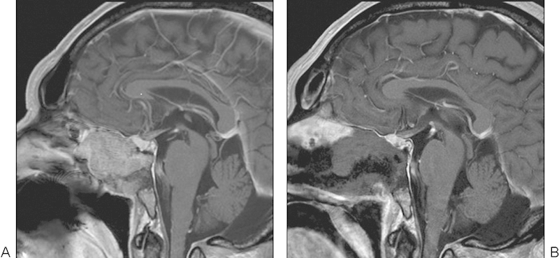
Patient 10 (A) preoperative and (B) postoperative T1 with contrast sagittal magnetic resonance imaging (MRI) scan demonstrating gross total resection (GTR) of a lesion involving the clivus following an expanded endoscopic endonasal approach (EEEA). Final diagnosis was consistent with chordoma.
Table 2. Location of Residual Tumor in Patients Who Underwent an Expanded Endoscopic Endonasal Approach (EEEA) for a Clival Lesion where GTR was Attempted but Not Achieved.
| Patient no. | Extent of resection | Location of residual tumor | Anatomic/surgical limitations |
|---|---|---|---|
| 1 | > 95% | Middle cranial fossa, dorsal/lateral resection cavity | CN V/Meckel cave lateral access |
| 4 | < 95% | Inferior/lateral petroclival region, medial jugular bulb/posterior carotid canal | Jugular bulb/posterior occipital condyle involvement, tumor extension posterior to petrous ICA |
| 9 | > 95% | Cavernous sinus, posterior paraclival ICA | Extensive cavernous sinus invasion, tumor involvement of paraclival ICA |
| 11 | < 95% | Anterior to midportion of dens, inferior resection cavity | Inferior tumor extension at mid to lower dens, inferior view limited by soft palate |
| 16 | < 95% | Cavernous sinus | Recurrent tumor in cavernous sinus with scarring and adherence to ICA/CNs |
| 17 | > 95% | Cavernous sinus/posterior clinoid | Recurrent tumor adherent to dura and ICA |
| 18 | > 95% | Inferior clivus | Recurrent tumor with extensive clival dura involvement, invasion posterior to clival ICA |
| 19 | > 95% | Sella/suprasellar region | Tumor adherence to optic chiasm |
Abbreviations: CN, cranial nerve; GTR, gross total resection; ICA, internal carotid canal.
Complications included six intraoperative CSF leaks for which a primary repair was performed during surgery. Three patients experienced failure of their repair and presented with a postoperative leak recurrence. Two of these cases required reoperation and one was treated successfully with lumbar drainage. Two patients developed postoperative CN VI deficits, one patient had temporary worsening of a CN VI deficit, one patient had a new CN V2 partial injury, and one patient developed a postoperative pulmonary embolus.
Illustrative Cases
Patient 1 presented with a heterogeneously enhancing mass in the left cerebellopontine angle (CPA) with extension to the middle cranial fossa and the Meckel cave. To access the lateral portion of the middle fossa component, we worked through small windows between the trigeminal nerve branches within the Meckel cave. Postoperative imaging revealed residual tumor at the dorsal, lateral extent of our resection cavity, indicating the difficulty we faced in visualizing tumor lateral to the trigeminal nerve, through an endonasal route (Fig. 2). The patient developed a partial CN V2 hypoesthesia postoperatively as a result of extensive manipulation of the nerve during the procedure.
Fig. 2.
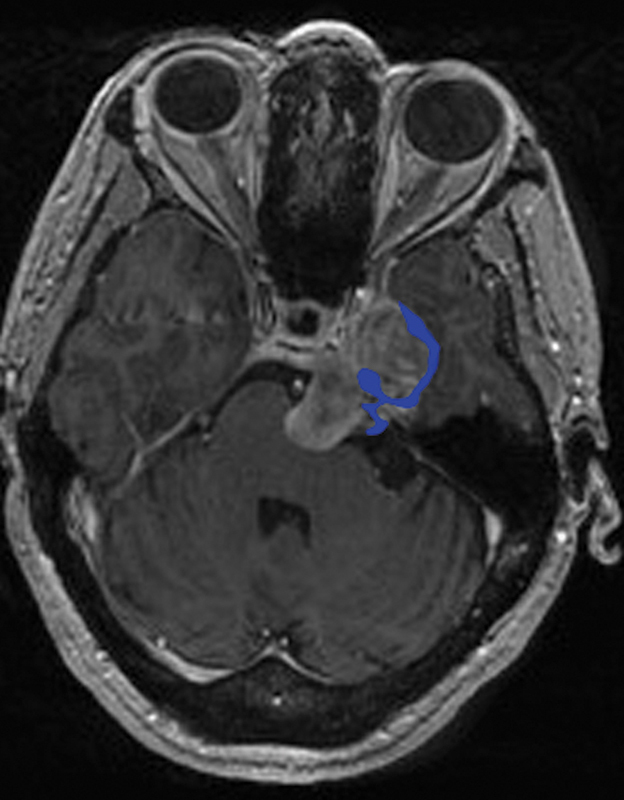
Patient 1 residual tumor (blue) superimposed over the initial lesion as seen on a preoperative T1 with contrast axial magnetic resonance imaging (MRI) scan.
Patient 4 presented with a heterogeneously enhancing lobulated mass centered within the right occipital condyle, lateral petroclival region, and jugular tubercle with lateral extension to the jugular bulb. The 45-degree endoscope allowed us to view what we believed to be the most inferior and lateral portions of the tumor. Tumor removal in this area, especially from within the occipital condyle, was challenging and required us to operate at an acute angle while using specially curved instruments. Postoperative imaging confirmed the difficulty we faced in accessing the far inferior and lateral extent of the resection cavity with residual tumor located adjacent to the jugular bulb, posterior to the carotid canal and within the occipital condyle (Fig. 3).
Fig. 3.
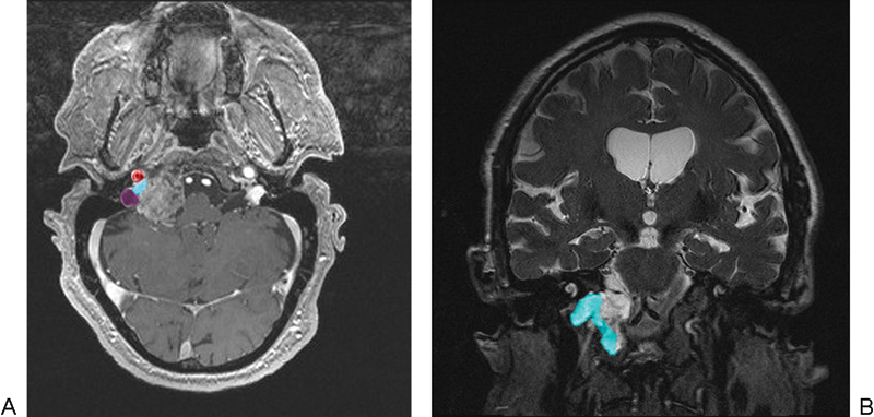
Patient 4 residual tumor (light blue) superimposed over the initial lesion as seen on a preoperative T1 with (A) contrast axial and (B) T2 coronal magnetic resonance imaging (MRI) scan. Location of the petrous internal carotid artery (ICA) (red) and jugular bulb (purple) are demonstrated.
Patient 9 presented with Cushing disease and was found on MRI to have a pituitary macroadenoma with extensive clival invasion. Although we were confident that near GTR had been achieved following surgery, the patient's cortisol level did not normalize. Residual tumor was noted on postoperative imaging to be located in the posterior cavernous sinus with small inferior extension behind the paraclival ICA segment (Fig. 4).
Fig. 4.
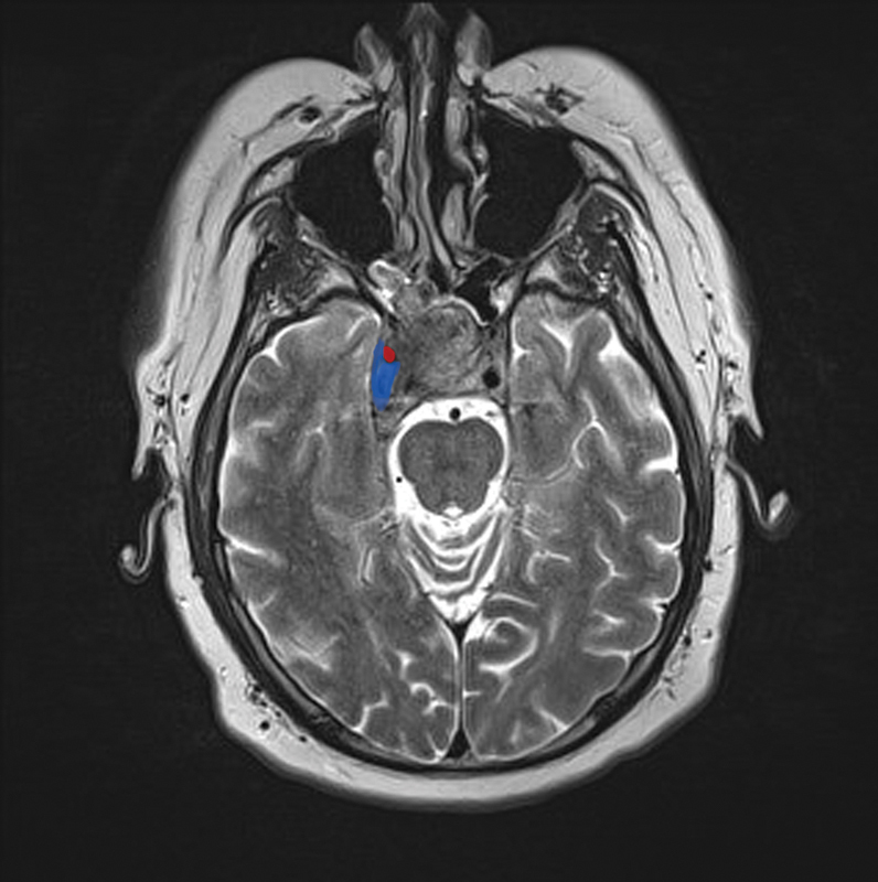
Patient 9 residual tumor (blue) superimposed over the initial lesion as seen on a preoperative T2 axial magnetic resonance imaging (MRI) scan. Location of the internal carotid artery (ICA) (red) is indicated.
Patient 11 was found to have a chordoma extending from the midportion of the clivus superiorly down to the level of the body of the dens. The tumor tissue was densely adherent to the nasopharyngeal fascia and required rather vigorous curetting to remove it. Although we had adequate visualization with angled endoscopes and felt that a GTR had been obtained intraoperatively, the postoperative imaging demonstrated a suspicion of residual tumor at the most inferior part of our resection cavity (Fig. 5).
Fig. 5.
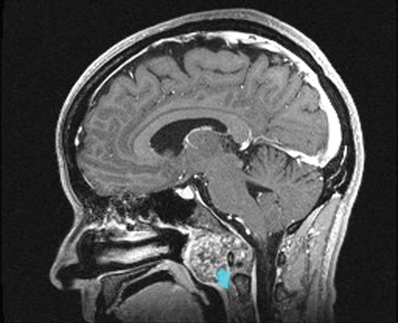
Patient 11 residual tumor (light blue) superimposed over the initial lesion as seen on a preoperative T1 with contrast sagittal magnetic resonance imaging (MRI) scan.
Patient 16 had a large recurrent nonfunctional pituitary adenoma with invasion of the right cavernous sinus. The decision was made intraoperatively not to remove the intracavernous portion of the tumor because of extensive scarring and adherence of the tumor to the ICA. Anatomic limitations were not a factor in the subtotal resection of this tumor.
Discussion
Our case series compares favorably with the established data on clival lesion resections utilizing either a transcranial skull base, microscopic transsphenoidal, or EEEA.12,14,15,16,20,25,26,27,28,29,30 Our experience with clival lesions accessed using the EEEA complements the experience of others in that this approach can be highly effective in removing these surgically challenging lesions. We were able to achieve > 95% resection rate in 81% of the cases for which a GTR was attempted. This rate agrees well with that of Fraser et al,16 who reported an 87% success rate in achieving > 95% resection of clival chordomas with an endoscopic endonasal transclival technique. They cited tumor extension beyond the ICAs as the most important limitation to obtaining a GTR.
To investigate the anatomic and surgical limitations of the EEEA to clival lesions, we performed a detailed radiographic analysis of the postoperative imaging on all of our patients where a GTR was not achieved. We found that residual tumor was most likely to be present with tumor invasion lateral or posterior to the intracavernous and paraclival ICA segments, with lateral tumor extension to the jugular bulb and carotid canal, with tumor at the foramen magnum extending into the occipital condyle, with inferior extension to the lower portion of the dens, with extensive cavernous sinus involvement, and in the presence of intradural tumor invasion with spread lateral to the CNs. These anatomic limitations are demonstrated in Fig. 6. We also found that the average tumor volume in patients who underwent subtotal resection in our series was significantly larger as compared with those where a near-total resection (> 95%) or GTR was achieved. Based on these results along with what has previously been reported in the literature,14,31 we can conclude that the portion of a clival tumor that may be difficult to access with an EEEA can be identified fairly easily on preoperative imaging. Whether or not and to what extent residual tumor is present postoperatively will therefore follow a fairly predictable pattern.
Fig. 6.
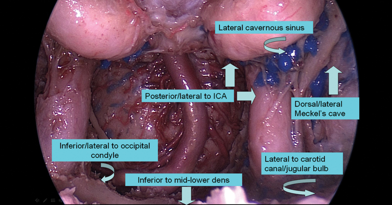
Areas of tumor extension that are associated with anatomic barriers to achieving gross total tumor resection (GTR) when using an expanded endoscopic endonasal approach (EEEA) to clival lesions. ICA, internal carotid artery.
Kassam et al23 and Morera et al31 indicated that when critical neurovascular structures are located primarily ventral and medial to a clival lesion, they require extensive manipulation prior to accessing the tumor and an EEEA in these cases is usually contraindicated. Dehdashti et al14 achieved GTR in 58% of clival chordoma patients using an EEEA. In addition to lateral extension, they mentioned tumor spread to the inferior clivus and occipital condyle as common barriers to complete tumor removal. Carrabba et al20 performed an EEEA on 17 patients with clival lesions. They reported a GTR rate of 59% and a subtotal resection rate, defined as > 80% tumor removal, of 41%. Among the most common reasons they cited for not achieving GTR was in cases of extensive lateral invasion of the lower portion of the tumor. Solares et al15 reported on their experience performing an EEEA to clival lesions. They concluded that the ideal tumors for this approach were those primarily centered in the midline with limited lateral extension. Finally, Kassam et al32 in describing the EEEA to the Meckel cave acknowledge the difficulty accessing tumor on the lateral side of the trigeminal nerve via the endonasal route. This is especially true in patients with normal sensory function when the nerve cannot be sacrificed.
Size also appears to play a role in tumor resectability with the endoscope. According to Stippler et al,25 tumors with diameters > 4 cm had a higher rate of radical resection with an open skull base approach as compared with a transsphenoidal endoscopic technique. Fraser et al16 found initial tumor volumes between 4.1 and 15.9 cm3 in patients who had > 95% tumor resection via an EEEA as opposed to volumes between 80.6 and 124.3 cm3 in patients with < 95% tumor resection. They concluded that tumors with volumes > 80 cm3 may be more suited for an open skull base or combined approach. These findings are not entirely unexpected, as larger tumors tend to have more significant lateral neurovascular invasion. When adherent to these structures, the tumor is unlikely to descend into the operative field of view once the center has been removed. In addition, the borders of larger tumors are often beyond the field of exposure provided by even an angled endoscope. In these cases, aggressive removal while attempting to achieve GTR often results in neurovascular injury. In our series as well as other published cases, longer follow-up data will be necessary to better assess long-term cure rates when using an endoscopic approach.
With regards to other measures of clinical efficacy, 6/8 (75%) patients with preoperative sixth nerve palsies showed at least partial resolution following surgery, and all patients with preoperative facial numbness or visual complaints improved postoperatively.
The EEEA to clival lesions is not without risk. Two patients developed new CN VI palsies, both with pituitary adenomas also involving the cavernous sinus. The partial CN V deficit associated with exploring the Meckel cave may have been prevented with less vigorous resection. Our postoperative CSF leak rate of 19% (3/19 patients) is similar to what has previously been reported for an EEEA to clival lesions,14,20,25 with all occurring primarily early in our experience. These leaks occurred despite the use of a nasoseptal flap. Wide dural openings and a history of prior radiation treatment increased the risk of repair failure.
Conclusion
The last decade has seen a significant change in the surgical management of clival lesions. Refinement of endoscopic surgical techniques and improvements in endoscopic equipment have allowed for clival lesions to be successfully treated via an EEEA with high rates of local tumor control while limiting surgical morbidity. Despite the advantages over traditional skull base approaches, however, the anatomic limitations to the EEEA must be recognized and acknowledged. This will allow surgeons to determine preoperatively which clival lesions are best suited for an endoscopic endonasal route and which may be more appropriate for an open or combined technique. Longer follow-up data will be necessary to more accurately compare the EEEA with traditional microscope-based approaches.
Footnotes
Disclosure The authors have no personal financial or institutional interest in any of the materials, devices, or techniques described in this article. Sources of Funding None. Conflict of Interest None.
References
- 1.al-Mefty O, Borba L A. Skull base chordomas: a management challenge. J Neurosurg. 1997;86(2):182–189. doi: 10.3171/jns.1997.86.2.0182. [DOI] [PubMed] [Google Scholar]
- 2.Tamaki N, Nagashima T, Ehara K, Motooka Y, Barua K K. Surgical approaches and strategies for skull base chordomas. Neurosurg Focus. 2001;10(3):E9. doi: 10.3171/foc.2001.10.3.10. [DOI] [PubMed] [Google Scholar]
- 3.Sekhar L N, Nanda A, Sen C N, Snyderman C N, Janecka I P. The extended frontal approach to tumors of the anterior, middle, and posterior skull base. J Neurosurg. 1992;76(2):198–206. doi: 10.3171/jns.1992.76.2.0198. [DOI] [PubMed] [Google Scholar]
- 4.Sekhar L N, Janecka I P, Jones N F. Subtemporal-infratemporal and basal subfrontal approach to extensive cranial base tumours. Acta Neurochir (Wien) 1988;92(1-4):83–92. doi: 10.1007/BF01401977. [DOI] [PubMed] [Google Scholar]
- 5.House W F, De la Cruz A, Hitselberger W E. Surgery of the skull base: transcochlear approach to the petrous apex and clivus. Otolaryngology. 1978;86(5):ORL-770–ORL-779. doi: 10.1177/019459987808600522. [DOI] [PubMed] [Google Scholar]
- 6.Babu R P, Sekhar L N, Wright D C. Extreme lateral transcondylar approach: technical improvements and lessons learned. J Neurosurg. 1994;81(1):49–59. doi: 10.3171/jns.1994.81.1.0049. [DOI] [PubMed] [Google Scholar]
- 7.Harsh G, Ojemann R, Varvares M, Swearingen B, Cheney M, Joseph M. Pedicled rhinotomy for clival chordomas invaginating the brainstem. Neurosurg Focus. 2001;10(3):E8. doi: 10.3171/foc.2001.10.3.9. [DOI] [PubMed] [Google Scholar]
- 8.Delgado T E, Garrido E, Harwick R D. Labiomandibular, transoral approach to chordomas in the clivus and upper cervical spine. Neurosurgery. 1981;8(6):675–679. doi: 10.1227/00006123-198106000-00007. [DOI] [PubMed] [Google Scholar]
- 9.DeMonte F, Diaz E Jr, Callender D, Suk I. Transmandibular, circumglossal, retropharyngeal approach for chordomas of the clivus and upper cervical spine. Technical note. Neurosurg Focus. 2001;10(3):E10. doi: 10.3171/foc.2001.10.3.11. [DOI] [PubMed] [Google Scholar]
- 10.Stevenson G C, Stoney R J, Perkins R K, Adams J E. A transcervical transclival approach to the ventral surface of the brain stem for removal of a clivus chordoma. J Neurosurg. 1966;24(2):544–551. doi: 10.3171/jns.1966.24.2.0544. [DOI] [PubMed] [Google Scholar]
- 11.Sekhar L N, Sen C, Snyderman C H, Janecka I P. New York: RavenPress; 1993. Anterior, anterolateral, and lateral approaches to extradural petroclival tumors; pp. 157–224. [Google Scholar]
- 12.Laws E R Jr. Transsphenoidal surgery for tumors of the clivus. Otolaryngol Head Neck Surg. 1984;92(1):100–101. doi: 10.1177/019459988409200121. [DOI] [PubMed] [Google Scholar]
- 13.Maira G, Pallini R, Anile C. et al. Surgical treatment of clival chordomas: the transsphenoidal approach revisited. J Neurosurg. 1996;85(5):784–792. doi: 10.3171/jns.1996.85.5.0784. [DOI] [PubMed] [Google Scholar]
- 14.Dehdashti A R Karabatsou K Ganna A Witterick I Gentili F Expanded endoscopic endonasal approach for treatment of clival chordomas: early results in 12 patients Neurosurgery 2008632299–307., discussion 307-309 [DOI] [PubMed] [Google Scholar]
- 15.Solares C A, Fakhri S, Batra P S, Lee J, Lanza D C. Transnasal endoscopic resection of lesions of the clivus: a preliminary report. Laryngoscope. 2005;115(11):1917–1922. doi: 10.1097/01.mlg.0000172070.93173.92. [DOI] [PubMed] [Google Scholar]
- 16.Fraser J F, Nyquist G G, Moore N, Anand V K, Schwartz T H. Endoscopic endonasal transclival resection of chordomas: operative technique, clinical outcome, and review of the literature. J Neurosurg. 2010;112(5):1061–1069. doi: 10.3171/2009.7.JNS081504. [DOI] [PubMed] [Google Scholar]
- 17.Hwang P Y Ho C L Neuronavigation using an image-guided endoscopic transnasal-sphenoethmoidal approach to clival chordomas Neurosurgery 200761502212–217., discussion 217-218 [DOI] [PubMed] [Google Scholar]
- 18.Schwartz T H Fraser J F Brown S Tabaee A Kacker A Anand V K Endoscopic cranial base surgery: classification of operative approaches Neurosurgery 2008625991–1002., discussion 1002-1005 [DOI] [PubMed] [Google Scholar]
- 19.Jho H D, Ha H G. Endoscopic endonasal skull base surgery: Part 3—The clivus and posterior fossa. Minim Invasive Neurosurg. 2004;47(1):16–23. doi: 10.1055/s-2004-818347. [DOI] [PubMed] [Google Scholar]
- 20.Carrabba G, Dehdashti A R, Gentili F. Surgery for clival lesions: open resection versus the expanded endoscopic endonasal approach. Neurosurg Focus. 2008;25(6):E7. doi: 10.3171/FOC.2008.25.12.E7. [DOI] [PubMed] [Google Scholar]
- 21.de Notaris M Cavallo L M Prats-Galino A et al. Endoscopic endonasal transclival approach and retrosigmoid approach to the clival and petroclival regions Neurosurgery 200965(6, Suppl):42–50., discussion 50-52 [DOI] [PubMed] [Google Scholar]
- 22.Cavallo L M, Messina A, Cappabianca P. et al. Endoscopic endonasal surgery of the midline skull base: anatomical study and clinical considerations. Neurosurg Focus. 2005;19(1):E2. [PubMed] [Google Scholar]
- 23.Kassam A B, Gardner P, Snyderman C, Mintz A, Carrau R. Expanded endonasal approach: fully endoscopic, completely transnasal approach to the middle third of the clivus, petrous bone, middle cranial fossa, and infratemporal fossa. Neurosurg Focus. 2005;19(1):E6. [PubMed] [Google Scholar]
- 24.Hadad G, Bassagasteguy L, Carrau R L. et al. A novel reconstructive technique after endoscopic expanded endonasal approaches: vascular pedicle nasoseptal flap. Laryngoscope. 2006;116(10):1882–1886. doi: 10.1097/01.mlg.0000234933.37779.e4. [DOI] [PubMed] [Google Scholar]
- 25.Stippler M Gardner P A Snyderman C H Carrau R L Prevedello D M Kassam A B Endoscopic endonasal approach for clival chordomas Neurosurgery 2009642268–277., discussion 277-278 [DOI] [PubMed] [Google Scholar]
- 26.Frank G Sciarretta V Calbucci F Farneti G Mazzatenta D Pasquini E The endoscopic transnasal transsphenoidal approach for the treatment of cranial base chordomas and chondrosarcomas Neurosurgery 200659101ONS50–ONS57., discussion ONS50-ONS57 [DOI] [PubMed] [Google Scholar]
- 27.Couldwell W T Weiss M H Rabb C Liu J K Apfelbaum R I Fukushima T Variations on the standard transsphenoidal approach to the sellar region, with emphasis on the extended approaches and parasellar approaches: surgical experience in 105 cases Neurosurgery 2004553539–547., discussion 547-550 [DOI] [PubMed] [Google Scholar]
- 28.Cappabianca P, Cavallo L M, Esposito F, De Divitiis O, Messina A, De Divitiis E. Extended endoscopic endonasal approach to the midline skull base: the evolving role of transsphenoidal surgery. Adv Tech Stand Neurosurg. 2008;33:151–199. doi: 10.1007/978-3-211-72283-1_4. [DOI] [PubMed] [Google Scholar]
- 29.Al-Mefty O, Kadri P A, Hasan D M, Isolan G R, Pravdenkova S. Anterior clivectomy: surgical technique and clinical applications. J Neurosurg. 2008;109(5):783–793. doi: 10.3171/JNS/2008/109/11/0783. [DOI] [PubMed] [Google Scholar]
- 30.Sekhar L N, Pranatartiharan R, Chanda A, Wright D C. Chordomas and chondrosarcomas of the skull base: results and complications of surgical management. Neurosurg Focus. 2001;10(3):E2. doi: 10.3171/foc.2001.10.3.3. [DOI] [PubMed] [Google Scholar]
- 31.Morera V A Fernandez-Miranda J C Prevedello D M et al. “Far-medial” expanded endonasal approach to the inferior third of the clivus: the transcondylar and transjugular tubercle approaches Neurosurgery 201066(6, Suppl Operative):211–219., discussion 219-220 [DOI] [PubMed] [Google Scholar]
- 32.Kassam A B Prevedello D M Carrau R L et al. The front door to meckel's cave: an anteromedial corridor via expanded endoscopic endonasal approach- technical considerations and clinical series Neurosurgery 200964:(3, Suppl):ons71–ons82., discussion ons82-ons83 [DOI] [PubMed] [Google Scholar]


