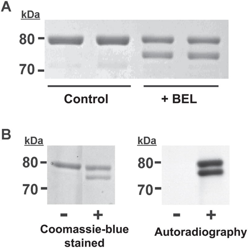
Figure 1. SDS-PAGE autoradiographic analyses of BEL-treated iPLA2 β.

A, Resolution of BEL-treated iPLA2β into two bands by SDS-PAGE. iPLA2β was incubated with racemic BEL at 22°C for 10 min and electrophoresed on a 7% SDS-PAGE gel. The upper band has an apparent molecular weight similar to native iPLA2β (80 kDa) while the lower band has an apparent molecular weight of approximately 75 kDa. B, Radiolabeling of iPLA2β with racemic [3H]-BEL. iPLA2β was incubated with racemic [3H]-BEL at room temperature for 5 min followed by separation on a 7% SDS-PAGE gel stained with Coomassie Blue (left) followed by visualization of the radiolabeled bands by autoradiography (right). Two bands are present in the lane containing [3H]-BEL-treated iPLA2β indicating that both bands are modified by BEL. “−“ indicates protein treated with ethanol vehicle alone; “+” indicates protein treated with [3H]-BEL.
