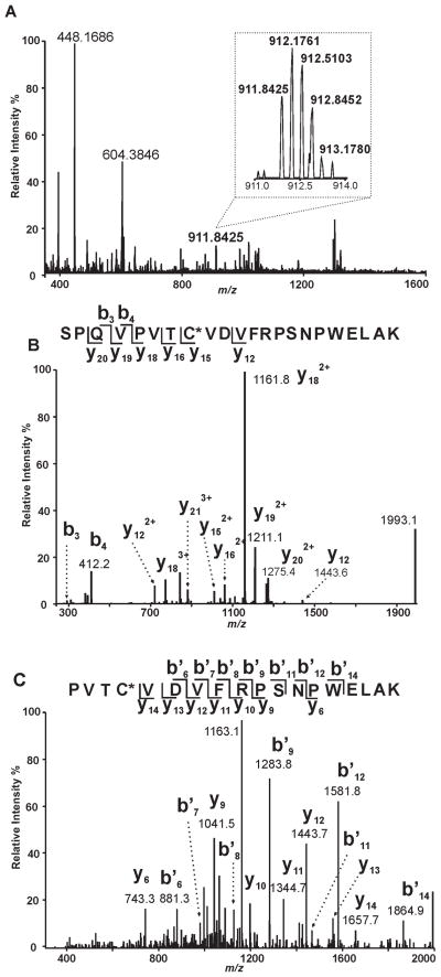
Figure 8. Identification of Covalent Oleoylation of C651 in iPLA2β by Oleoyl-CoA.
A, Full scan mass spectrum (m/z 350 – 1600) at 112.4 min of the LC-MS/MS analysis of the tryptic digest from iPLA2β treated with oleoyl-CoA in vitro. A triply-charged ion of m/z 911.8425 was observed with a resolution power of 30000 at m/z 400. The calculated mass of this ion (2732.5041 Th) matches the mass of the oleoylated 644SPQVPVTCVDVFRPSNPWELAK665 with a mass accuracy of 3 ppm. B, the ion of m/z 911.8425 was selected for CID (collision-induced dissociation) fragmentation. The resulting fragments (y and b ions as labeled) confirm the sequence of the peptide as 644SPQVPVTCVDVFRPSNPWELAK665 and localize the modification site as C651. C, the ion of m/z 1161.8 (the base peak in panel B) from the fragment of 911.8425 was selected for further fragmentation by CID (MS3). The fragmentation pattern of this ion substantiates the identity of the oleoylated peptide and the modification site.

