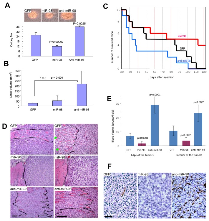
Figure 3. Anti-miR-98 promotes tumor formation, invasion, and angiogenesis.
(A) 4T1 cells (103) were mixed with 0.3% low melting agarose containing 10% FBS and plated on 0.66% agarose-coated 6-well plates. Four weeks after cell inoculation, colonies were examined and photographed. Large colonies and smaller colonies within the cell complexes were counted. Typical large colonies from each group are shown (Lower Panel). (B) The cells were injected subcutaneously into Balb/c regular mice. Tumor growth was monitored. Expression of anti-miR-98 promoted tumor growth. (C) Animal viability was analyzed. (D) Tumors formed by the cells were subjected to H&E staining. Invasion of the tumor cells with stromal muscles (marked by dotted lines) occurred extensively in the anti-miR-98 cells than GFP cells. The miR-98 cells showed little invasive activity. scale bars, 100 μm. (E) The tumor sections were subjected to immunohistochemistry probed with anti-CD34 antibody to detect blood vessels. The number of blood vessels was counted in 10 randomly selected imaging fields and statistical analysis. Expression of miR-98 inhibited blood formation while expression of anti-miR-98 promoted this process. n=10. (F) At higher magnification, large number of vacuoles, sign of unhealthy and dead cells, could be detected in the miR-98 tumor, but not in the other two groups. scale bar, 40 μm.

