NEUROBIOLOGY Correction for “High-resolution structure of hair-cell tip links,” by Bechara Kachar, Marianne Parakkal, Mauricio Kurc, Yi-dong Zhao, and Peter G. Gillespie, which appeared in issue 24, November 21, 2000, of Proc Natl Acad Sci USA (97:13336–13341; 10.1073/pnas.97.24.13336).
The authors note that Figure 3 appeared incorrectly. The corrected figure and its legend appear below. This error does not affect the conclusions of the article.
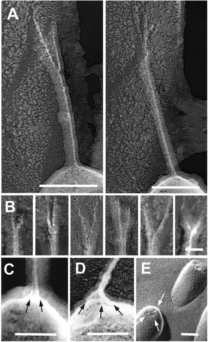
Fig. 3.
Upper and lower attachments of the tip link. (A and B) Freeze-etch images of tip-link upper insertions in guinea pig cochlea (A) and (left to right) two from guinea pig cochlea, two from bullfrog sacculus, and two from guinea pig utriculus (B). Each example shows pronounced branching. (C and D) Freeze-etch images of the tip-link lower insertion in stereocilia from bullfrog sacculus (C) and guinea pig utriculus (D); multiple strands (arrows) arise from the stereociliary tip. (E) Freeze-fracture image of stereociliary tips from bullfrog sacculus; indentations at tips are indicated by arrows. (Scale bars: A = 100 nm, B = 25 nm; C–E = 100 nm.)


