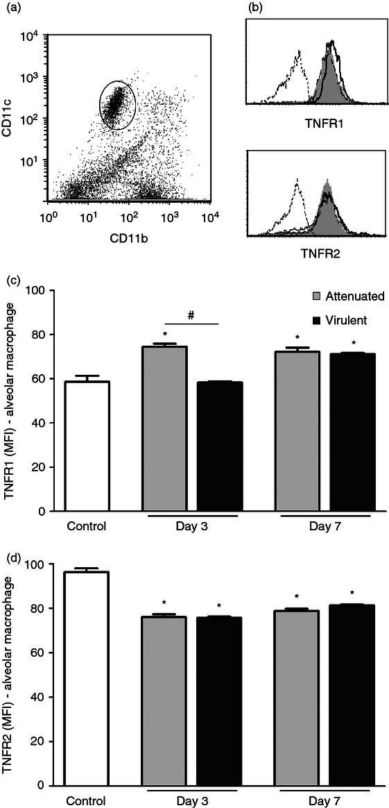
Figure 2.

Surface expression of tumour necrosis factor receptor 1 (TNFR1) and TNFR2 on alveolar macrophages after infection with attenuated and virulent Mycobacterium bovis. C57BL/6 mice were intratracheally infected with attenuated (bacillus Calmette–Guérin Moreau) or virulent (ATCC19274) M. bovis. Control mice were inoculated with PBS. After 3 and 7 days, cells were obtained by bronchoalveolar lavage. Alveolar macrophages were identified as CD11b− CD11c+/high (a) and cell surface expression of TNFR1 and TNFR2 was assessed by FACS, as shown by representative histograms (b). Results of specific staining for TNFR1 (c) and TNFR2 (d) were expressed as the mean channel fluorescence intensity from 10 000 events per sample. Each bar represents mean ± SE of one representative experiment of three with similar results. *P < 0.05 versus control, #P < 0.05.
