Abstract
Currently, the monophyletic lineage of anther smuts on Caryophyllaceae includes 22 species classified in the genus Microbotryum. They are model organisms studied in many disciplines of fungal biology. A molecular phylogenetic approach was used to resolve species boundaries within the caryophyllaceous anther smuts, as species delimitation based solely on phenotypic characters was problematic. Several cryptic species were found amongst the anther smuts on Caryophyllaceae, although some morphologically distinct species were discernible, and most species were characterized by high host-specificity. In this study, anther smut specimens infecting Silene saxifraga were analysed using rDNA sequences (ITS and LSU) and morphology to resolve their specific status and to discuss their phylogenetic position within the lineage of caryophyllaceous anther smuts. The molecular phylogeny revealed that all specimens form a monophyletic lineage that is supported by the morphological trait of reticulate spores with tuberculate interspaces (observed in certain spores). This lineage cannot be attributed to any of the previously described species, and the anther smut on Silene saxifraga is described and illustrated here as a new species, Microbotryum silenes-saxifragae. This species clusters in a clade that includes Microbotryum species, which infect both closely and distantly related host plants growing in diverse ecological habitats. It appears possible that host shifts combined with changes to ecological host niches drove the evolution of Microbotryum species within this clade.
Keywords: Anther smuts, Caryophyllaceae, Microbotryales, Microbotryum violaceum complex, Molecular phylogenetics, Plant pathogens, Pseudo-cryptic species
INTRODUCTION
Plant parasitic fungi sporulating in the anthers of their hosts evolved independently in several genera/species of two major phylogenetic basidiomycetous lineages, including the pucciniomycotinous genera Bauerago (Vánky 1999, 2012) and Microbotryum (Vánky 1998, 2012, Kemler et al. 2006, 2009), and the ustilaginomycotinous genera Antherospora (Bauer et al. 2008, Piątek et al. 2011, 2013), Thecaphora (Roets et al. 2008, 2012, Vánky & Lutz 2007) and Urocystis (Vánky 2012). The anther smuts of Caryophyllaceae, commonly referred to as the Microbotryum violaceum complex, form a monophyletic lineage within the genus Microbotryum. They are model organisms studied in many disciplines of fungal biology, for example, ecology (Thrall et al. 1993), genomics (Hood 2005, Yockteng et al. 2007), population studies (Lee 1981, Alexander & Antonovics 1995, Alexander et al. 1996), life cycle studies (Schäfer et al. 2010), geographic distribution (Hood et al. 2010, Fontaine et al. 2013), phylogeography (Vercken et al. 2010), evolutionary history (López-Villavicencio et al. 2005, Refrégier et al. 2008), speciation (Devier et al. 2010, Gladieux et al. 2011), and phylogeny and systematics (Lutz et al. 2005, 2008, Denchev et al. 2009, Piątek et al. 2012, Kemler et al. 2013).
The assignment of organisms to the appropriate species is critically important in every biological discipline, however challenging as in cases of complexes of morphologically similar species. Delimitation of species within the Microbotryum violaceum complex is a good example where morphology (of spores) alone is inadequate. The vast majority of species and specimens within this complex have reticulate spores of similar size, with only a few exceptions from this general morphological pattern (Vánky 2004, 2012). The oldest available species name for anther smuts on Caryophyllaceae, that is, Microbotryum violaceum (syn. Ustilago violacea), has been usually assigned to morphologically similar specimens on diverse host plants worldwide. However, cross infection experiments (Zillig 1921a, Liro 1924) and molecular analyses (Bucheli et al. 2000, Freeman et al. 2002, Le Gac et al. 2007) have indicated that many of such specimens are biologically and genetically distinct.
In order to break the limitation of phenotype-based species identification, Lutz et al. (2005) provided a robust phylogenetic framework for species delimitation based on the nuclear ribosomal ITS region, which recently was proposed as barcode marker for Fungi (Schoch et al. 2012). ITS well resolves species boundaries in the Microbotryum violaceum complex, and the obtained resolution agrees well with that obtained with other phylogenetic markers (ß-tub, γ-tub, Ef1α, pheromone receptors pr-MatA1, pr-MatA2) (Le Gac et al. 2007, Refrégier et al. 2008, Devier et al. 2010). This phylogenetic framework has subsequently been improved by adding further species and specimens from diverse host plants and incorporating the nuclear LSU rDNA, combined with the ITS, as an additional phylogenetic marker (Lutz et al. 2008, Piątek et al. 2012). The resultant molecular phylogeny and genetic divergences between specimens on different hosts, together with ecological and, if available, morphological data confirm that multiple species are hidden within the Microbotryum violaceum morphotype, with most specific to single host species. ITS and LSU sequences are available for 18 out of 22 recognized Microbotryum species in the anthers of caryophyllaceous plants. One species (Microbotryum savilei) is not sequenced yet, and for three species (M. carthusianorum, M. coronariae, M. dianthorum s. str.) sequences are available for some nuclear DNA regions (ß-tub, γ-tub, Ef1α, ITS, pheromone receptors pr-MatA1, pr-MatA2) (Lutz et al. 2005, Le Gac et al. 2007, Refrégier et al. 2008, Denchev et al. 2009, Devier et al. 2010, Kemler et al. 2013), but not for the LSU. Microbotryum violaceum s. str. is currently restricted to Silene nutans and its taxonomy is stabilized by the sequenced neotype specimen (ITS and LSU) from material collected in Germany (Lutz et al. 2008). It is likely that many undescribed species of anther smuts remain to be discovered amongst the large number of specimens reported from different hosts worldwide, especially considering that anther smuts from 108 different caryophyllaceous hosts listed in recent smut world monograph (Vánky 2012) are still not analysed with molecular methods. The re-collection of fresh materials is desirable since many of herbarium materials are too old for effective isolation of DNA.
The anther smut on Silene saxifraga (incl. S. hayekiana, Tutin et al. 1993) reported from several European countries (Zillig 1921b, Zundel 1953, Scholz & Scholz 1988, Vánky 1994, 2012, Almaraz & Durrieu 1997, Zwetko & Blanz 2004, Lutz & Vánky 2009, as Microbotryum violaceum, M. violaceum s. l. or Ustilago violacea) is a putative distinct species. In the molecular studies of Lutz et al. (2005, 2008), the sequences from two specimens of the anther smut on Silene saxifraga (as S. saxifraga subsp. hayekiana) clustered together in a sister position to the lineage containing Microbotryum silenes-inflatae on Silene maritima and S. vulgaris, and M. aff. violaceum on Lychnis flos-cuculi (as S. flos-cuculi) and S. dioica, the latter smut now referred to as M. coronariae. The limited number of samples was the main reason why the new species for the anther smut on Silene saxifraga was not described at that time. Additionally, only LM morphology was assessed, and SEM studies were not conducted for these two specimens.
The present study aims to resolve the specific status of the anther smut on Silene saxifraga using molecular phylogenetic analyses of concatenated ITS + LSU rDNA sequences as well as light and scanning electron microscope examination of specimens from several populations. A further aim is to discuss the phylogenetic position of the anther smut on Silene saxifraga within the lineage of anther smuts on Caryophyllaceae, and to expand the number of ITS and LSU sequences available for genetic analyses and comparisons.
MATERIALS AND METHODS
Host plant nomenclature, specimen sampling and documentation
In accordance with Tutin et al. (1993), and supported by molecular phylogenetic data (Kemler et al. 2013), the host species names Silene saxifraga and S. hayekiana are accepted as single species Silene saxifraga. All examined host plant specimens were assigned to this species.
This study is based on phylogenetical and/or morphological analyses of specimens of Microbotryum sp. on Silene saxifraga originating from nine populations in three main European high mountain ranges, namely the Alps, the Dinaric Alps, and the Pyrenees. Six specimens were freshly collected in the field, three were received from fungal herbaria, and three were found by screening the sheets with Silene saxifraga (eight sheets labelled as S. hayekiana – none infected, 40 sheets labelled as S. saxifraga – three infected) preserved in the phanerogamic Herbarium of the W. Szafer Institute of Botany, Polish Academy of Sciences, Kraków, Poland (KRAM). Additionally, the LSU and ITS + LSU, respectively, of two specimens of Microbotryum coronariae on Lychnis flos-cuculi were newly sequenced for phylogenetic analyses. The voucher specimens are deposited in KR-M, KRAM, KRAM F, TUB, and H.U.V. (Table 1). The latter abbreviation refers to the personal collection of Kálmán Vánky, “Herbarium Ustilaginales Vánky” currently held at his home (Gabriel-Biel-Straße 5, D-72076 Tübingen, Germany). Nomenclatural novelty was registered in MycoBank (www.MycoBank.org, Crous et al. 2004). The genetype concept follows the proposal of Chakrabarty (2010).
Table 1.List of examined Microbotryum specimens, with host plants, GenBank accession numbers, spore size range, mean spore sizes with standard deviation and reference specimens.
| Species | Host plants | GenBank acc. no. | Spore size range (μm) | Mean spore size with standard deviation (μm) | Reference specimens1 |
|---|---|---|---|---|---|
| Microbotryum coronariae | Lychnis flos-cuculi | ITS: KC684887 LSU: KC684886 |
Not analysed | Not analysed | Germany, Bayern, Allgäu, Ks. Oberallgäu, Oberjoch, Kematsriedmoos, Westteil, ca. 1150 m a.s.l., 25 Jun. 2008, M. Scholler, KR-M-23797 |
| Microbotryum coronariae | Lychnis flos-cuculi | 2ITS: AY877417 LSU: KC684885 | Not analysed | Not analysed | Norway, Kristiansund, Farstad, 14 Aug. 2002, M. Lutz, TUB 012115 |
| Microbotryum silenes-saxifragae | Silene saxifraga | – | 6.5–9.5(−10.5) × 6.0–8.5 | 7.6 ± 0.9 × 7.1 ± 0.6 | Austria, Carinthia, Karawanken, 7 km WSW of Bad Eisenkappel, Trögern valley, 22 Jul. 1962, H. Teppner, KR-M-34470 (Dupla Graecensia Fungorum 237) |
| Microbotryum silenes-saxifragae | Silene saxifraga | ITS: AY588102 LSU: JN000077 |
5.5–8.5(−9.5) × 5.0–6.5(−7.0) | 6.7 ± 0.8 × 6.0 ± 0.5 | Austria, Carinthia, Villach, Finkenstein, Kanzianiberg, 18 Jun. 2003, M. Lutz, TUB 11791 |
| Microbotryum silenes-saxifragae | Silene saxifraga | ITS: JN000073 LSU: JN000079 |
5.0–8.5 × 5.0–8.0 | 6.7 ± 0.8 × 6.2 ± 0.6 | Austria, Carinthia, Villach, Finkenstein, nort of Kanzianiberg, 7 Jul. 2005, M. Lutz, KR-M-23889 |
| Microbotryum silenes-saxifragae | Silene saxifraga | ITS: JN000071 LSU: JN000075 |
5.0–8.0(−9.0) × (4.5−)5.0–7.5 | 6.6 ± 0.8 × 6.1 ± 0.8 | Austria, Carinthia, Villach, Finkenstein, southern part of the Kanzianiberg, near the church, 24 Jun. 2006, M. Lutz, KR-M-23890 – holotype |
| Microbotryum silenes-saxifragae | Silene saxifraga | – | (5.5−)6.0–7.5(−8.5) × 5.0–7.5 | 6.8 ± 0.6 × 6.2 ± 0.6 | France, Central Pyrenees, rocks between Gavarnie village and Cirque de Gavarnie, 11 Jul. 1961, S. Batko, KRAM 1762 |
| Microbotryum silenes-saxifragae | Silene saxifraga | ITS: JN000074 LSU: JN000078 |
5.5–7.5 × 5.0–6.5(−7.0) | 6.4 ± 0.5 × 5.8 ± 0.5 | Germany, Baden-Württemberg, Tübingen, Botanical Garden, cultivated (originating from Slovenia, Bovec, Vas na Skali, 17 Jul. 1994), 11 Jun. 1999, C. Vánky & K. Vánky, H.U.V. 19570 |
| Microbotryum silenes-saxifragae | Silene saxifraga | – | 6.0–7.5 × 5.5–7.5 | 6.7 ± 0.4 × 6.3 ± 0.5 | Germany, Baden-Württemberg, Tübingen, Botanical Garden, cultivated (originating from Slovenia, Bovec, Vas na Skali, 17 Jul. 1994), 24 May 2011, M. Lutz, KRAM F-49439 |
| Microbotryum silenes-saxifragae | Silene saxifraga | – | 6.5–8.5(−9.5) × 6.0–8.0 | 7.4 ± 0.7 × 6.9 ± 0.5 | Italy, Tridentum, Doss Trento, 25 May 1893, Evers, KRAM 1760 |
| Microbotryum silenes-saxifragae | Silene saxifraga | – | 6.0–8.0(−8.5) × 6.0–7.5 | 7.0 ± 0.5 × 6.5 ± 0.5 | Italy, Alpi Maritime, Valle de Gosso, 7 Jun. 1992, M. Schubert, KR-M-23949 |
| Microbotryum silenes-saxifragae | Silene saxifraga | ITS: JN000072 LSU: JN000080 |
6.0–8.5(−9.5) × 5.5–8.5(−9.0) | 7.0 ± 0.8 × 6.5 ± 0.8 | Montenegro, Dinaric Alps, Durmitor Mts, along trail Sedlo-Bobotov Kuk, Surutka valley, 10 Aug. 2009, A. Ronikier & M. Ronikier, KRAM F-49440 |
| Microbotryum silenes-saxifragae | Silene saxifraga | – | 6.5–8.5(−9.5) × (5.5−)6.0–7.5(−9.0) | 7.3 ± 0.7 × 6.8 ± 0.6 | Slovenia, Carniola, “Schibeneggergraben bei Ratschach”, 3 Jun. 1885, J.C. Eques Pittoni a Dannenfeldt, KRAM 108297 |
| Microbotryum silenes-saxifragae | Silene saxifraga | ITS: AY588101 LSU: JN000076 |
5.5–7.5 × (4.5−)5.0–6.5(−7.0) | 6.7 ± 0.5 × 6.2 ± 0.5 | Slovenia, Bovec, Trenta, Juliana Alpine Botanical Garden, cultivated, 7 Aug. 2001, D. Begerow & M. Lutz, TUB 11790 |
1H.U.V. – Herbarium Ustilaginales Vánky, Gabriel-Biel-Str. 5, D-72076 Tübingen, Germany; KR-M – Mycological Herbarium of the Staatliches Museum für Naturkunde Karlsruhe, Germany; KRAM – Phanerogamic Herbarium of the W. Szafer Institute of Botany, Polish Academy of Sciences, Kraków, Poland; KRAM F – Mycological Herbarium of the W. Szafer Institute of Botany, Polish Academy of Sciences, Kraków, Poland; TUB – Herbarium of the Eberhard-Karls-Universität Tübingen, Germany.
2Taken from Lutz et al. (2005).
Morphological examination
Dried fungal spores of the investigated specimens were mounted in lactic acid, heated to boiling point, and then examined under a Nikon Eclipse 80i light microscope at a magnification of ×1000, using Nomarski optics (DIC). Spores were measured using NIS-Elements BR 3.0 imaging software. The extreme measurements were adjusted to the nearest 0.5 μm. The spore size range, mean and standard deviation of 50 spore measurements from each specimen are shown in Table 1. The species description includes combined values from all measured specimens. LM micrographs were taken with a Nikon DS-Fi1 camera. The infected anthers of Silene saxifraga (KRAM F-49440), and the spore ornamentation in specimens from different populations (H.U.V. 19570, KR-M-23890, KRAM 1760, KRAM F-49439, 49440, TUB 11790) were analysed using scanning electron microscopy (SEM). For this purpose, infected anthers and dry spores were mounted on carbon tabs and fixed to an aluminium stub with double-sided transparent tape. The tabs were sputter-coated with carbon using a Cressington sputter-coater and viewed with a Hitachi S-4700 scanning electron microscope, with a working distance of ca. 12 mm. SEM micrographs were taken in the Laboratory of Field Emission Scanning Electron Microscopy and Microanalysis at the Institute of Geological Sciences, Jagiellonian University, Kraków (Poland).
DNA extraction, PCR, and sequencing
The methods of isolation of fungal material, DNA extraction, amplification of the ITS 1 and ITS 2 regions of the rDNA including the 5.8S rDNA (ITS, about 690 bp) and the 5’-end of the nuclear large subunit ribosomal DNA (LSU, about 625 bp), purification of PCR products, sequencing, and processing of the raw data followed Lutz et al. (2004) and Piątek et al. (2012). DNA sequences determined for this study were deposited in GenBank. GenBank accession numbers are given in Table 1 and Fig. 1.
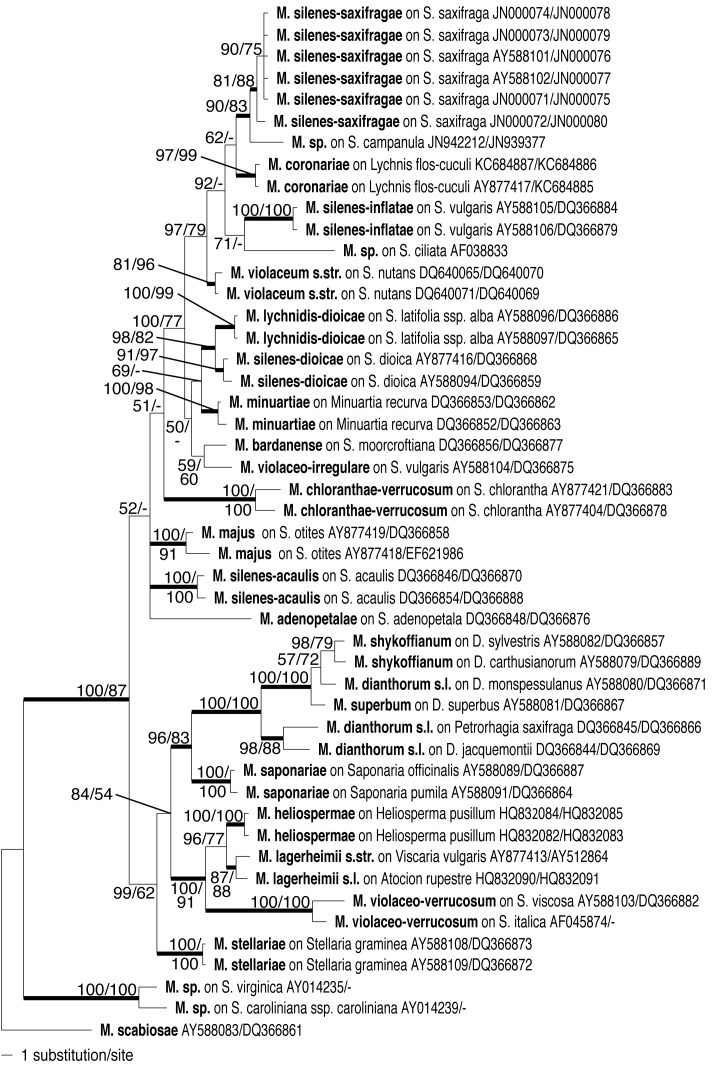
Fig. 1.
Bayesian inference of phylogenetic relationships between the sampled Microbotryum species: Markov chain Monte Carlo analysis of an alignment of concatenated ITS + LSU base sequences using the GTR+I+G model of DNA substitution with gamma distributed substitution rates and estimation of invariant sites, random starting trees and default starting parameters of the DNA substitution model. A 50% majority-rule consensus tree is shown computed from 75 000 trees that were sampled after the process had reached stationarity. The topology was rooted with Microbotryum scabiosae. Bold branches indicate support values higher than 80 in all analyses. Numbers on branches before slashes are estimates for a posteriori probabilities; numbers on branches after slashes are ML bootstrap support values. Branch lengths were averaged over the sampled trees. They are scaled in terms of expected numbers of nucleotide substitutions per site. D. = Dianthus, M. = Microbotryum, S. = Silene.
Phylogenetic analyses
The Microbotryum specimens examined in this study are listed in Table 1. To elucidate the phylogenetic position of the Microbotryum specimens on Silene saxifraga their concatenated ITS + LSU sequences were analysed within a dataset that covered all caryophyllaceous anther smut species of which ITS and LSU sequences were available (Freeman et al. 2002, Lutz et al. 2005, 2008, Hood et al. 2010, Sloan et al. 2008, Piątek et al. 2012) or that were sequenced in the present study (Microbotryum coronariae, Table 1), comprising 19 of the 22 currently recognized species. For the final analysis the dataset was reduced to a maximum of two sequences per species. All sequences available in GenBank that clustered both within the Microbotryum sp. on Silene saxifraga clade and its sister clade (M. coronariae, M. silenes-inflatae) and which could not be assigned to any Microbotryum species (compare Fig. 1) were kept.
Sequence alignment was obtained using MAFFT 6.845b (Katoh et al. 2002, Katoh & Toh 2008) and the L-INS-I option. In the final alignment we retained conserved alignment positions using GBlocks (Castresana 2000) with the following options: ‘Minimum Number of Sequences for a Conserved Position’: 25, ‘Minimum Number of Sequences for a Flank Position’: 25, ‘Maximum Number of Contiguous Non-conserved Positions’: 8, ‘Minimum Length of a Block’: 5 and ‘Allowed Gap Positions’ to ‘With half’.
The resulting alignment [new number of positions: 1276 (57% of the original 2232 positions) variable sites: 212] was used for phylogenetic analyses. Bayesian Analysis (BA) was performed using MrBayes 3.1.2 (Huelsenbeck & Ronquist 2001, Ronquist & Huelsenbeck 2003) applying the same settings as in Piątek et al. (2012). Four incrementally heated chains were run for 10,000,000 generations, sampled every 100th generation, thereby resulting in 100,001 trees of which the first 25,001 sampled trees were discarded. Maximum Likelihood (ML) was performed using RAxML 7.2.6 (Stamatakis 2006) via the raxmlGUI (Silvestro & Michalak 2012). We used the GTRGAMMA and rapid bootstrap option (Stamatakis et al. 2008). Trees were rooted with Microbotryum scabiosae following Kemler et al. (2006).
RESULTS
Morphology
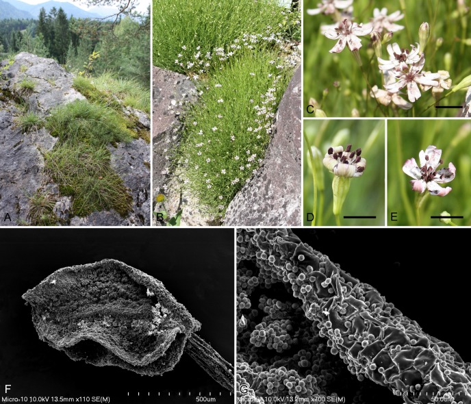
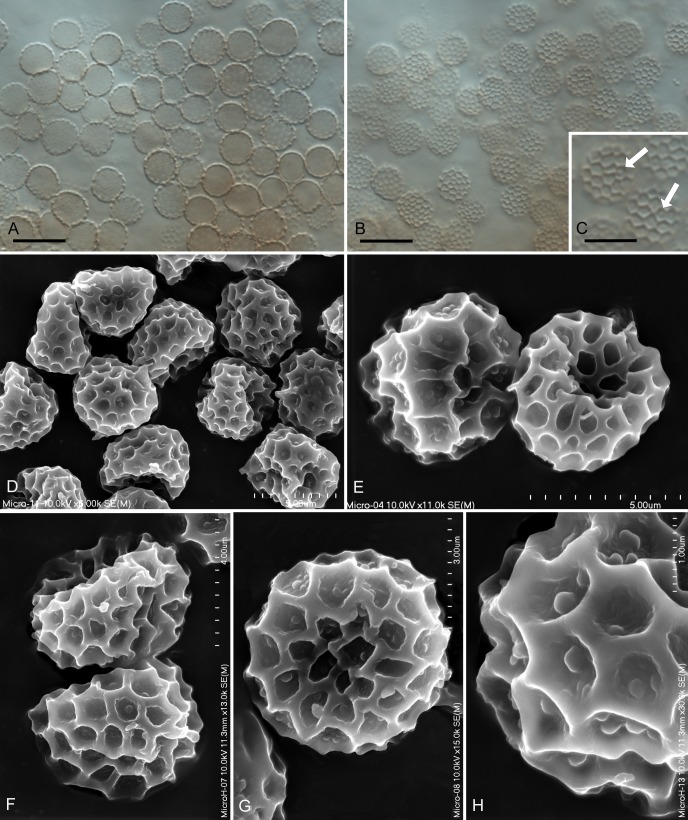
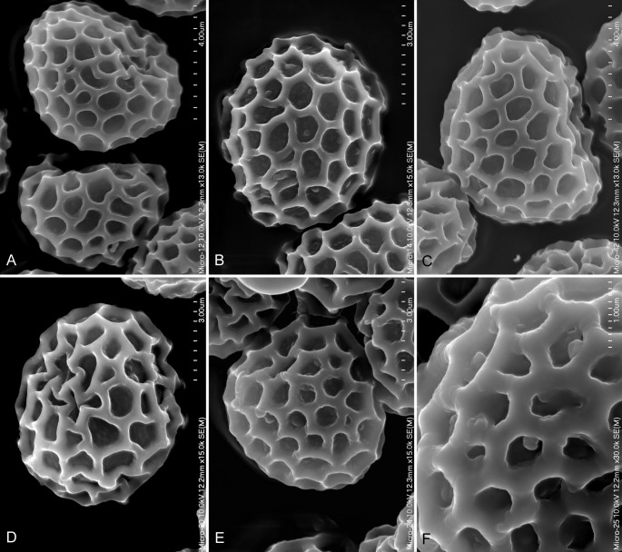
The specimens on Silene saxifraga developed sori in all anthers of an inflorescence and within a plant clump most (but not all) flowers contained anthers with smut spores (Fig. 2). The smut sporulated inside the pollen sacs, which at first were completely covered by the anther’s epidermis and later split longitudinally by the stomia revealing a dark brownish violet, powdery mass of spores. Pollen was not produced by infected anthers (Fig. 2). The spores in all specimens were reticulate under the light microscope, regular in shape and uniform in size within each collection, and highly uniform in shape, size range and average size between different collections (Fig. 3, Table 1). In scanning electron microscope, the spores were reticulate with variably ornamented interspaces. The interspaces usually ranged from almost smooth to rough or verruculose, but certain spores had more or less well-developed tuberculate warts on the interspaces or lower parts of the muri (Figs 3–4). The spores with tuberculate warts constituted a small but regular fraction of spores. Tuberculate warts were most apparent in the material from Kanzianiberg (Austria) that is designated here as holotype of the new species.
Fig. 2.
Microbotryum silenes-saxifragae sp. nov. on Silene saxifraga. A. The type locality at the Kanzianiberg, Austria. B. The clump of Silene saxifraga with infected flowers in the Botanical Garden of Tübingen, Germany. C–E. Infected inflorescences, with the fungus sporulating in the anthers in the Botanical Garden of Tübingen, Germany. F. Infected anther: an open pollen sac filled with teliospores seen at the foreground, made in SEM (KRAM F-49440). G. Teliospores inside the pollen sac and the anther’s epidermis, seen by SEM. (KRAM F-49440). Bars: C–E = 5 mm, F = 500 μm, G = 50 μm.
Fig. 3.
Microbotryum silenes-saxifragae sp. nov. on Silene saxifraga (KR-M-23890 – holotype). A–B. Spores seen by LM, median and superficial views. C. Hardly visible tubercles in LM at very high magnification using Nomarski optics, indicated by arrows. D–G. Spores with tuberculate, rough and verruculose interspaces seen by SEM. H. Close-up of spore ornamentation seen by SEM. Bars: A–B = 10 μm, C–E = 5 μm, F = 4 μm, G = 3 μm, H = 1 μm.
Fig. 4.
Variability of interspaces ornamentation in different spores and specimens of Microbotryum silenes-saxifragae sp. nov. seen by SEM. A–B. From KRAM F-49439. C–D. From KRAM F-49440. E–F. From KRAM 1760. Bars: A, C = 4 μm, B, D–E = 3 μm, F = 1 μm.
Phylogenetic analyses
For both the ITS and LSU, the sequences of the Silene saxifraga anther smut specimen from Montenegro (KRAM F-49440) differed in one position from the remaining sequences, which were identical among each other.
The different runs of the BA that were performed and the ML analyses yielded consistent topologies. To illustrate the results, the consensus tree of one run of the BA is presented (Fig. 1).
In all analyses, the known species were inferred with high support values except for Microbotryum dianthorum s. l., for which the sequences clustered in two different lineages. With high to moderate support in all analyses the sequences of anther smut specimens on Silene saxifraga clustered together, forming the sister lineage to Microbotryum sp. on S. campanula. That clade formed a monophylum with M. coronariae, M. silenes-inflatae, M. sp. on S. ciliata, and M. violaceum s. str. However the phylogenetic relations between those taxa received only low support values. Within the cluster of anther smut on Silene saxifraga, the specimen from Montenegro (KRAM F-49440) was revealed in a sister position to the remaining S. saxifraga anther smut specimens, which were identical among each other.
Considering groups that received considerable support in all analyses the phylogenetic relationships between the species inferred here were in contrast to the results discussed by Piątek et al. (2012) in two aspects: Microbotryum violaceo-verrucosum clustered as sister taxon to M. heliospermae and M. lagerheimii, and M. saponariae was revealed as sister taxon to the Dianthus and Petrorhagia anther smuts.
TAXONOMY
Microbotryum silenes-saxifragae M. Lutz, M. Piątek & Kemler, sp. nov.
MycoBank MB800823
Etymology: The name of the species refers to the host plant species, Silene saxifraga.
Description: Parasitic on Silene saxifraga. Sori in anthers; all anthers of the inflorescence infected, and most flowers in a clump contain smut spores; spore mass powdery, dark brownish violet. Spores pale violaceous or violaceous in transmitted light, regular in shape and size, globose, subglobose, broadly ellipsoidal, or rarely ovoid, 5.0–8.5(−10.5) × (4.5−)5.0–7.5(−9.0) μm; wall reticulate, ca. 0.5–0.7 μm high, meshes more or less polyhedral, usually irregular, rarely regular, 5–8 (usually 6–7) meshes per spore diameter, interspaces usually smooth as observed by LM (sometimes weak tubercles are visible at very high magnification using Nomarski optics), almost smooth, rough, verruculose or tuberculate as observed by SEM.
Type: Austria: Carinthia: Villach, Finkenstein, southern part of the Kanzianiberg, near the church, [630 m a.s.l.], on Silene saxifraga, 24 June 2006, M. Lutz (KR-M-23890 – holotype). The ITS/LSU hologenetype sequences are deposited in GenBank as JN000071/JN000075, respectively.
Additional specimens examined (paratypes): Austria: Carinthia: Karawanken, 7 km WSW of Bad Eisenkappel, Trögern valley, 700–800 m a.s.l., on Silene saxifraga, 22 July 1962, H. Teppner (KR-M-34470, Dupla Graecensia Fungorum 237); Villach, Finkenstein, Kanzianiberg, [630 m a.s.l.], on Silene saxifraga, 18 June 2003, M. Lutz (TUB 11791); Villach, Finkenstein, north of Kanzianiberg, [630 m a.s.l.], on Silene saxifraga, 7 July 2005, M. Lutz (KR-M-23889); France: Central Pyrenees: rocks between Gavarnie village and Cirque de Gavarnie, [between 1400 and 1600 m a.s.l.], on Silene saxifraga, 11 July 1961, S. Batko (KRAM 1762). – Germany: Baden-Württemberg: Tübingen, Botanical Garden (originating from Slovenia, Bovec, Vas na Skali, 17 July 1994), [440 m a.s.l.], on cultivated Silene saxifraga, 11 June 1999, C. Vánky & K. Vánky (H.U.V. 19570); Tübingen, Botanical Garden (originating from Slovenia, Bovec, Vas na Skali, 17 July 1994), [440 m a.s.l.], on cultivated Silene saxifraga, 24 May 2011, M. Lutz (KRAM F-49439). – Italy: Tridentum: Doss Trento, [300 m a.s.l.], on Silene saxifraga, 25 May 1893, Evers (KRAM 1760); Alpi Maritime: Valle de Gosso, [? – site not located in Google Earth], on Silene saxifraga, 7 June 1992, M. Schubert (KR-M-23949). – Montenegro: Dinaric Alps: Durmitor Mts, along trail Sedlo-Bobotov Kuk, Surutka valley, ca. 2090 m a.s.l., on Silene saxifraga, 10 August 2009, A. Ronikier & M. Ronikier (KRAM F-49440). – Slovenia: Carniola: “Schibeneggergraben bei Ratschach”, [Ratschach - 870 m a.s.l., Schibeneggergraben not located in Google Earth], on Silene saxifraga, 3 June 1885, J.C. Eques Pittoni a Dannenfeldt (KRAM 108297); Bovec, Trenta, Juliana Alpine Botanical Garden, [600 m a.s.l.], on cultivated Silene saxifraga, 7 August 2001, D. Begerow & M. Lutz (TUB 11790).
Host range and distribution: On Silene saxifraga (Caryophyllaceae subfam. Silenoideae); Europe (Austria, France, Germany, Italy, Montenegro, Slovenia). The localities in Austria, Italy and Slovenia are placed in the Alps, the locality in France in the Pyrenees and the locality in Montenegro in the Dinaric Alps. The locality in Germany is artificial as plants were cultivated.
Ecology: The infected plants were found from May to August in the natural localities, in May and June cultivated in the Botanical Garden in Tübingen (Germany), and in August cultivated in the Juliana Alpine Botanical Garden in Trenta (Slovenia). At the type locality, the Kanzianiberg (Austria), large populations of infected plants were observed between 2003 and 2006 in different places on the limestone hill. In other localities for which data on the habitat are available, Microbotryum silenes-saxifragae occurred on plants growing on rocks, together with Rhamnus pumila (France: between Gavarnie and Cirque de Gavarnie), between limestone rocks (Austria: Karawanken) and on grassland (Montenegro: Surutka valley). Altitude data were available for few of the examined specimens, and the approximate altitude data (included in square brackets in the list of specimens examined) for most of the remaining specimens were obtained through Google Earth (google.earth.com). The lowest locality was recorded at 300 m a.s.l., and the highest locality at 2090 m a.s.l., which indicates that Microbotryum silenes-saxifragae is a submontane-montane species with wide altitudinal amplitude.
DISCUSSION
Silene saxifraga is widely distributed on rocks and screes in southern European mountains, extending northwards to West Austria (Tutin et al. 1993). The anther smut of Silene saxifraga, although reported in the literature and referred to the catch-all name Microbotryum violaceum (syn. Ustilago violacea), is poorly known and has never been critically examined using samples from different populations. In this study, molecular phylogenetic analyses and morphological data were used to resolve the systematic position of the anther smut on Silene saxifraga.
The phylogenetic analyses of the concatenated ITS + LSU dataset showed that the analysed specimens on Silene saxifraga from three geographically distinct populations (Austria, Slovenia, and Montenegro; the German specimen derived from a Slovenian population) form a well supported independent evolutionary lineage of caryophyllaceous anther smuts. The genetic divergence of the lineage is comparable to genetic distances between the remaining anther smuts on caryophyllaceous hosts (Lutz et al. 2005, 2008, Piątek et al. 2012) or between Microbotryum species on non-caryophyllaceous hosts (Kemler et al. 2006, 2009). Within this lineage, the anther smut specimen on Silene saxifraga from Montenegro (Dinaric Alps) revealed some sequence divergence in comparison to the specimens from populations in Austria and Slovenia (Alps). A similar phylogeographic split between specimen from the Alps and specimens from the Carpathians and the Dinaric Alps has been observed in another montane species, namely Microbotryum heliospermae, a parasite on Heliosperma pusillum (Piątek et al. 2012). This is congruent with a similar phylogeographical pattern observed in several vascular plant species (Ronikier 2011). It may indicate long-term separation and different evolutionary histories of populations in the Alps and the Dinaric Alps (and South-East European mountains in general).
The closest phylogenetic relative of the analysed anther smut specimens on Silene saxifraga may be the anther smut on Silene campanula assigned by Schoch et al. (2012) to Microbotryum violaceum1. However, it does not belong to this species, which is restricted to Silene nutans. According to our molecular phylogenetic analyses, the anther smut on Silene campanula occupies a sister position to the anther smut on Silene saxifraga, well separated from Microbotryum violaceum s. str. (incl. neogenetype ITS/LSU sequences DQ640065/640070, Lutz et al. 2008).
The morphological examination of the anther smut specimens on Silene saxifraga revealed some characteristics, such as a reticulate ornamentation of spores, spore shape and size, that are shared with other Microbotryum species in anthers of caryophyllaceous hosts (Lutz et al. 2005, 2008, Denchev et al. 2009, Piątek et al. 2012, Vánky 2012). However, in contrast to other species of the caryophyllaceous anther smuts lineage, the interspaces in spores (= bottom of the muri) were variably ornamented from almost smooth to rough, verruculose or, in some spores, distinctly tuberculate. This feature is almost invisible in LM (sometimes weak tubercles are visible at very high magnification using Nomarski optics), but visible in SEM. The reason for morphological variation in different specimens is uncertain, but it might be related to different developmental stages or environmental conditions.
In conclusion, the genetic divergence, the host plant and the distinct spore morphology indicate that the anther smut on Silene saxifraga represents a new species that is described in this work as Microbotryum silenes-saxifragae. The genetic divergence and only one sequenced specimen of the anther smut on Silene campanula do not allow the possible assignment of this specimen to Microbotryum silenes-saxifragae.
The distinct morphological trait of tuberculate interspaces observed in a certain portion of reticulate spores of Microbotryum silenes-saxifragae is unique in the lineage of caryophyllaceous anther smuts. Most species of this clade have spores with reticulate ornamentation but with smooth interspaces (Lutz et al. 2005, 2008, Piątek et al. 2012, Vánky 2012), exceptionally rough or at most verruculose interspaces (Microbotryum adenopetalae, Lutz et al. 2008, M. carthusianorum, M. shykoffianum, Denchev et al. 2009). There are only four evident exceptions from this general pattern, namely two species have verrucose spores (Microbotryum chloranthae-verrucosum, M. violaceo-verrucosum, Lutz et al. 2005, Vánky 2012) and two species have irregularly verrucose-reticulate spores (M. bardanense, M. violaceo-irregulare, Deml & Oberwinkler 1983, Chlebicki & Suková 2005, Vánky 2012). Microbotryum silenes-saxifragae adds a new type of ornamentation to the lineage of caryophyllaceous anther smuts.
The development of reticulate spores having tuberculate interspaces is a common feature in other Microbotryum species, and it is especially widespread in many species infecting flowers of different plants from the family Polygonaceae that cluster in Microbotryum group II resolved in the phylogenetic study of Kemler et al. (2006). The reticulate spores with tuberculate interspaces are also produced by some anther smuts on non-caryophyllaceous hosts (Vánky 2012) and are developed in some Microbotryum species sporulating in the ovaries of caryophyllaceous plants (Piątek 2005, Vánky 2012). Most of these species cluster in Microbotryum group II (Kemler et al. 2006), though not for all of them sequence data are available. It seems that the feature of reticulate spores with tuberculate interspaces is a homoplasy that evolved convergently in different Microbotryum species.
Microbotryum silenes-saxifragae is embedded in a well supported clade composed of three other described species, M. coronariae on Lychnis flos-cuculi, M. silenes-inflatae on S. vulgaris and M. violaceum s. str. on Silene nutans, as well as the unresolved Microbotryum sp. on Silene campanula and Microbotryum sp. on S. ciliata. In accordance with the studies of Lutz et al. (2005, 2008), where only two sequences of anther smut on Silene saxifraga were available, Microbotryum violaceum s. str. is revealed as sister taxon to all other clades of this group. All other phylogenetic relations received only moderate to low support with one exception, the sister relation of Microbotryum sp. on Silene campanula and M. silenes-saxifragae. The host plant phylogenetic relations and ecology indicate a complex evolutionary history within this clade of caryophyllaceous anther smuts. While Silene campanula, S. nutans and S. saxifraga are phylogenetically closely related (Greenberg & Donoghue 2011, Kemler et al. 2013), S. ciliata and especially Lychnis flos-cuculi and Silene vulgaris have distant phylogenetic relations (Greenberg & Donoghue 2011, Kemler et al. 2013). Moreover, the host plants ocuppy different ecological niches, Silene campanula, S. ciliata, S. nutans and S. saxifraga occur on calcareous rocks, mostly in mountains, Lychnis flos-cuculi on wet meadows, in mountains and lowlands, and S. vulgaris on dry meadows or disturbed places (roadsides, fields) in mountains and lowlands. It appears possible that the evolution of Microbotryum species within this clade was driven by host shifts combined with the changes in the ecological niches of the hosts. Host shifts, both evolutionary ancient (Refrégier et al. 2008) or recent (Antonovics et al. 2002, López-Villavicencio et al. 2005, Kummer 2010), were often reported in caryophyllaceous anther smuts. Furthermore, artificial cross-inoculation experiments indicate that the potential host range of some Microbotryum species is larger than the actual host range observed in natural populations, but the ability to infect non-host plants is higher for plants that are phylogenetically closer related to the original host (de Vienne et al. 2009). This phenomenon could promote higher speciation rates in this group of plant parasites. In this respect, the clade warrants further study with increased sampling of anther smut specimens, especially on other hosts, likely to reveal other closely related species.
The lineage of caryophyllaceous anther smuts is predominantly composed of cryptic species, with only a few species differing morphologically (Lutz et al. 2005, 2008). In some species (Microbotryum adenopetalae, M. heliospermae, M. minuartiae) subtle morphological features were found after their initial detection by molecular methods (Lutz et al. 2008, Piątek et al. 2012). In the absence of molecular support or cross infection experiments, these features could easily be considered as phenotypic variation within a single species. In contrast, the tuberculate interspaces observed in a certain portion of reticulate spores of Microbotryum silenes-saxifragae are rather evident, although not previously noticed (probably due to the absence of SEM studies of anther smut specimens on Silene saxifraga). Species that have only been recognized as morphologically distinct after application of methods other than comparative morphology, usually methods of molecular biology, are called pseudo-cryptic species in different studies on systematically diverse organisms (Knowlton 1993, Amato & Montresor 2008, Luttikhuizen & Dekker 2010, Medina et al. 2012). The species mentioned in this paragraph, including Microbotryum silenes-saxifragae, match that concept very well.
The discovery of morphological species showing genetic differences that are comparable with genetic divergences between cryptic species embedded within the lineage of caryophyllaceous anther smuts supports the narrow species concept for this group of smut fungi advocated by Liro (1924) and confirmed by recent molecular studies (Lutz et al. 2005, 2008, Le Gac et al. 2007, Refrégier et al. 2008, Devier et al. 2010, Piątek et al. 2012). Furthermore, the strict correlation of Microbotryum silenes-saxifragae with its host species confirms the earlier conclusion that host-specific species delimitation might reflect the evolution of many anther smut parasites best (Lutz et al. 2005). It is likely that the uncharted species diversity of anther smuts is much higher and that, in addition to cryptic species, also pseudo-cryptic species still could be detected as forming part of caryophyllaceous anther smuts lineage.
Acknowledgments
We thank Michael Weiß, Sigisfredo Garnica, and Robert Bauer (Tübingen, Germany) for providing facilities for molecular analyses, Günther Deml (Braunschweig, Germany) for sending rare literature, the Curators of herbaria: Markus Scholler (KR-M), and Kálmán Vánky (H.U.V.) for loan of specimens, Anna and Michał Ronikier (Kraków, Poland) for collecting material, and Anna Łatkiewicz (Kraków, Poland) for her help with the SEM pictures.
Footnotes
1see: http://www.fungalbarcoding.org/BioloMICS.aspx?Table=Fungal%20barcodes&Fields=All&Rec=2110 – data retrieved on 1 March 2013; the specimen is deposited in private herbarium and it was not possible to re-examine it during the course of this study.
REFERENCES
- Alexander HM, Antonovics J. (1995) Spread of anther-smut disease (Ustilago violacea) and character correlations in a genetically variable experimental population of Silene alba. Journal of Ecology 83: 783–794 [Google Scholar]
- Alexander HM, Thrall PH, Antonovics J, Jarosz AM, Oudemans PV. (1996) Population dynamics and genetics of plant disease: A case study of anther-smut disease. Ecology 77: 990–996 [Google Scholar]
- Almaraz T, Durrieu G. (1997) Ustilaginales from the Spanish Pyrenees and Andorra. Mycotaxon 65: 223–236 [Google Scholar]
- Amato A, Montresor M. (2008) Morphology, phylogeny, and sexual cycle of Pseudo-nitzschia mannii sp. nov. (Bacillariophyceae): a pseudo-cryptic species within the P. pseudodelicatissima complex. Phycologia 47: 487–497 [Google Scholar]
- Antonovics J, Hood ME, Partain JL. (2002) The ecology and genetics of a host shift: Microbotryum as a model system. The American Naturalist 160: S40–S53 [DOI] [PubMed] [Google Scholar]
- Bauer R, Lutz M, Begerow D, Piątek M, Vánky K, Bacigálová K, Oberwinkler F. (2008) Anther smut fungi on monocots. Mycological Research 112: 1297–1306 [DOI] [PubMed] [Google Scholar]
- Bucheli E, Gautschi B, Shykoff JA. (2000) Host-specific differentiation in the anther smut fungus Microbotryum violaceum as revealed by microsatellites. Journal of Evolutionary Biology 13: 188–198 [Google Scholar]
- Castresana J. (2000) Selection of conserved blocks from multiple alignments for their use in phylogenetic analysis. Molecular Biology and Evolution 17: 540–552 [DOI] [PubMed] [Google Scholar]
- Chakrabarty P. (2010) Genetypes: a concept to help integrate molecular phylogenetics and taxonomy. Zootaxa 2632: 67–68 [Google Scholar]
- Chlebicki A, Suková M. (2005) Two Microbotryum species from the Himalayas. Mycotaxon 93: 149–154 [Google Scholar]
- Crous PW, Gams W, Stalpers JA, Robert V, Stegehuis G. (2004) MycoBank: an online initiative to launch mycology into the 21st century. Studies in Mycology 50: 19–22 [Google Scholar]
- De Vienne DM, Hood ME, Giraud T.(2009) Phylogenetic determinants of potential host shifts in fungal pathogens. Journal of Evolutionary Biology 22: 2532–2541 [DOI] [PubMed] [Google Scholar]
- Deml G, Oberwinkler F. (1983) Studies in Heterobasidiomycetes, Part 31. On the anther smuts of Silene vulgaris (Moench) Garcke. Phytopathologische Zeitschrift 108: 61–70 [Google Scholar]
- Denchev CM, Giraud T, Hood ME. (2009) Three new species of anthericolous smut fungi on Caryophyllaceae. Mycologia Balcanica 6: 79–84 [Google Scholar]
- Devier B, Aguileta G, Hood ME, Giraud T. (2010) Using phylogenies of pheromone receptor genes in the Microbotryum violaceum species complex to investigate possible speciation by hybridization. Mycologia 102: 689–696 [DOI] [PubMed] [Google Scholar]
- Fontaine MC, Gladieux P, Hood ME, Giraud T. (2013) History of the invasion of the anther smut pathogen on Silene latifolia in North America. New Phytologist (in press). [DOI] [PubMed] [Google Scholar]
- Freeman AB, Doung KK, Shi T, Hughes CF, Perlin MH. (2002) Isolates of Microbotryum violaceum from North American host species are phylogenetically distinct from their European host-derived counterparts. Molecular Phylogenetics and Evolution 23: 158–170 [DOI] [PubMed] [Google Scholar]
- Gladieux P, Vercken E, Fontaine MC, Hood ME, Jonot O, Couloux A, Giraud T.(2011)Maintenance of fungal pathogen species that are specialized to different hosts: allopatric divergence and introgression through secondary contact. Molecular Biology and Evolution 28: 459–471 [DOI] [PubMed] [Google Scholar]
- Greenberg AK, Donoghue MJ. (2011) Molecular systematics and character evolution in Caryophyllaceae. Taxon 60: 1637–1652 [Google Scholar]
- Hood ME. (2005) Repetitive DNA in the automictic fungus Microbotryum violaceum. Genetica 124: 1–10 [DOI] [PubMed] [Google Scholar]
- Hood ME, Mena-Alí JI, Gibson AK, Oxelman B, Giraud T, Yockteng R, Arroyo MTK, Conti F, Pedersen AB, Gladieux P, Antonovics J. (2010) Distribution of the anther-smut pathogen Microbotryum on species of the Caryophyllaceae. New Phytologist 187: 217–229 [DOI] [PMC free article] [PubMed] [Google Scholar]
- Huelsenbeck JP, Ronquist F. (2001) MRBAYES: Bayesian inference of phylogenetic trees. Bioinformatics 17: 754–755 [DOI] [PubMed] [Google Scholar]
- Katoh K, Misawa K, Kuma K, Miyata T. (2002) MAFFT: a novel method for rapid multiple sequence alignment based on fast Fourier transform. Nucleic Acids Research 30: 3059–3066 [DOI] [PMC free article] [PubMed] [Google Scholar]
- Katoh K, Toh H. (2008) Recent developments in the MAFFT multiple sequence alignment program. Briefings in Bioinformatics 9: 286–298 [DOI] [PubMed] [Google Scholar]
- Kemler M, Göker M, Oberwinkler F, Begerow D. (2006) Implications of molecular characters for phylogeny of the Microbotryaceae (Basidiomycota: Urediniomycetes). BMC Evolutionary Biology 6: 35 [DOI] [PMC free article] [PubMed] [Google Scholar]
- Kemler M, Lutz M, Göker M, Oberwinkler F, Begerow D. (2009) Hidden diversity in the non-caryophyllaceous plant-parasitic members of Microbotryum (Pucciniomycotina: Microbotryales). Systematics and Biodiversity 7: 297–306 [Google Scholar]
- Kemler M, Martín MP, Tellería MT, Schäfer AM, Yurkov A, Begerow D. (2013) Contrasting phylogenetic patterns of anther smuts (Pucciniomycotina: Microbotryum) reflect phylogenetic patterns of their caryophyllaceous hosts. Organisms Diversity and Evolution (in press). [Google Scholar]
- Knowlton N. (1993) Sibling species in the sea. Annual Review of Ecology and Systematics 24: 189–216 [Google Scholar]
- Kummer V. (2010) Bemerkenswerte Pilzfunde auf der 38. Brandenburgischen Botanikertagung in Groß Pinnow/Uckermark 2007. Verhandlungen des Botanischen Vereins von Berlin und Brandenburg 142: 223–245 [Google Scholar]
- Le Gac M, Hood ME, Fournier E, Giraud T. (2007) Phylogenetic evidence of host-specific cryptic species in the anther smut fungus. Evolution 61: 15–26 [DOI] [PubMed] [Google Scholar]
- Lee JA. (1981) Variation in the infection of Silene dioica (L.) Clairv. by Ustilago violacea (Pers.) Fuckel in North West England. New Phytologist 87: 81–99 [Google Scholar]
- Liro JI. (1924) Die Ustilagineen Finnlands I. Annales Academiae Scientiarum Fennicae, Series A 17: 1–636 [Google Scholar]
- López-Villavicencio M, Enjalbert J, Hood ME, Shykoff JA, Raquin C, Giraud T. (2005) The anther smut disease on Gypsophila repens: a case of parasite sub-optimal performance following a recent shift? Journal of Evolutionary Biology 18: 1293–1303 [DOI] [PubMed] [Google Scholar]
- Luttikhuizen PC, Dekker R. (2010) Pseudo-cryptic species Arenicola defodiens and Arenicola marina (Polychaeta: Arenicolidae) in Wadden Sea, North Sea and Skagerrak: Morphological and molecular variation. Journal of Sea Research 63: 17–23 [Google Scholar]
- Lutz M, Bauer R, Begerow D, Oberwinkler F, Triebel D. (2004) Tuberculina, rust relatives attack rusts. Mycologia 96: 614–626 [PubMed] [Google Scholar]
- Lutz M, Göker M, Piątek M, Kemler M, Begerow D, Oberwinkler F. (2005) Anther smuts of Caryophyllaceae: molecular characters indicate host-dependent species delimitation. Mycological Progress 4: 225–238 [Google Scholar]
- Lutz M, Piątek M, Kemler M, Chlebicki A, Oberwinkler F. (2008) Anther smuts of Caryophyllaceae: molecular analyses reveal further new species. Mycological Research 112: 1280–1296 [DOI] [PubMed] [Google Scholar]
- Lutz M, Vánky K. (2009) An annotated checklist of smut fungi (Basidiomycota: Ustilaginomycotina and Microbotryales) in Slovenia. Lidia 7: 33–78 [Google Scholar]
- Medina R, Lara F, Goffinet B, Garilleti R, Mazimpaka V. (2012) Integrative taxonomy successfully resolves the pseudo-cryptic complex of the disjunct epiphytic moss Orthotrichum consimile s.l. (Orthotrichaceae). Taxon 61: 1180–1198 [Google Scholar]
- Piątek M. (2005) Taxonomic position of the smut fungus Ustilago alsines. Polish Botanical Journal 50: 7–10 [Google Scholar]
- Piątek M, Lutz M, Chater AO. (2013) Cryptic diversity in the Antherospora vaillantii complex on Muscari species. IMA Fungus 4: 5–19 [DOI] [PMC free article] [PubMed] [Google Scholar]
- Piątek M, Lutz M, Ronikier A, Kemler M, Świderska-Burek U. (2012) Microbotryum heliospermae, a new anther smut fungus parasitic on Heliosperma pusillum in the mountains of the European Alpine System. Fungal Biology 116: 185–195 [DOI] [PubMed] [Google Scholar]
- Piątek M, Lutz M, Smith PA, Chater AO. (2011) A new species of Antherospora supports the systematic placement of its host plant. IMA Fungus 2: 135–142 [DOI] [PMC free article] [PubMed] [Google Scholar]
- Refrégier G, Le Gac M, Jabbour F, Widmer A, Shykoff JA, Yockteng R, Hood ME, Giraud T. (2008) Cophylogeny of the anther smut fungi and their caryophyllaceous hosts: prevalence of host shifts and importance of delimiting parasite species for inferring cospeciation. BMC Evolutionary Biology 8: 100 [DOI] [PMC free article] [PubMed] [Google Scholar]
- Roets F, Curran H, Dreyer LL. (2012) Morphological and reproductive consequences of an anther smut fungus on Oxalis. Sydowia 64: 267–280 [Google Scholar]
- Roets F, Dreyer LL, Wingfield MJ, Begerow D. (2008) Thecaphora capensis sp. nov., an unusual new anther smut on Oxalis in South Africa. Persoonia 21: 147–152 [DOI] [PMC free article] [PubMed] [Google Scholar]
- Ronikier M. (2011) Biogeography of high-mountain plants in the Carpathians: An emerging phylogeographical perspective. Taxon 60: 373–389 [Google Scholar]
- Ronquist F, Huelsenbeck JP. (2003) MRBAYES 3: Bayesian phylogenetic inference under mixed models. Bioinformatics 19: 1572–1574 [DOI] [PubMed] [Google Scholar]
- Schäfer AM, Kemler M, Bauer R, Begerow D. (2010) The illustrated life cycle of Microbotryum on the host plant Silene latifolia. Botany 88: 875–885 [Google Scholar]
- Schoch CL, Seifert KA, Huhndorf S, Robert V, Spouge JL, Levesque CA, Chen W, Fungal Barcoding Consortium (2012) Nuclear ribosomal internal transcribed spacer (ITS) region as a universal DNA barcode marker for Fungi. Proceedings of the National Academy of Sciences, USA 109: 6241–6246 [DOI] [PMC free article] [PubMed] [Google Scholar]
- Scholz H, Scholz I. (1988) Die Brandpilze Deutschlands (Ustilaginales). Englera 8: 1–691 [Google Scholar]
- Silvestro D, Michalak I. (2012) raxmlGUI: a graphical front-end for RAxML. Organisms Diversity and Evolution 12: 335–337 [Google Scholar]
- Sloan DB, Giraud T, Hood ME. (2008) Maximized virulence in a sterilizing pathogen: the anther-smut fungus and its co-evolved hosts. Journal of Evolutionary Biology 21: 1544–1554 [DOI] [PubMed] [Google Scholar]
- Stamatakis A. (2006) RAxML-VI-HPC: maximum likelihood-based phylogenetic analysis with thousands of taxa and mixed models. Bioinformatics 22: 2688–2690 [DOI] [PubMed] [Google Scholar]
- Stamatakis A, Hoover P, Rougemont J. (2008) A rapid bootstrap algorithm for the RAxML web servers. Systematic Biology 57: 758–771 [DOI] [PubMed] [Google Scholar]
- Thrall PH, Biere A, Antonovics J. (1993) Plant life-history and disease susceptibility – the occurrence of Ustilago violacea on different species within the Caryophyllaceae. Journal of Ecology 81: 489–498 [Google Scholar]
- Tutin TG, Burges NA, Chater AO, Edmondson JR, Heywood VH, Moore DM, Valentine DH, Walters SM, Webb DA. (1993) Flora Europaea 1: Psilotaceae to Platanacae. Ed. 2.Cambridge: Cambridge University Press; [Google Scholar]
- Vánky K. (1994) European Smut Fungi. G. Fischer Verlag, Stuttgart–Jena–New York. [Google Scholar]
- Vánky K. (1998) The genus Microbotryum (smut fungi). Mycotaxon 67: 33–60 [Google Scholar]
- Vánky K. (1999) The new classificatory system for smut fungi, and two new genera. Mycotaxon 70: 35-49 [Google Scholar]
- Vánky K. (2004) Anther smuts of Caryophyllaceae. Taxonomy, nomenclature, problems in species delimitation. Mycologia Balcanica 1: 189–191 [Google Scholar]
- Vánky K. (2012) Smut Fungi of the World. American Phytopathological Society, St Paul, Minnesota, USA: [Google Scholar]
- Vánky K, Lutz M. (2007) Revision of some Thecaphora species (Ustilaginomycotina) on Caryophyllaceae. Mycological Research 111: 1207–1219 [DOI] [PubMed] [Google Scholar]
- Vercken E, Fontaine MC, Gladieux P, Hood ME, Jonot O, Giraud T. (2010) Glacial refugia in pathogens: European genetic structure of anther smut pathogens on Silene latifolia and Silene dioica. PloS Pathogens 6e1001229. 10.1371/journal.ppat.1001229 [DOI] [PMC free article] [PubMed] [Google Scholar]
- Yockteng R, Marthey S, Chiapello H, Gendrault A, Hood ME, Rodolphe F, Devier B, Wincker P, Dossat C, Giraud T. (2007) Expressed sequences tags of the anther smut fungus, Microbotryum violaceum, identify mating and pathogenicity gene. BMC Genomics 8: 272 [DOI] [PMC free article] [PubMed] [Google Scholar]
- Zillig H. (1921a) Über spezialisierte Formen beim Antherenbrand, Ustilago violacea (Pers.) Fuck. Zentralblatt für Bakteriologie II 53: 33–74 [Google Scholar]
- Zillig H. (1921b) Unsere heutigen Kenntnisse von der Verbreitung des Antherenbrandes (Ustilago violacea (Pers.) Fuck.). Annales Mycologici 18: 136–153 [Google Scholar]
- Zundel GL. (1953) The Ustilaginales of the world. Pennsylvania State College School of Agriculture Department of Botany Contribution 176: xi+1–410 [Google Scholar]
- Zwetko P, Blanz P. (2004) Die Brandpilze Österreichs. Catalogus Florae Austriae III/3. Österreichische Akademie der Wissenschaften, Wien. [Google Scholar]


