Abstract
Introduction
In mammalian system, spermatozoa are not able to fertilize the oocyte immediately upon ejaculation, thus they undergo a series of biochemical and molecular changes which is termed capacitation. During sperm capacitation, signal transduction pathways are activated which lead to protein tyrosine phosphorylation. Tyrosine phosphorylated proteins have an important role in sperm capacitation such as hyperactive motility, interaction with zona pellucida and acrosome reaction. Evaluation of tyrosine phosphorylation pattern is important for further understanding of molecular mechanisms of fertilization and the etiology of sperm dysfunctions and abnormalities such as teratospermia. The goal of this study is to characterize tyrosine phosphorylation pattern in sperm proteins isolated from normospermic and teratospermic infertile men attending Avicenna Infertility Clinic in Tehran.
Materials and Methods
Semen samples were collected and the spermatozoa were isolated using Percoll gradient centrifugation. Then the spermatozoa were incubated up to 6h at 37°C with 5% CO2 in 3% Bovine Serum Albumin-supplemented Ham's F-10 for capacitation to take place. The total proteins from spermatozoa were extracted and were subjected to SDS-PAGE before and after capacitation. To evaluate protein tyrosine phosphorylation pattern, western blotting with specific antibody against phosphorylated tyrosines was performed.
Results
The results upon western blotting showed: 1) at least six protein bands were detected before capacitation in the spermatozoa from normospermic samples. However, comparable levels of tyrosine phosphorylation was not observed in the spermatozoa from teratospermic samples. 2) The intensity of protein tyrosine phosphorylation appears to have been increased during capacitation in the normospermic relative to the teratospermic group.
Conclusion
For the first time, these findings demonstrate and suggest that the differences in the types of proteins and diminished tyrosine phosphorylation efficiency in sperm from teratospermic men may be responsible for their compromised capacitation and low fertilization success rates.
Keywords: Capacitation, Male infertility, Normospermia, Sperm, Spermatozoa, Teratospermia, Tyrosine phosphorylation
Introduction
In various species, including human beings, spermatozoa must undergo a series of biochemical, molecular and functional changes before fertilization. These changes enable spermatozoa to gain hyperactive motility and undergo acrosome reaction to initiate oocyte plasma membrane fusion. This process is collectively known as capacitation (1). Capacitation occurs in female genital tract but it can be induced in vitro by media containing a source of metabolic energy such as glucose (2), electrolytes such as Ca+, NaHCO3 − (3, 4), and serum albumin serving as the cholesterol receptor (5). Capacitation includes cholesterol efflux, ion fluxes and increase in membrane fluidity, which causes an increase in tyrosine phosphorylation, induction of hyperactive motility, interaction with zona pellucida and acrosome reaction (6, 7).
Increase in the level of tyrosine phosphorylation is an important aspect of sperm capacitation during which signal transduction pathways are activated. Capacitation is associated with increases in adenyl cyclase activity (8), intracellular cyclic AMP (cAMP) concentration (9) and protein kinase A (PKA) activity (10). Activation of signal transduction cascade causing induction of tyrosine kinase activity and protein tyrosine phosphorylation occurs uniquely in male germ line (11).
Protein tyrosine phosphorylation in sperm tail during capacitation has been demonstrated in a number of species (12–14). Protein tyrosine phosphorylation in sperm tail has been shown to have an important role in sperm hyperactive motility (12–15) which is one of the important aspects of capacitation. Spermatozoa from asthenospermic males may be compromised in their ability to phosphorylate proteins (5, 15). Tyrosine phosphorylation of plasma membrane proteins during capacitation has been reported in boar spermatozoa (16). It has been shown that during capacitation sperm plasma membrane proteins become phosphorylated and play a role in the interaction with zona pellucida (11).
Teratospermia is a condition in which more than 70% of the sperm in the ejaculates have abnormal morphologies. This condition is relatively common among numerous species (17). Such abnormal spermatozoa from teratospermic ejaculates have diminished capacitation and acrosomal reaction (18).
In addition, protein tyrosine phosphorylation is compromised (19) even in normal appearing spermatozoa from teratospermic ejaculates in comparison with spermatozoa from normospermic cats. Thus, the etiology of dysfunctional spermatozoa from teratospermic subjects involves an array of biochemical, molecular and regulatory factors at cellular levels.
No reports are available in humans regarding the tyrosine phosphorylation pattern in spermatozoa from teratospermic male. In the present study, we investigated the etiology of compromised sperm capacitation in teratospermic and normospermic groups by analyzing tyrosine phosphorylated proteins before and after capacitation. This study is the first to examine the status of tyrosine phosphorylated proteins in the sperm of terato-spermic individuals.
Materials and Methods
Semen collection and analysis
The research project was approved by the bioethics committee of Avicenna Research Institute and informed consents were obtained from the patients attending Avicenna Infertility Clinic in Tehran, Iran. Semen samples were collected after 3-5 days of sexual abstinence. The samples were liquefied at room temperature for 1 hour. The staff at Andrology Department of Avicenna Iinfertility Clinic assessed semen parameters such as sperm count, motility and morphology according to the World Health Organization (WHO) manual. After preliminary semen analysis, the remnants of the samples were used for the rest of the study. Twenty normospermic sperm samples (with sperm concentrations >20×106 spermatozoa/ml, percentage of motile cells >50%, percentage of viable spermatozoa >80%, and percentage of spermatozoa with normal morphology >30%) and 20 teratospermic sperm samples (with sperm concentrations >20×106 spermatozoa/ml, percentage of motile cells >50%, percentage of viable spermatozoa >80%, and percentage of spermatozoa with normal morphology <30%) were included in the study.
Preparation of spermatozoa
Sperm cells from each sample were isolated from seminal plasma by Percoll (Sigma, USA) gradient centrifugation. Semen samples were overlaid on a two-layer Percoll density gradient that included a 90% and a 45% isotonic Percoll solutions, prepared in Ham's F-10 medium (20) and then centrifuged at 300×g for 30min at room temperature. After centrifugation, the sperm pellet was observed at the bottom of the 90% layer. This pellet was diluted with Ham's F-10 medium (Sigma, USA) and centrifuged at 450×g for 5min (three times). The final pellet was resuspended in 1ml of BSA-supplemented Ham's F-10 (3mg/ml) (BSA; Sigma, USA) that was considered the first incubation (T0). Sperm concentration was adjusted to approximately 10×106 spermatozoa/ ml and incubated (T6) at 37° C in 5% CO2 for six hours (5). At the end of each sperm sample preparation cycle, the samples were washed with 1ml of phosphate-buffered saline (PBS), pH 7.4, and centrifuged at 450×g for 5min at room temperature. Then the pellet was resuspended in PBS at an average of 30×106 spermatozoa/ml and stored at −70°C. Due to the low yield of sperm per sample, the collected spermatozoa from all twenty sperm samples from each group were pooled as needed prior to use.
Solubilization of spermatozoa
Proteins from the entire spermatozoa (head, neck and tail regions) were isolated as described by Li et al (20). Briefly, the spermatozoa were washed by 1ml of phosphate-buffered saline (PBS), pH 7.4, centrifuged at 450×g for 5min at room temperature and solubilized in lysis buffer containing 4% CHAPS (BioRad, USA), 40mM tris-base (Sigma, USA), 75mM DDT (BioRad, USA), 1mM PMSF (Sigma, USA), 1mM EDTA (Sigma, USA), 7mM urea (USB, UK), 2mM thiourea (Sigma, USA), 1mM sodium orthovanadate (Sigma, USA) and a protease inhibitor cocktail (Roche; Mannheim, Germany). Then, the mixture was incubated at room temperature for 60 minutes, followed by centrifugation at 10,000×g for 30 min at 4°C. After centrifugation, the supernatant was separated and stored at −70°C for further use. Protein concentration was determined by Bradford method (21).
SDS-PAGE and Western blotting
Spermatozoa proteins were analyzed by using SDS-PAGE method and Western blotting. Extracted proteins were resuspended in Laemmli (22) sample buffer (25mM Tris, 0.5% SDS and 5% glycerol, and a pH of 6.8) and later centrifuged at 6000×g for 5 min. The supernatants were recovered and heated at 100°C for 5min at the presence of 70mM 2β-mercaptoethanol. SDS-PAGE was performed using solubilized proteins obtained from 2×10⁶ spermatozoa (~5μg per lane) and then were separated on 12% polyacrylamide gels. Prestained molecular weight markers were used parallel with samples and the gels were later stained by silver. Sperm proteins were electro-blotted and transferred to PVDF membrane (Millipore, Bedford, USA) at 4°C for 75min. The PVDF membrane was incubated in 2% dry skimmed milk in PBS-0.1% Tween-20 (a blocking solution) for blocking non-specific binding sites overnight. Then, it was incubated with monoclonal anti-phosphotyrosine antibody (PY-99) (Santa Cruz, CA) as the primary antibody (diluted 1:1000 in blocking solution) for 1 h at room temperature. After washing (PBS-0.1% Tween-20) for four times, PVDF membrane was incubated with rabbit anti-mouse peroxidase–conjugated Ig as the secondary antibody (diluted 1:1000 in blocking solution) for 1 h at room temperature. Following incubation, the membrane was washed four times [PBS–Tween20 (0.1%)] and reactive bands were detected by enhanced chemiluminescence using ECL kit (Amersham Bioscience, Uppsala, Sweden) according to the manufacturer's instructions (5, 23).
To quantify changes in protein tyrosine phosphorrylation, rectangular boxes were drawn around bands on scanned digital images from ECL contact photographs of the Western blots, and adjusted optical densities for each lane were obtained using Kodak software 4.0.5V.
Results
Seminal characteristics
Semen analysis results in normospermic and teratospermic groups including sperm concentration, counts, motility (a, b, c, and d degrees), morphology and viability are shown in Table 1.
Table 1.
Comparison of semen analysis parameters between normo-spermic and teratospermic samples (Values are M±SD)
| Sperm parameters | Normospermia (n = 20) | Teratospermia (n = 20) |
|---|---|---|
| Concentration (×10 6 / ml) | 121.1±59.49 | 62.31±34.57 |
| Total count (×10 6 / ml) | 367.13±214.68 | 182.12±106.34 |
| Motility ( % ) | ||
| a degree (%) | 20±9 | 10.3±7 |
| b degree (%) | 27±7 | 22.1±5 |
| c degree (%) | 11.5±5 | 22.5±3 |
| d degree (%) | 41.5±1 | 45.1±10 |
| Normal morphology ( % ) | 31±6 | 7±1 |
| Viability ( % ) | 87±5 | 85± 6 |
Detection of sperm phosphotyrosine proteins
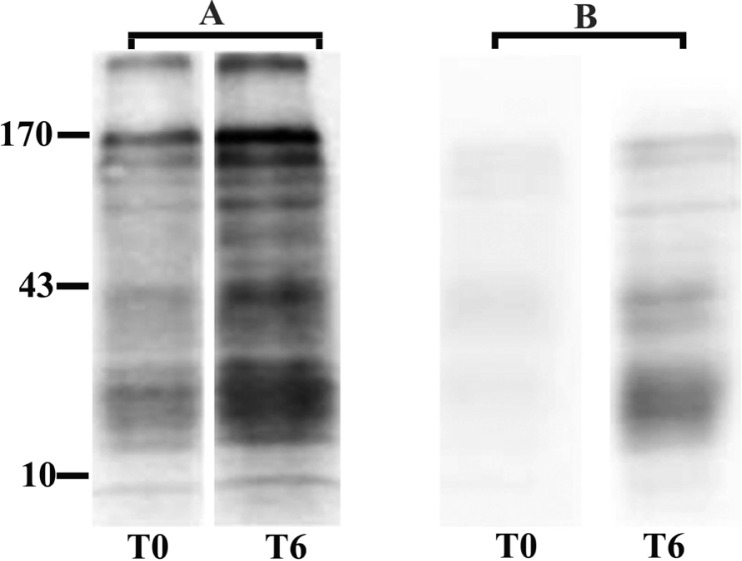
The anti-phosphotyrosine antibody(PY-99) recognized protein bands in the spermatozoa from normo-spermic ejaculates. Western blotting results (Fig. 1-A) depicted the presence of at least six proteins with molecular weights of 170, 130, 95, 70, 43 and 25kDa before capacitation in the spermatozoa from normospermic samples (T0). However, comparable levels of tyrosine phos-phorylation were not observed in the spermatozoa from teratospermic samples. As shown in Fig. 1-B, no significant levels of tyrosine phosphoryla-tion is detectable in proteins before capacitation (T0).
Figure 1.
Western blotting of sperm tyrosine phosphorylated proteins extracted immediately after washing (T0) and after 6 h (T6) of incubation under capacitating conditions. A: Normospermia; B: Teratospermia.
Changes in tyrosine phosphorylation following capacitation
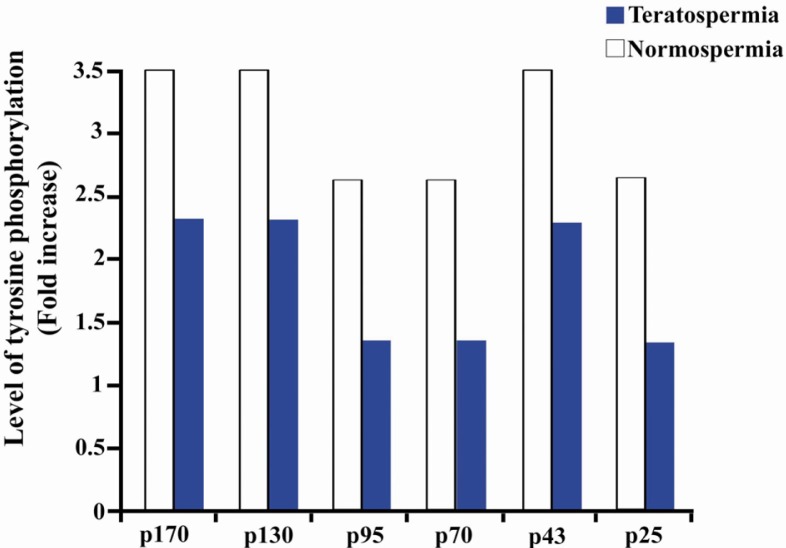
Capacitation causes an increase in the level of tyrosine phosphorylation in the spermatozoa from normospermic (Fig. 1-A-T6) and teratospermic (Fig. 1.B-T6) samples. Densitometric scanning (Fig. 2) shows that capacitation has resulted in a 3.5-fold increase in tyrosine phosphorylation of 170kDa, 130kDa and 43kDa proteins in the spermatozoa from normospermic samples, compared to uncapacitated samples. Only a 2.3-fold increase was seen in teratospermic men, which was different from normospermic counterparts. Likewise, phosphorylation of 95 kDa, 70kDa and 25kDa proteins increased 2.6-fold in normospermic subjects compared to 1.3-fold in teratospermic males.
Figure 2.
Quantitation of changes in protein phosphorylation following sperm capacitation in normospermic and teratospermic samples. Changes in the level of phosphorylation were expressed as a fold increase over the controls (the uncapacitated samples).
Discussion
Phosphorylation is an important post-translational modification that regulates various cellular functions such as cell cycle control, cellular growth, ionic current modulation and receptor regulation (24, 25). Mature spermatozoa are highly specialized cells that are not very active transcriptionally. Thus, sperm rely on post-translational modifications such as phosphorylation to regulate important events such as capacitation, hyper-activation and acrosome reaction, which are required for spermatozoa to reach, bind, penetrate and fuse oocytes (1). Tyrosine and serine/threonine phosphorylation of proteins has been reported in spermatozoa (26) and tyrosine phosphorylation has been found to be more important as an indicator of a signal transduction pathway relative to serine/threonine phosphorylation.
Teratospermia is a common cause of male infertility and it is characterized by the presence of abnormal sperm morphology in semen (>70% of spermatozoa are morphologically abnormal). It has been reported that capacitated sperm from teratospermic ejaculates are less able to complete acrosome reaction at the presence of calcium ionophore (27) or progesterone (28) compared to sperm from normospermic ejaculates. Sperm capacitation and acrosome reaction have been shown to be compromised in the spermatozoa from teratospermic cats (18). Additionally, malformed sperm are not able to penetrate the zona pellucida (29). The etiology of dysfunctional spermatozoa from teratospermic ejaculates involves an array of biochemical, molecular and regulatory factors at cellular levels. Tyrosine phosphorylation is one of the key biochemical and molecular factors which is less active in the spermatozoa from teratospermic ejaculates even under capacitating conditions (19).
In the present study, the status of tyrosine-phosphorylated proteins in spermatozoa from infertile men with teratospermic condition was evaluated compared with that of the normo-spermic men before and after capacitation. This should advance our knowledge and contribute to a deeper understanding of the basic molecular and cellular mechanisms regulating sperm functions in vitro. To do so, the presence of at least six tyrosine phosphorylated proteins (170, 130, 95, 70, 43 and 25 kilo Daltons) was determined in human spermatozoa. Second, it was demonstrated that spermatozoa from teratospermic males had a compromised ability to undergo tyrosine phosphorylation following capacitation when compareed with normospermic counterparts.
Phosphotyrosine–containing proteins have been reported to be present in the spermatozoa of numerous species and they are believed to have an important role in sperm functions such as hyper-active motility, interaction with zona pellucida and acrosome reaction. In mouse sperm, anti-phosphotyrosine antibody was used to study capacitation and three proteins with molecular weights of 52kDa, 75kDa and 95kDa were identified. The 95kDa protein was reported to have enhanced immunoreactivity after sperm capacitation (30). Tyrosine phosphorylation has been demonstrated in the sperm of several mammalian species including humans, rats, rabbit, and mice. In human sperm, four sets of tyrosine-phosphorylated protein with molecular weights ranging form 95/94±3kDa, 46±3kDa, 25±7kDa to 12±2kDa have been reported (26). Some of these phosphorylated proteins have been shown to bear an important role in sperm motility (26). In another study two plasma membrane proteins (35kDa and 46kDa), isolated from capacitated boar sperm cells, were reported to have high binding affinity with zona pellucida (16).
In the present study we have demonstrated that tyrosine phosphorylation of several proteins from human spermatozoa are similar to that of mice (30) and boars (16) after capacitation. Specifically, tyrosine phosphorylation of six sets of sperm proteins (170kDa, 130kDa, 95kDa, 70kDa, 43kDa and 25kDa) appeared to be compromised in teratospermic samples when compared with normospermic ones. Thus, it is proposed that diminished protein tyrosine phos-phorylation may constitute one of the factors responsible for compromised sperm functions in teratospermic males.
Furthermore, it was demonstrated that tyrosine phosphorylation of the 95kDa sperm protein from teratospermic males was compromised which is similar to the findings reported in wild felids (31) and domestic cats (19). This protein is localized to the acrosomal region in the spermatozoa from domestic cats and it is known to interact with zona pellucida protein(s) (19, 31). Similar findings in humans and cats indicate that the 95kDa protein may play a role in one or more steps associated with sperm-oocyte interaction, which enables zona penetration (19, 31).
In human sperm, a 46±3kDa protein is phos-phorylated at tyrosine residues during capacitation (26). More recently, a 46kDa protein was reported in boar sperm (16), which was phosphorylated in tyrosine residues during capacitation. This 46kDa protein is located in plasma membrane of sperm head and it has been demonstrated to have an important role in interacting with zona pellucid. In this study, it was shown that tyrosine phos-phorylation of a 43kDa protein is compromised during capacitation in teratospermic males. The size of this protein is very similar to the protein previously reported in human (26) and boar sperm (16). These observations suggest that this 43kDa plasma membrane protein may be involved in the interaction with zona Pellucida.
For a better understanding of capacitation process in human sperm, it is important to continue studying post-translational modifications of proteins because of their role in sperm functions. To accomplish this goal, studies are underway in our laboratories to identify the tyrosine-phosphorylated proteins that might play a role in the capacitation of sperm. These findings can help the characterization of molecular mechanisms of sperm functions and can further explain the causes of male infertility.
Conclusion
Tyrosine phosphorylation of sperm proteins may have an important role in the function of sperm and defects in the level of tyrosine-phosphorylated proteins may contribute to low fertilization rates in teratospermic men.
Acknowledgement
The authors thank Ms. Mahshid Hodjat, Ms. Zahra Ghaempanah, Ms. Elham Savadi Shiraz and Ms. Sima Binafar for their assistance. We also thank the staff in the embryology and andrology departments at Avicenna Infertility Clinic for their help. This study was supported by a grant from ACECR (grant number 1634-33) awarded to Ali M. Ardekani.
To cite this article: Jabbari S, Sadeghi MR, Akhondi MM, Ebrahimi Habibi A, Amirjanati N, Lakpour N, et al. Tyrosine Phosphorylation Pattern in Sperm Proteins Isolated from Normospermic and Teratospermic Men. J Reprod Infertil. 2009;10(3):185-191.
References
- 1.Yanagimachi R, editor. Mammalian fertilization. New York: Raven Press; c1994. p. 189. (Knobil E, Neill J, editors. The physiology of reproduction; vol.1). [Google Scholar]
- 2.Urner F, Leppens-Luisier G, Sakkas D. Protein tyrosine phosphorylation in sperm during gamete interaction in the mouse: the influence of glucose. Biol Reprod. 2001;64(5):1350–7. doi: 10.1095/biolreprod64.5.1350. [DOI] [PubMed] [Google Scholar]
- 3.Marín-Briggiler CI, Gonzalez-Echeverría F, Buffone M, Calamera JC, Tezón JG, Vazquez-Levin MH. Calcium requirements for human sperm function in vitro. Fertil Steril. 2003;79(6):1396–403. doi: 10.1016/s0015-0282(03)00267-x. [DOI] [PubMed] [Google Scholar]
- 4.Da Ros VG, Munuce MJ, Cohen DJ, Marín-Briggiler CI, Busso D, Visconti PE, et al. Bicarbonate is required for migration of sperm epididymal protein DE (CRISP-1) to the equatorial segment and expression of rat sperm fusion ability. Biol Reprod. 2004;70(5):1325–32. doi: 10.1095/biolreprod.103.022822. [DOI] [PubMed] [Google Scholar]
- 5.Buffone MG, Calamera JC, Verstraeten SV, Doncel GF. Capacitation-associated protein tyrosine phosphorylation and membrane fluidity changes are impaired in the spermatozoa of asthenozoospermic patients. Reproduction. 2005;129(6):697–705. doi: 10.1530/rep.1.00584. [DOI] [PubMed] [Google Scholar]
- 6.Buffone MG, Verstraeten SV, Calamera JC, Doncel GF. High cholesterol content and decreased membrane fluidity in human spermatozoa are associated with protein tyrosine phosphorylation and functional deficiencies. J Androl. 2009;30(5):552–8. doi: 10.2164/jandrol.108.006551. [DOI] [PubMed] [Google Scholar]
- 7.Buffone MG, Doncel GF, Calamera JC, Verstraeten SV. Capacitation-associated changes in membrane fluidity in asthenozoospermic human spermatozoa. Int J Androl. 2009;32(4):360–75. doi: 10.1111/j.1365-2605.2008.00874.x. [DOI] [PubMed] [Google Scholar]
- 8.Morton B, Albagli L. Modification of hamster sperm adenyl cyclase by capacitation in vitro. Biochem Biophys Res Commun. 1973;50(3):697–703. doi: 10.1016/0006-291x(73)91300-4. [DOI] [PubMed] [Google Scholar]
- 9.Hyne RV, Garbers DL. Calcium-dependent increase in adenosine 3’,5’-monophosphate and induction of the acrosome reaction in guinea pig spermatozoa. Proc Natl Acad Sci U S A. 1979;76(11):5699–703. doi: 10.1073/pnas.76.11.5699. [DOI] [PMC free article] [PubMed] [Google Scholar]
- 10.Visconti PE, Johnson LR, Oyaski M, Fornés M, Moss SB, Gerton GL, et al. Regulation, localization, and anchoring of protein kinase A subunits during mouse sperm capacitation. Dev Biol. 1997;192(2):351–63. doi: 10.1006/dbio.1997.8768. [DOI] [PubMed] [Google Scholar]
- 11.Asquith KL, Baleato RM, McLaughlin EA, Nixon B, Aitken RJ. Tyrosine phosphorylation activates surface chaperones facilitating sperm-zona recognition. J Cell Sci. 2004;117(Pt 16):3645–57. doi: 10.1242/jcs.01214. [DOI] [PubMed] [Google Scholar]
- 12.Ecroyd H, Jones RC, Aitken RJ. Tyrosine phosphorylation of HSP-90 during mammalian sperm capacitation. Biol Reprod. 2003;69(6):1801–7. doi: 10.1095/biolreprod.103.017350. [DOI] [PubMed] [Google Scholar]
- 13.Ficarro S, Chertihin O, Westbrook VA, White F, Jayes F, Kalab P, et al. Phosphoproteome analysis of capacitated human sperm. Evidence of tyrosine phosphorylation of a kinase-anchoring protein 3 and valosin-containing protein/p97 during capacitation. J Biol Chem. 2003;278(13):11579–89. doi: 10.1074/jbc.M202325200. [DOI] [PubMed] [Google Scholar]
- 14.Arcelay E, Salicioni AM, Wertheimer E, Visconti PE. Identification of proteins undergoing tyrosine phosphorylation during mouse sperm capacitation. Int J Dev Biol. 2008;52(5-6):463–72. doi: 10.1387/ijdb.072555ea. [DOI] [PubMed] [Google Scholar]
- 15.Yunes R, Doncel GF, Acosta AA. Incidence of sperm-tail tyrosine phosphorylation and hyper-activated motility in normozoospermic and asthenozoospermic human sperm samples. Biocell. 2003;27(1):29–36. [PubMed] [Google Scholar]
- 16.Flesch FM, Wijnand E, van de Lest CH, Colenbrander B, van Golde LM, Gadella BM. Capacitation dependent activation of tyrosine phos-phorylation generates two sperm head plasma membrane proteins with high primary binding affinity for the zona pellucida. Mol Reprod Dev. 2001;60(1):107–15. doi: 10.1002/mrd.1067. [DOI] [PubMed] [Google Scholar]
- 17.Howard JG, Brown JL, Bush M, Wildt DE. Teratospermic and normospermic domestic cats: ejaculate traits, pituitary-gonadal hormones, and improvement of spermatozoal motility and morphology after swim-up processing. J Androl. 1990;11(3):204–15. [PubMed] [Google Scholar]
- 18.Long JA, Wildt DE, Wolfe BA, Critser JK, DeRossi RV, Howard J. Sperm capacitation and the acrosome reaction are compromised in terato-spermic domestic cats. Biol Reprod. 1996;54(3):638–46. doi: 10.1095/biolreprod54.3.638. [DOI] [PubMed] [Google Scholar]
- 19.Pukazhenthi BS, Wildt DE, Ottinger MA, Howard J. Compromised sperm protein phosphorylation after capacitation, swim-up, and zona pellucida exposure in teratospermic domestic cats. J Androl. 1996;17(4):409–19. [PubMed] [Google Scholar]
- 20.Li LW, Fan LQ, Zhu WB, Nien HC, Sun BL, Luo KL, et al. Establishment of a high-resolution 2-D reference map of human spermatozoal proteins from 12 fertile sperm-bank donors. Asian J Androl. 2007;9(3):321–9. doi: 10.1111/j.1745-7262.2007.00261.x. [DOI] [PubMed] [Google Scholar]
- 21.Bradford MM. A rapid and sensitive method for the quantitation of microgram quantities of protein utilizing the principle of protein-day binding. Anal Biochem. 1976;72:248–54. doi: 10.1016/0003-2697(76)90527-3. [DOI] [PubMed] [Google Scholar]
- 22.Laemmli UK. Cleavage of structural proteins during the assembly of the head of bacteriophage T4. Nature. 1970;227(5259):680–5. doi: 10.1038/227680a0. [DOI] [PubMed] [Google Scholar]
- 23.Buffone MG, Brugo-Olmedo S, Calamera JC, Verstraeten SV, Urrutia F, Grippo L, et al. Decreased protein tyrosine phosphorylation and membrane fluidity in spermatozoa from infertile men with varicocele. Mol Reprod Dev. 2006;73(12):1591–9. doi: 10.1002/mrd.20611. [DOI] [PubMed] [Google Scholar]
- 24.Pawson T. Specificity in signal transduction: from phosphotyrosine-SH2 domain interactions to complex cellular systems. Cell. 2004;116(2):191–203. doi: 10.1016/s0092-8674(03)01077-8. [DOI] [PubMed] [Google Scholar]
- 25.Liu BA, Jablonowski K, Raina M, Arcé M, Pawson T, Nash PD. The human and mouse complement of SH2 domain proteins-establishing the boundaries of phosphotyrosine signaling. Mol Cell. 2006;22(6):851–68. doi: 10.1016/j.molcel.2006.06.001. [DOI] [PubMed] [Google Scholar]
- 26.Visconti PE, Kopf GS. Regulation of protein phosphorylation during sperm capacitation. Biol Reprod. 1998;59(1):1–6. doi: 10.1095/biolreprod59.1.1. [DOI] [PubMed] [Google Scholar]
- 27.Kholkute SD, Meherji P, Puri CP. Capacitation and the acrosome reaction in sperm from men with various semen profiles monitored by a chlortetra-cycline fluorescence assay. Int J Androl. 1992;15(1):43–53. doi: 10.1111/j.1365-2605.1992.tb01113.x. [DOI] [PubMed] [Google Scholar]
- 28.Oehninger S, Blackmore P, Morshedi M, Sueldo C, Acosta AA, Alexander NJ. Defective calcium influx and acrosome reaction (spontaneous and progesterone-induced) in spermatozoa of infertile men with severe teratozoospermia. Fertil Steril. 1994;61(2):349–54. doi: 10.1016/s0015-0282(16)56530-3. [DOI] [PubMed] [Google Scholar]
- 29.Howard J, Bush M, Wildt DE. Teratospermia in domestic cats compromises penetration of zona-free hamster ova and cat zonae pellucidae. J Androl. 1991;12(1):36–45. [PubMed] [Google Scholar]
- 30.Leyton L, Saling P. 95 kd sperm proteins bind ZP3 and serve as tyrosine kinase substrates in response to zona binding. Cell. 1989;57(7):1123–30. doi: 10.1016/0092-8674(89)90049-4. [DOI] [PubMed] [Google Scholar]
- 31.Pukazhenthi BS, Long JA, Wildt DE, Ottinger MA, Armstrong DL, Howard J. Regulation of sperm function by protein tyrosine phosphorylation in diverse wild felid species. J Androl. 1998;19(6):675–85. [PubMed] [Google Scholar]


