

Abstract
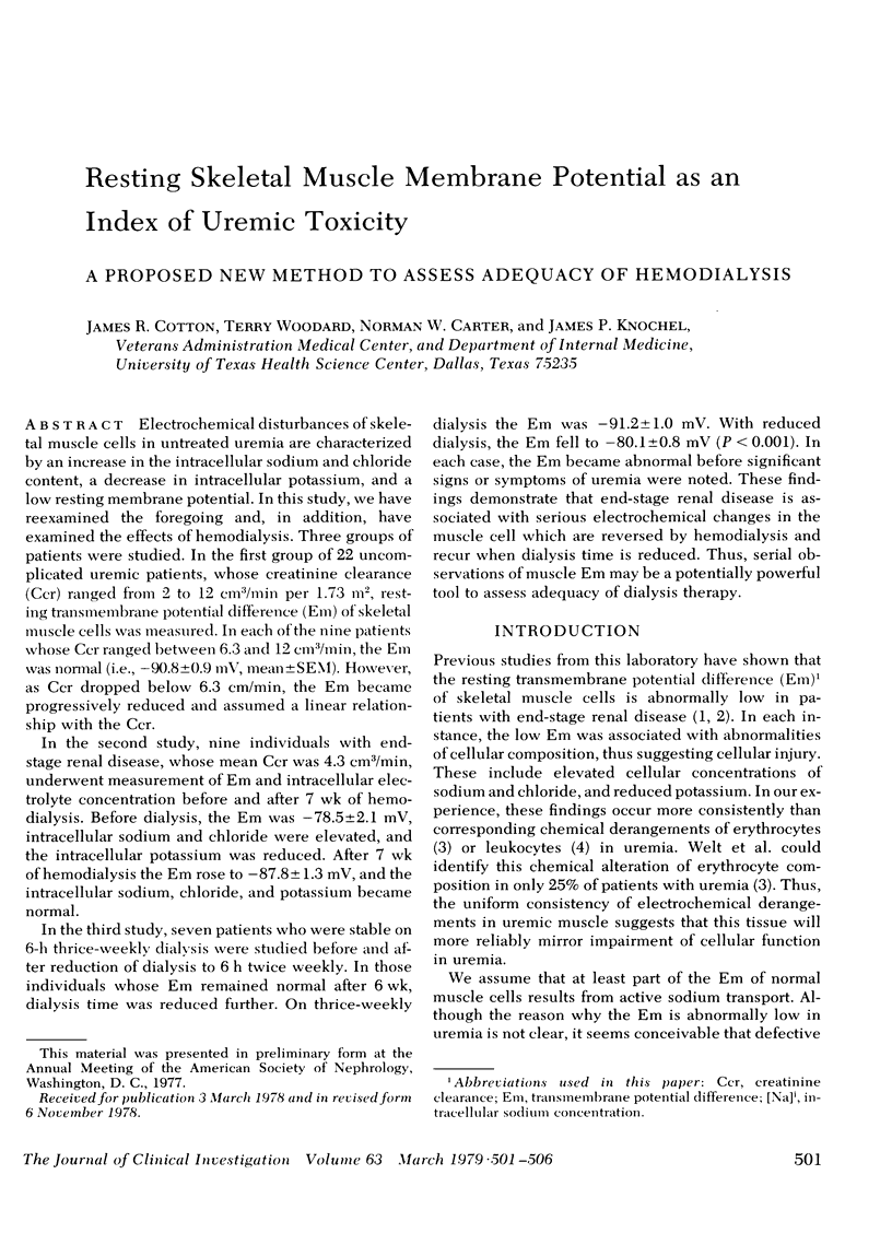
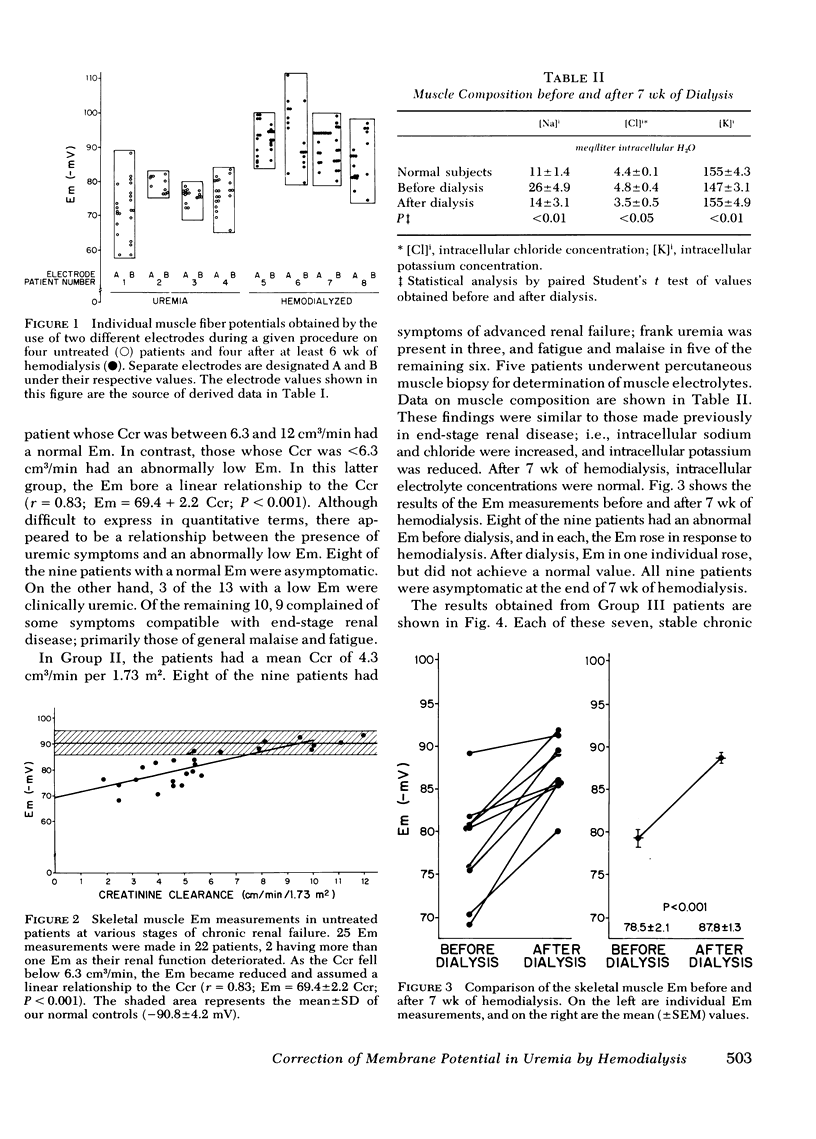
Electrochemical disturbances of skeletal muscle cells in untreated uremia are characterized by an increase in the intracellular sodium and chloride content, a decrease in intracellular potassium, and a low resting membrane potential. In this study, we have reexamined the foregoing and, in addition, have examined the effects of hemodialysis. Three groups of patients were studied. In the first group of 22 uncomplicated uremic patients, whose creatinine clearance (Ccr) ranged from 2 to 12 cm3/min per 1.73 m2, resting transmembrane potential difference (Em) of skeletal muscle cells was measured. In each of the nine patients whose Ccr ranged between 6.3 and 12 cm3/min, the Em was normal (i.e., −90.8±0.9 mV, mean±SEM). However, as Ccr dropped below 6.3 cm/min, the Em became progressively reduced and assumed a linear relationship with the Ccr.
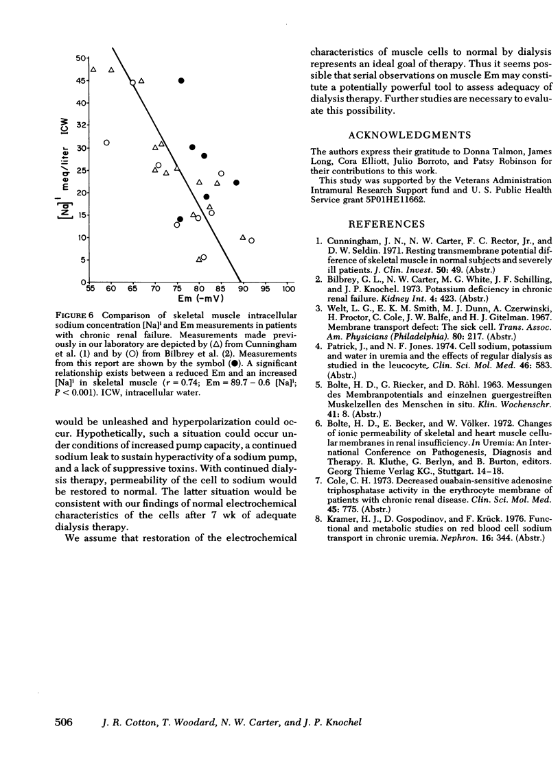
In the second study, nine individuals with end-stage renal disease, whose mean Ccr was 4.3 cm3/min, underwent measurement of Em and intracellular electrolyte concentration before and after 7 wk of hemodialysis. Before dialysis, the Em was −78.5±2.1 mV, intracellular sodium and chloride were elevated, and the intracellular potassium was reduced. After 7 wk of hemodialysis the Em rose to −87.8±1.3 mV, and the intracellular sodium, chloride, and potassium became normal.
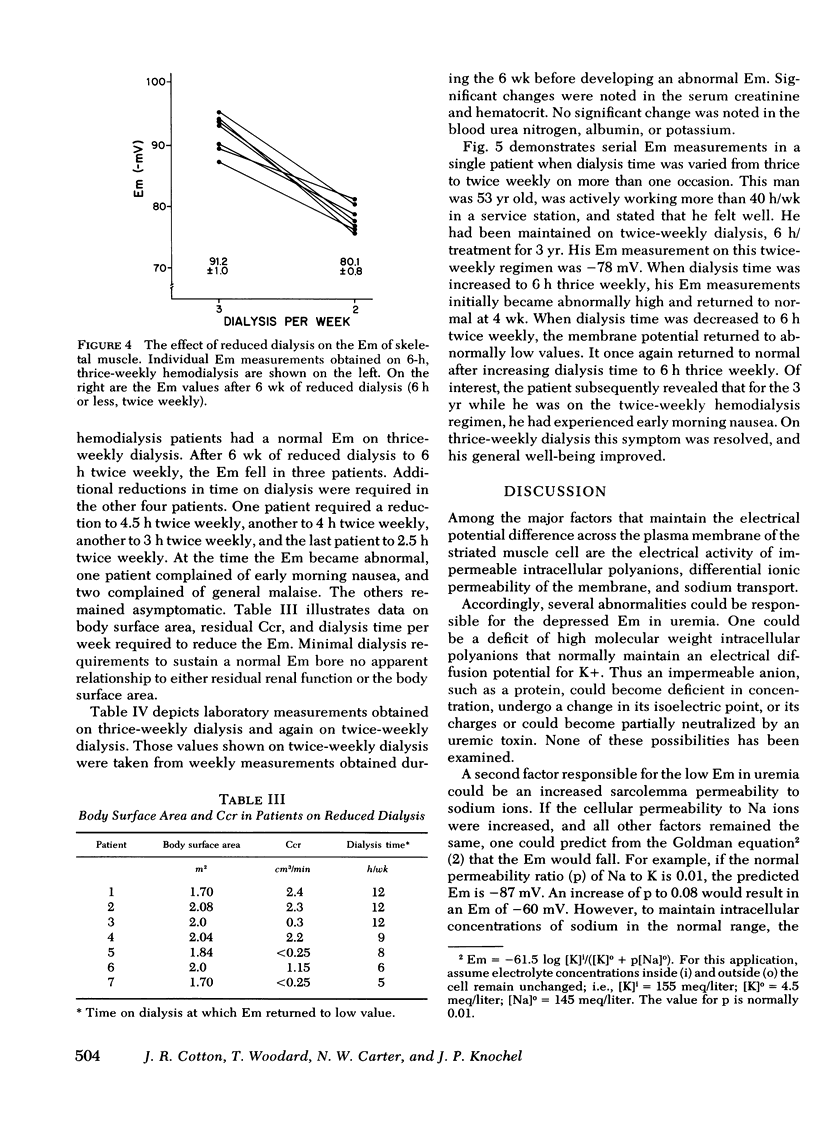
In the third study, seven patients who were stable on 6-h thrice-weekly dialysis were studied before and after reduction of dialysis to 6 h twice weekly. In those individuals whose Em remained normal after 6 wk, dialysis time was reduced further. On thrice-weekly dialysis the Em was −91.2±1.0 mV. With reduced dialysis, the Em fell to −80.1±0.8 mV (P < 0.001). In each case, the Em became abnormal before significant signs or symptoms of uremia were noted. These findings demonstrate that end-stage renal disease is associated with serious electrochemical changes in the muscle cell which are reversed by hemodialysis and recur when dialysis time is reduced. Thus, serial observations of muscle Em may be a potentially powerful tool to assess adequacy of dialysis therapy.
Full text
PDF

Selected References
These references are in PubMed. This may not be the complete list of references from this article.
- Bilbrey G. L., Carter N. W., White M. G., Schilling J. F., Knochel J. P. Potassium deficiency in chronic renal failure. Kidney Int. 1973 Dec;4(6):423–430. doi: 10.1038/ki.1973.138. [DOI] [PubMed] [Google Scholar]
- Cole C. H. Decreased ouabain-sensitive adenosine triphosphatase activity in the erythrocyte membrame of patients with chronic renal disease. Clin Sci Mol Med. 1973 Dec;45(6):775–784. doi: 10.1042/cs0450775. [DOI] [PubMed] [Google Scholar]
- Kramer H. J., Gospodinov D., Krück F. Functional and metabolic studies on red blood cell sodium transport in chronic uremia. Nephron. 1976;16(5):344–358. doi: 10.1159/000180621. [DOI] [PubMed] [Google Scholar]
- Patrick J., Jones N. F. Cell sodium, potassium and water in uraemia and the effects of regular dialysis as studied in the leucocyte. Clin Sci Mol Med. 1974 May;46(5):583–590. doi: 10.1042/cs0460583. [DOI] [PubMed] [Google Scholar]