Abstract
Objective
To demonstrate whether the preoperative integrity of the inner segment/outer segment (IS/OS) junction of photoreceptors studied by spectral-domain optical coherence tomography (SD-OCT) is a prognostic factor in epiretinal membrane surgery.
Methods
We retrospectively studied patients with an idiopathic epiretinal membrane who underwent a 23-gauge vitrectomy to remove this membrane. Best-corrected visual acuity (BCVA) and SD-OCT scans were examined before and 6 months after the surgery. We studied the retinal microstructure, especially the IS/OS junction of the photoreceptors, and evaluated the intergroup differences between patients with an intact layer and those with an irregular or disrupted layer. We applied both the Wilcoxon and Mann–Whitney tests for statistical analysis.
Results
In total, 51 eyes from 51 enrolled patients were examined in this study. The postoperative BCVA was significantly better for eyes that had an intact IS/OS junction than for eyes that had an irregular or disrupted IS/OS junction, as preoperatively observed with SD-OCT scans (P < 0.001). We also observed an important association between disrupted IS/OS junctions and the presence of cystic macular edema (P < 0.01).
Conclusion
The presence of an intact IS/OS junction on the preoperative SD-OCT scan was an important predictor of better visual recovery after epiretinal membrane surgery.
Keywords: epiretinal membrane, photoreceptors, inner segment/outer segment junction, spectral-domain optical coherence tomography
Introduction
Epiretinal membrane (ERM) is a condition with 7% prevalence in the general population.1 This condition is associated with a number of ocular problems, such as vascular and inflammatory disease, trauma, tumors, and retinal dystrophies. We consider this condition to be idiopathic when it occurs in healthy eyes with no history of ocular problems.
Optical coherence tomography (OCT) has been used for some time to classify ERMs.2 More recently, new-generation technology, such as spectral-domain OCT (SD-OCT), has been utilized to define all of the factors that could determine the postoperative result after surgical membrane removal.3,4 There is a report that discusses preoperative prognostic factors, such as the onset time of symptoms, cystoid macular edema, macular hole, or poor visual acuity.5
More recent studies have focused on the layer of union of the inner segment and outer segment (IS/OS) of the photoreceptors as an important prognostic factor in ERM surgery and other ocular problems.6–9
For the authors of studies previously mentioned, the integrity of the IS/OS junction is essential for improving visual acuity after surgically removing the ERMs. When an interruption in this layer is detected by OCT, visual acuity before and after surgery is worse. Several of these studies were performed with low-resolution OCT.6
This topic is of interest because it is important to know, preoperatively, whether surgery will result in an increase in visual acuity.
In this study, we used SD-OCT (3D OCT-2000 Spectral Domain OCT; Topcon Medical Systems, Oakland, NJ, USA) to evaluate retinal microstructure features, such as presence of cystic macular edema and integrity of the IS/OS junction of photoreceptors. Scans were performed on a considerable number of patients with ERMs before and after vitreoretinal surgery to see if surgery outcomes could be predicted.
Materials and methods
51 eyes of 51 patients who underwent a 23-gauge vitrectomy to remove an idiopathic ERM between 2008 and 2010 were enrolled in this study. Patients with nonidiopathic ERM or other ocular diseases were excluded. Surgery was assisted by the use of Trypan blue to stain the ERM. All patients were followed for at least 6 months after their procedure. Surgical procedures were performed by two experienced vitreoretinal surgeons (LA and JMRM) at Bellvitge University Hospital, Barcelona, Spain and Alicante Institute of Ophthalmology, Alicante, Spain.
A complete ophthalmic examination, which included a best-corrected visual acuity (BCVA) test that was measured on a Snellen chart, slit-lamp biomicroscopy with a fundus examination, and macular examinations with the Topcon SD-OCT, were performed before and 6 months after surgery.
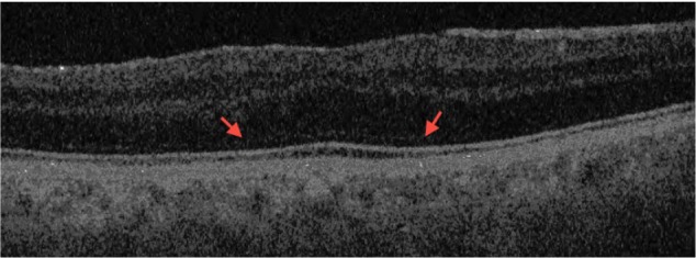
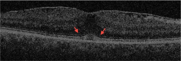
The collected patient data included central foveal thickness and retinal microstructure integrity, especially of the IS/OS junction. We evaluated whether the IS/OS layer was intact (hyperreflective continuous line [Figure 1]) or disrupted (hyporeflective irregularities along the hyperreflective line [Figure 2]).
Figure 1.

Intact inner segment/outer segment junction. A continuous layer with no disruption is visible.
Figure 2.

Disrupted inner segment/outer segment layer. Subfoveal disruption in the inner segment/outer segment junction is visible.
Wilcoxon and Mann–Whitney tests were used for the statistical analyses.
Results
The baseline characteristics of the patients are shown in Table 1. Fifty-one eyes (28 right eyes and 23 left eyes) of 51 patients (28 men and 23 women) were studied. The mean age was 66.82 years (standard deviation [SD] ± 7.3; range: 55–85 years). There were 21 pseudophakic and 30 phakic eyes.
Table 1.
Patient characteristics
| Eyes, n | 51 |
| Patients | 51 |
| Men, n (%) | 28 (54.90) |
| Women, n (%) | 23 (45.09) |
| Mean age ± SD (range) | 66.82 ± 7.3 (55–85) |
| BCVA ± SD | 0.25 ± 0.14 |
| Pseudophakic eyes, n (%) | 21 (41.17) |
| IS/OS junction | |
| Intact, n (%) | 29 (56.86) |
| Irregular, n (%) | 22 (43.13) |
| Mean CFT ± SD (μm) | 413 ± 73,26 |
| Cystic macular edema, n (%) | 14 (27.4) |
| Pseudohole, n (%) | 3 (5.88) |
Abbreviations: BCVA, best-corrected visual acuity; CFT, central foveal thickness; IS/OS, inner segment/outer segment; SD, standard deviation.
The mean BCVA before surgery was 0.25 ± 0.14. An intact IS/OS junction was observed in 29 patients (56.86%), and 22 (43.13%) patients had a disrupted or irregular layer. The mean central foveal thickness was 413 μm before surgery (SD ± 73.26).
Cystic macular edema was observed in 14 eyes (27.40%), and a macular pseudohole was observed in three eyes (5.88%). There was a strong association between patients with cystic macular edema and a disrupted IS/OS layer before as well as after surgery (P < 0.001). This data is presented in Table 1.
All patients had their ERMs successfully removed with no relevant side effects. The procedure to remove the ERM was a 23-gauge vitrectomy, which was assisted by the use of Trypan blue. In six of the cases, the surgery was combined with cataract surgery. No other procedures, such as cataract removal, were performed during the follow-up period.
Six months after surgery, the mean BCVA was 0.53 ± 0.37. The difference between the BCVA before and after surgery was statistically significant (P < 0.001). Change in visual acuity was greater in the intact IS/OS layer (0.44 ± 0.24) patient group compared with the disrupted IS/OS layer (0.08 ± 0.18) patient group (P < 0.001).
There were no patients with an intact IS/OS layer before surgery and a disrupted IS/OS layer after surgery. Nevertheless, there was one patient with a disrupted IS/OS layer who had an intact IS/OS layer after surgery. This patient’s visual acuity improved from 0.05 to 0.6. Comparison data of the two groups is shown in Table 2.
Table 2.
Intergroup comparison of patients with an intact IS/OS layer and patients with an irregular IS/OS layer
| Intact IS/OS | Irregular IS/OS | P* | |
|---|---|---|---|
| Number of eyes | 29 | 22 | |
| BCVA pre-surgery ± SD | 0.30 ± 0.04 | 0.19 ± 0.02 | 0.042 |
| Mean CFT pre-surgery ± SD | 404 ± 72.15 | 422 ± 76.30 | 0.555 |
| Cystic macular edema, n (%) | 0 (0) | 14 (63.63) | 0.001 |
| Macular pseudohole, n (%) | 1 (3.44) | 2 (9.09) | 0.571 |
| Mean CFT post-surgery ± SD | 312.67 ± 59.89 | 344.08 ± 41.95 | 0.126 |
| BCVA post-surgery | 0.69 ± 0.07 | 0.32 ± 0.07 | 0.002 |
| Change in BCVA ± SD | 0.44 ± 0.24 | 0.08 ± 0.18 | 0.001 |
Notes: Change in BCVA = BCVA after surgery – BCVA before surgery.
Mann–Whitney test, Wilcoxon test.
Abbreviations: BCVA, best-corrected visual acuity; CFT, central foveal thickness; IS/OS, inner segment/outer segment; SD, standard deviation.
Discussion
These results demonstrate that the integrity of the IS/OS junction, studied by SD-OCT, is a statistically significant predictor of postoperative visual acuity in patients with ERM.
It is well known that some patients who undergo surgery for ERMs experience no improvement in visual acuity. This fact is an important point of interest and concern for retinal surgeons.
The issue of predictor factors in surgery has been addressed in published reports concerning ERM and other conditions, such as macular hole.10,11 Niwa et al utilized preoperative electroretinograms to predict which patients could have poor results after surgery.12
Other technology, such as OCT, has been used similarly. Several studies6,13 report the use of time-domain OCT, which offers low-resolution images. Mitamura et al demonstrated that the IS/OS junction is important in predicting postoperative visual acuity and that the retinal microstructure could improve after surgery.13 However, SD-OCT has a higher resolution and better correlation with actual retinal anatomy and permits the simultaneous evaluation of the IS/OS junction and external limiting membrane.14
In our study, all patients experienced increased visual acuity after ERM surgery. If we analyze only the patients in the intact IS/OS junction group by SD-OCT, the increase in visual acuity is greater, and the difference between this group and the group with a disrupted layer is statistically significant. Inoue et al reported corroborating results and demonstrated an important correlation between postoperative visual acuity and the integrity of photoreceptors.9
There are other prognostic factors in epiretinal surgery, such as macular edema.15,16 In our study, macular edema showed a significant association with IS/OS junction intactness. Therefore, upon observing cystic macular edema, a disrupted IS/OS junction was also observed. Both conditions are associated with a long-term disease and thus increased damage to the photoreceptor cells and worsened functionality.
There were some limitations to our study, including the small number of patients enrolled. Moreover, the follow-up period was limited and, because this was not a prospective study, the groups were unequal in personal characteristics such as lens state.
Conclusion
The preoperative study of the IS/OS junction by SD-OCT should be performed in every patient with ERM to determine those who will have a greater probability of improved visual acuity after the procedure. Large, multicenter studies are warranted to corroborate our findings.
Footnotes
Disclosure
The authors report no conflicts of interest in this work.
References
- 1.Fraser-Bell S, Guzowski M, Rochtchina E, Wang JJ, Mitchell P. Five-year cumulative incidence and progression of epiretinal membranes: the Blue Mountains Eye Study. Ophthalmology. 2003;110(1):34–40. doi: 10.1016/s0161-6420(02)01443-4. [DOI] [PubMed] [Google Scholar]
- 2.Watanabe A, Arimoto S, Nishi O. Correlation between metamorphopsia and epiretinal membrane optical coherence tomography findings. Ophthalmology. 2009;116(9):1788–1793. doi: 10.1016/j.ophtha.2009.04.046. [DOI] [PubMed] [Google Scholar]
- 3.Trese MT, Chandler DB, Machemer R. Macular pucker. I. Prognostic criteria. Graefes Arch Clin Exp Ophthalmol. 1983;221:12–15. doi: 10.1007/BF02171725. [DOI] [PubMed] [Google Scholar]
- 4.Wong JG, Sachdev N, Beaumont PE, Chang AA. Visual outcomes following vitrectomy and peeling of epiretinal membrane. Clin Experiment Ophthalmol. 2005;33:373–378. doi: 10.1111/j.1442-9071.2005.01025.x. [DOI] [PubMed] [Google Scholar]
- 5.Michalewski J, Michalewska Z, Cisiecki S, Nawrocki J. Morphologically functional correlations of macular pathology connected with epiretinal membrane formation in spectral optical coherence tomography (SOCT) Graefes Arch Clin Exp Ophthalmol. 2007;245(11):1623–1631. doi: 10.1007/s00417-007-0579-4. [DOI] [PubMed] [Google Scholar]
- 6.Suh MH, Seo JM, Park KH, Yu HG. Associations between macular findings by optical coherence tomography and visual outcomes after epiretinal membrane removal. Am J Ophthalmol. 2009;147:473–480. doi: 10.1016/j.ajo.2008.09.020. [DOI] [PubMed] [Google Scholar]
- 7.Oster SF, Mojana F, Brar M, Yuson RM, Cheng L, Freeman WR. Disruption of the photoreceptor inner segment/outer segment layer on spectral domain-optical coherence tomography is a predictor of poor visual acuity in patients with epiretinal membranes. Retina. 2010;30(5):713–718. doi: 10.1097/IAE.0b013e3181c596e3. [DOI] [PubMed] [Google Scholar]
- 8.Inoue M, Morita S, Watanabe Y, et al. Preoperative inner segment/outer segment junction in spectral-domain optical coherence tomography as a prognostic factor in epiretinal membrane surgery. Retina. 2011;31(7):1366–1372. doi: 10.1097/IAE.0b013e318203c156. [DOI] [PubMed] [Google Scholar]
- 9.Inoue M, Morita S, Watanabe Y, et al. Inner segment/outer segment junction assessed by spectral-domain optical coherence tomography in patients with idiopathic epiretinal membrane. Am J Ophthalmol. 2010;150(6):834–839. doi: 10.1016/j.ajo.2010.06.006. [DOI] [PubMed] [Google Scholar]
- 10.Ko TH, Fujimoto JG, Duker JS, et al. Comparison of ultrahigh- and standard-resolution optical coherence tomography for imaging macular hole pathology and repair. Ophthalmology. 2004;111:2033–2043. doi: 10.1016/j.ophtha.2004.05.021. [DOI] [PMC free article] [PubMed] [Google Scholar]
- 11.Baba T, Yamamoto S, Arai M. Correlation of visual recovery and presence of photoreceptor inner/outer segment junction in optical coherence images after successful macular hole repair. Retina. 2008;28:453–458. doi: 10.1097/IAE.0b013e3181571398. [DOI] [PubMed] [Google Scholar]
- 12.Niwa T, Terasaki H, Kondo M, Piao CH, Suzuki T, Miyake Y. Function and morphology of macula before and after removal of idiopathic epiretinal membrane. Invest Ophthalmol Vis Sci. 2003;44:1652–1656. doi: 10.1167/iovs.02-0404. [DOI] [PubMed] [Google Scholar]
- 13.Mitamura Y, Hirano K, Baba T, Yamamoto S. Correlation of visual recovery with presence of photoreceptor inner/outer segment junction in optical coherence images after epiretinal membrane surgery. Br J Ophthalmol. 2009;93:171–175. doi: 10.1136/bjo.2008.146381. [DOI] [PubMed] [Google Scholar]
- 14.Schmidt-Erfurth U, Leitgeb RA, Michels S, et al. Three-dimensional ultrahigh-resolution optical coherence tomography of macular diseases. Invest Ophthalmol Vis Sci. 2005;46:3393–3402. doi: 10.1167/iovs.05-0370. [DOI] [PubMed] [Google Scholar]
- 15.Brar M, Yuson R, Kozak I, et al. Correlation between morphologic features on spectral-domain optical coherence tomography and angiographic leakage patterns in macular edema. Retina. 2010;30:383–389. doi: 10.1097/IAE.0b013e3181cd4803. [DOI] [PMC free article] [PubMed] [Google Scholar]
- 16.Massin P, Allouch C, Haouchine B, et al. Optical coherence tomography of idiopathic macular epiretinal membranes before and after surgery. Am J Ophthalmol. 2000;130:732–739. doi: 10.1016/s0002-9394(00)00574-2. [DOI] [PubMed] [Google Scholar]


