Abstract
Purpose The first autologous adipose tissue grafting was performed by Neuber in 1893 with an open approach. In the early 1980s, Illouz and Fournier introduced closed liposuction. In the 1990s, Coleman published a new method of atraumatic fat transplantation. Recently, immunohistochemical studies of the extracellular matrix of the lipoaspirate showed the presence of adipose-derived stem cells. The purpose of this study is to describe the role of fat grafting in the management of posttraumatic facial deformities.
Methods The study population was composed of all patients who underwent facial fat grafting between March 2008 and November 2010 as a secondary reconstructive procedure after an initial unsatisfactory treatment of the skeletal fractures. We analyzed the postoperative morphological changes by comparing the grafted side of the face to the contralateral side with the aid of a software package.
Results Nineteen patients were surgically treated with fat transplantation for facial asymmetry due to a pathological postoperative healing of the soft tissue. Clinical examination and software analysis showed adequate postoperative facial balance without major complications.
Conclusion Fat grafting is a very powerful tool to correct posttraumatic maxillofacial deformities and to ensure a long-term follow-up. Although we have achieved excellent clinical results in our reconstructive clinical cases, we are convinced that more complex prospective studies, enriched by long-term radiological controls, are needed to fully understand the biological behavior of the transplanted fat in the posttraumatic face.
Keywords: fat grafting, maxillofacial fracture, posttraumatic facial deformities
The first autologous adipose tissue grafting was performed in 1893 with an open approach by a German surgeon named Neuber,1 who successfully managed an orbitopalpebral scar secondary to tubercular osteomyelitis with fat harvested from the arm.
In early 1980s Illouz introduced liposuction performed by a syringe and Fournier introduced liposuction performed by a suction pump.2,3,4,5 They described a closed technique of unpurified fat injection that was called “lipofilling.” The inconsistent results of their work characterized by a complete or almost complete absorption of the injected materials did not attract the attention of the scientific literature.
In the 1990s Coleman published a new method of fat transplantation by pioneering the concept of structural fat grafting.6,7 He codified atraumatic surgical steps of harvesting and centrifugation and transfer, stating that the fat must not be compressed, filtered, washed, or manipulated with vacuum or high pressure. Originally developed for reconstructive purposes, this technique has spread to various areas of plastic and aesthetic surgery.
Recently, immunohistochemical studies of the extracellular matrix of the lipoaspirate showed the presence of adipose-derived stem cells that are capable of differentiating into many types of tissue such as muscle, nerve, blood vessel, cartilage, and bone. Experimental and clinical studies both in humans and animals are encouraging.8,9,10
The purpose of this study was to introduce our experience in managing posttraumatic maxillofacial deformities by fat grafting. The specific aims of the study were to analyze the outcome of this surgical procedure by measuring the morphological changes of the face with a software package. Moreover we analyzed the postoperative complications and the patient's satisfaction.
Materials and Methods
To address the research purpose, we designed a retrospective case series. The study population was composed of all patients who underwent fat transplantation after surgical repair of maxillofacial fractures between March 2008 and November 2010.
The inclusion criteria were facial asymmetry after surgical repair of facial fracture, loss of tissue after soft tissue trauma, and retracting scars. The exclusion criteria were important facial defects that needed implants, extremely thin patients, poor general health, and psychic disorders (Table 1).
Table 1. Characteristics of the patients who underwent structural fat grafting at our institution.
| Patients | Gender | Age (y) | Bones | Soft tissue | Preoperative defects | Donor sites | Procedures | Anesthesia | Complications |
|---|---|---|---|---|---|---|---|---|---|
| A.R. | F | 28 | Mx, Fr | Yes | MF, UF | Ab | One | G.A. | None |
| M.N. | M | 36 | Md, Oz, No, Fr | Yes | LF, MF, UF | Ab | One | L.A. | None |
| P.G. | M | 17 | Oz, Fr | No | UF | Ab | One | L.A. | Abdominal hematoma |
| F.F. | M | 19 | Oz, No, Fr | Yes | UF | Th | One | L.A. | None |
| N.P. | F | 27 | Oz, No | Yes | UF | Ab | One | L.A. | None |
| S.F. | M | 31 | Mx, No, Fr | No | MF, UF | Ab | One | L.A. | None |
| T.M. | M | 29 | Md, Mx | No | LF, MF | Ab | Two | G.A. | None |
| P.L. | M | 30 | Md, Mx, Oz | No | LF, MF | Ab | One | G.A. | None |
| T.Y. | F | 36 | No, Fr | Yes | UF | Ab | One | L.A. | None |
| G.F. | F | 19 | Oz | No | MF | Th | One | L.A. | None |
| G.F. | M | 41 | Md, Mx, Oz, No | Yes | LF, MF, UF | Ab | One | L.A. | None |
| C.D. | F | 28 | Md, No | Yes | LF, UF | Th, Ab | Two | L.A. | None |
| S.H. | M | 36 | Mx, Oz, No | No | MF, UF | Ab | One | L.A. | None |
| C.R. | F | 37 | Oz, No, Fr | No | MF, UF | Ab | One | G.A. | None |
| G.L. | M | 28 | Md, Mx, Oz, No, Fr | Yes | LF, MF, UF | Ab | One | L.A. | None |
| P.C. | M | 43 | Md, Oz | No | LF, MF | Ab | One | G.A. | None |
| T.A. | M | 31 | Md, Mx, Oz | Yes | LF, MF | Ab | Three | L.A. | None |
| S.L. | F | 34 | Mx, Oz, No | Yes | MF, UF | Ab | One | L.A. | None |
| S.P. | M | 28 | Mx, Oz, No, Fr | No | LF, MF, UF | Ab | One | L.A. | None |
Abbreviations: Ab: abdomen; Fr, frontal; G.A., general anesthesia; L.A., local anesthesia; LF, lower face; Md, mandible; MF, midface; Mx, maxilla; No, nose; Oz, orbitozygomatic; Th, thigh; UF, upper face.
Surgical Technique
Under general anesthesia (26%) or local anesthesia (74%) we injected plain saline solution and a mixture of 1% lidocaine with 1:100,000 epinephrine with blunt cannulas into the donor sites (abdomen, 85%, and thigh, 15%). We harvested the fat and we processed it by centrifugation for 3 minutes at 3,000 rpm in a sterile environment; we eliminated the infranatant (blood and lidocaine) and the supernatant (lysed fat cells). Finally we transferred the purified fat graft into 1-mL Luer-Lok syringes. We injected with blunt cannulas different facial structures in a decrescendo sequence of frequency: zygoma, temporal area, periorbital complex, mandibular contour, nose, and lips. The plan of injection was strictly in the subcutaneous space to avoid vascular and/or neuronal damage; the type of scar was depressed due to the absorption of the subcutaneous tissue. The average length of stay was 2 days and all patients recovered.
After surgery, the patients were clinically followed (at 1, 3, and 6 months and at 1 year) to observe fat absorption, graft stability, and patient's satisfaction. We evaluated the outcome of these surgical procedures by measuring the morphological change of the face with a software package (Adobe Photoshop, Microsoft Corporation, CA). Preoperatively, the photograph of the healthy side of the face has been reflected with the software to obtain the virtual ideal face (VIF). Postoperatively, we compared the VIF with the postoperative photograph. For each patient, VIF and postoperative images have been analyzed by six blinded clinicians (maxillofacial surgeons) to establish the equality of the photographs. A score of 1 to 5 was assigned to determine the outcome of this procedure: (1) very different, (2) different, (3) similar, (4) very similar, (5) identical.
Results
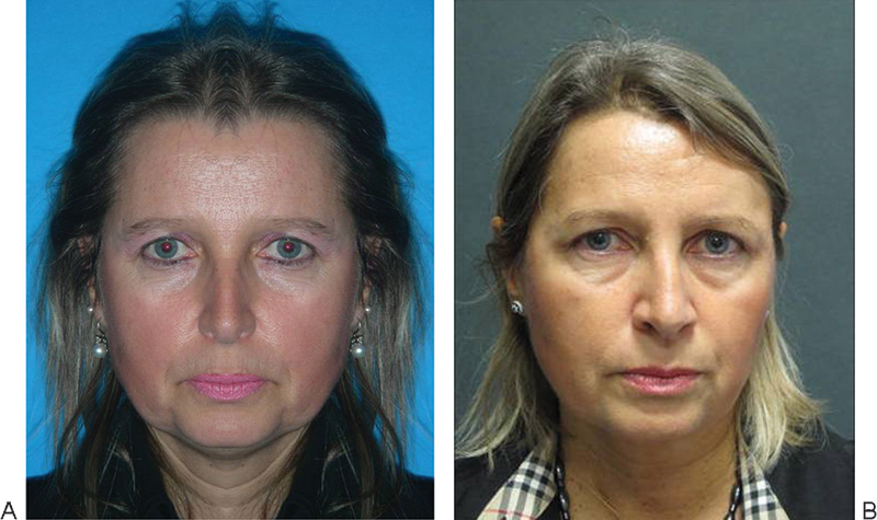
The postoperative facial defects of 19 patients (mean age, 30.4 years; range, 17 to 53 years; 12 males and 7 females) were managed by facial fat grafting. The objective evaluation was possible by comparing the VIF to the postoperative face with the aid of a software package. We obtained a similar facial morphology in 16 patients (84%), who received scores of ≥ 3 (Figs. 12345).
Figure 1.
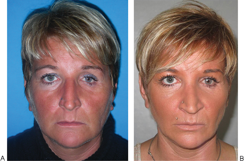
(A) Patient surgically treated for a complex left zygomatic-orbital fracture, which led to an “operated look” due to the deep hollowness around the left eye and the atrophy of the malar projection. (B) Postoperative face at 1-year follow-up characterized by a full and convex left upper eyelid with a direct flow from the lower lid to a well projected malar area.
Figure 2.
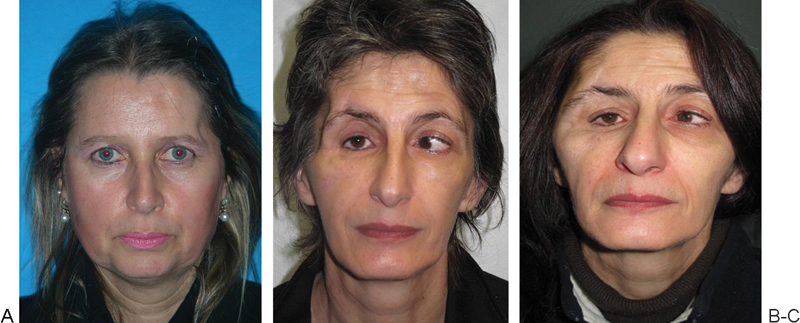
(A) Patient treated by open reduction and internal fixation for a left fronto-naso-orbital fracture by a coronal approach, which led to a concave temporal area and a visible orbital ridge. (B) Virtual ideal face obtained by preoperatively mirroring the healthy side of the face. (C) Nine months postoperatively showing the full area over the temporal muscle with an imperceptible orbital ridge.
Figure 3.
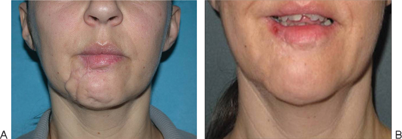
(A) Patient treated surgically by open reduction and internal fixation for a complex orbitozygomatic fracture that resulted in a skeletonized facial profile. (B) Ten months postoperatively demonstrating the correction of the defect.
Figure 4.
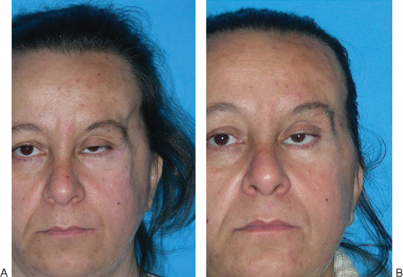
(A) Posttraumatic deformity of the lower lip after a soft tissue injury associated to dentoalveolar fracture. (B) Managed successfully by fat grafting.
Figure 5.
(A) Complex craniofacial injury that resulted in a concave temporal area (B) corrected by fat transplantation.
Three patients were scored as different (< 3); due to fat absorption, two patients underwent a second operation and one patient had a third touch-up procedure. In the latter cited cases, the loss of facial soft tissue caused by the trauma was significant and the patients refused our preoperative indication of facial implant due to the high costs of alloplastic materials; thus we opted for a large amount of fat grafting, conscious of the limitations of this procedure for large facial defects.
Apart from the standard postoperative discomforts such as bruising, edema, and pain, we did not observe major complications except for a superficial abdominal hematoma, which needed a percutaneous surgical drainage at 2 days postoperatively.
All patients were satisfied with the postoperative facial morphology and with the fast recovery time; we did not have any complaints.
Discussion
We described a retrospective study of fat grafting used to correct peculiar facial deformities after trauma. The healing of the soft tissue on an operated skeleton is unpredictable; we used fat grafting to ensure a proper long-term follow-up. We analyzed our results with the aid of a software package that enabled us to make quantitative measurements. This method was similar to the technique developed by Magro-Filho et al and Giangreco et al to analyze photographs of orthognathic patients.11,12
Coleman's technique of fat transplantation is characterized by the placement of parcels of purified fat by multiple passes with blunt cannulas; at the beginning it was introduced to improve facial aesthetics, and recently it was converted to perform reconstructive procedures.6,7 It is currently used to manage posttraumatic deformities, to refine craniofacial anomalies, and as an ancillary reconstructive procedure after tumor resections. Clinical studies in the field of fat grafting are very rare. However, scientific literature describes an absorption rate ranging from a 20 to 60%.13,14,15
Fat grafting is generally considered as a safe procedure with low morbidity when compared with conventional open surgical techniques; however, apart from bruising and swelling, postoperative complications can occur with a wide range of severity: aesthetic irregularities, infection, damage to nerves and vessels, and intravascular embolization. At the beginning of the transplantation process, fat grafting does not maintain any vascular connections with the blood vessels and the fat cells draw nourishment only with direct contact. It is therefore clear that if the fat is injected in large quantities, not all fat cells are in contact with the vascularized tissue, but rather the majority will be isolated within the “bolus,” infiltrated without the possibility of survival.11,12,13
The current concept regarding the adipose tissue of the face consists of two points; it resides in well-defined regions of the deep and superficial planes bounded by septa that support a blood supply. This experimental concept based on the clinical observation of the morphological changes between adjacent areas during the aging process leads to the idea of anatomic facial subunits that act differently and independently.17,18
These studies can be the key to determine the role of fat grafting as a fundamental step in treating posttraumatic maxillofacial deformities and to establish future protocols of total, subtotal, and segmental fat grafting for facial rehabilitation during a long-term posttraumatic follow-up. Although we could not perform statistical evaluation, this retrospective clinical study highlights the potentiality of fat grafting and enriches the field of cosmetic/reconstructive literature.
Conclusion
Fat grafting can be considered a very powerful tool to correct posttraumatic maxillofacial deformities and it permits a long-term clinical follow-up. Moreover, it can avoid other conventional reconstructive procedures with less morbidity for the patients and less costs for the society.
To the best of our knowledge, fat grafting is well discussed by the scientific literature in the fields of craniofacial anomalies and facial rejuvenation. However, poor attention is given to the pathological changes of the soft tissue of a posttraumatic face although it is a proper osteosynthesis of the facial bones. More complex prospective studies, enriched by sophisticated radiological controls, are needed to fully understand the biological behavior of the transplanted fat in the posttraumatic face.
References
- 1.Neuber G A Fett trasplantation Verl Dtsch Ges Chir 1893666 Long Vern [Google Scholar]
- 2.Illouz Y G. The fat cell “graft”: a new technique to fill depressions. Plast Reconstr Surg. 1986;6:122–123. [PubMed] [Google Scholar]
- 3.Illouz Y G. Present results of fat injection. Aesthetic Plast Surg. 1988;6:175–181. doi: 10.1007/BF01570929. [DOI] [PubMed] [Google Scholar]
- 4.Fournier P F. Facial recontouring with fat grafting. Dermatol Clin. 1990;6:523–537. [PubMed] [Google Scholar]
- 5.Fournier P. Microlipoextraction et microlipoinjection. Rev Chir Esthet Lang Fr. 1985;6:40. [Google Scholar]
- 6.Coleman S R. Long-term survival of fat transplants: controlled demonstrations. Aesthetic Plast Surg. 1995;6:421–425. doi: 10.1007/BF00453875. [DOI] [PubMed] [Google Scholar]
- 7.Coleman S R. Facial recontouring with lipostructure. Clin Plast Surg. 1997;6:347–367. [PubMed] [Google Scholar]
- 8.Zuk P A, Zhu M, Mizuno H. et al. Multilineage cells from human adipose tissue: implications for cell-based therapies. Tissue Eng. 2001;6:211–228. doi: 10.1089/107632701300062859. [DOI] [PubMed] [Google Scholar]
- 9.Yuksel E, Weinfeld A B, Cleek R. et al. De novo adipose tissue generation through long-term, local delivery of insulin and insulin-like growth factor-1 by PLGA/PEG microspheres in an in vivo rat model: a novel concept and capability. Plast Reconstr Surg. 2000;6:1721–1729. doi: 10.1097/00006534-200004050-00018. [DOI] [PubMed] [Google Scholar]
- 10.Suga H, Eto H, Aoi N. et al. Adipose tissue remodeling under ischemia: death of adipocytes and activation of stem/progenitor cells. Plast Reconstr Surg. 2010;6:1911–1923. doi: 10.1097/PRS.0b013e3181f4468b. [DOI] [PubMed] [Google Scholar]
- 11.Magro-Filho O Magro-Ernica N Queiroz T P Aranega A M Garcia I R Jr Comparative study of 2 software programs for predicting profile changes in class III patients having double-jaw orthognathic surgery Am J Orthod Dentofacial Orthop 20106e1–e5.; discussion 452-453 [DOI] [PubMed] [Google Scholar]
- 12.Giangreco T A, Forbes D P, Jacobson R S, Kallal R H, Moretti R J, Marshall S D. Subjective evaluation of profile prediction using video imaging. Int J Adult Orthodon Orthognath Surg. 1995;6:211–217. [PubMed] [Google Scholar]
- 13.Lam S M. Fat transfer for the management of soft tissue trauma: the do's and the don'ts. Facial Plast Surg. 2010;6:488–493. doi: 10.1055/s-0030-1267723. [DOI] [PubMed] [Google Scholar]
- 14.Clauser L, Polito J, Mandrioli S, Tieghi R, Denes S A, Galiè M. Structural fat grafting in complex reconstructive surgery. J Craniofac Surg. 2008;6:187–191. doi: 10.1097/scs.0b013e31815c94f5. [DOI] [PubMed] [Google Scholar]
- 15.Ducic Y Fat grafting in trauma and reconstructive surgery Facial Plast Surg Clin North Am 20086409–416., v-vi [DOI] [PubMed] [Google Scholar]
- 16.Schaverien M V, Pessa J E, Rohrich R J. Vascularized membranes determine the anatomical boundaries of the subcutaneous fat compartments. Plast Reconstr Surg. 2009;6:695–700. doi: 10.1097/PRS.0b013e31817d53fc. [DOI] [PubMed] [Google Scholar]
- 17.Lambros V Models of facial aging and implications for treatment Clin Plast Surg 20086319–327.; discussion 317 [DOI] [PubMed] [Google Scholar]
- 18.Mendelson B C, Freeman M E, Wu W, Huggins R J. Surgical anatomy of the lower face: the premasseter space, the jowl, and the labiomandibular fold. Aesthetic Plast Surg. 2008;6:185–195. doi: 10.1007/s00266-007-9060-3. [DOI] [PubMed] [Google Scholar]


