Abstract
The p53 tumor suppressor is a key transcription factor regulating cellular pathways such as DNA repair, cell cycle, apoptosis, angiogenesis, and senescence. It acts as an important defense mechanism against cancer onset and progression, and is negatively regulated by interaction with the oncoprotein MDM2. In human cancers, the TP53 gene is frequently mutated or deleted, or the wild-type p53 function is inhibited by high levels of MDM2, leading to downregulation of tumor suppressive p53 pathways. Thus, the inhibition of MDM2-p53 interaction presents an appealing therapeutic strategy for the treatment of cancer. However, recent studies have revealed the MDM2-p53 interaction to be more complex involving multiple levels of regulation by numerous cellular proteins and epigenetic mechanisms, making it imperative to reexamine this intricate interplay from a holistic viewpoint. This review aims to highlight the multifaceted network of molecules regulating the MDM2-p53 axis to better understand the pathway and exploit it for anticancer therapy.
Keywords: oncogene, tumor suppressor, MDM2-p53 interaction, cancer therapy
INTRODUCTION
Malignant transformation of a cell is attributed to a series of genetic and epigenetic events involving alterations in several oncogenes, tumor-suppressor genes, or microRNA genes, typically, in somatic cells[1]–[3]. The genomic instability resulting from the accumulation of multiple lesions leads to changes in cell signaling, gene expression and cell cycle progression culminating in the malignant phenotype which is characterized by sustained proliferative potential, evasion of growth suppressors, resistance to cell death and replicative mortality, increased angiogenesis, and activation of invasion and metastasis[4].
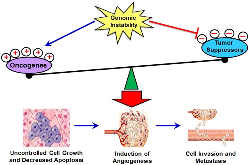
Oncogene activation and tumor suppressor gene inactivation are the most widely studied mechanisms for cancer development and progression (Fig. 1), and as such, oncogenes and tumor suppressor genes have been identified and validated as viable therapeutic targets[1]–[3]. Oncogenes typically encode cell proliferation and apoptosis controlling proteins, and are usually activated by mutation or gene fusion, by association with enhancer elements, or by amplification. On the other hand, tumor suppressor genes typically activate antiproliferative and pro-apoptotic pathways, thus protecting the cell from advancing on the path to cancer. When such a gene is mutated causing a partial or total loss of function, the cell can progress to cancer, usually in combination with other genetic changes[5]–[7]. Typically, hematopoietic tumors or soft tissue sarcomas are initiated by oncogene activation followed by tumor suppressor inactivation and other genetic changes while the reverse sequence is seen in carcinomas[1].
Fig. 1. Oncogenes, tumor suppressor, and cancer.
Genomic instability caused by various factors such as viruses, cytotoxic drugs, and ionizing radiation triggers mutations in oncogenes or tumor suppressor genes and perpetuates the unstable genome on the way to malignancy. Besides mutations, other genetic alterations responsible for oncogene activation include amplification (egfr, mdm2, myc), translocation (bcr/abl), protein overexpression (MDM2, Ras) and increased protein stability (Ras). Alterations leading to tumor suppressor inactivation include loss-of-function mutations (Rb, p53), deletions (p53, DCC). Epigenetic changes such as promoter methylation can also lead to tumor suppressor inactivation (IL-2Rγ).
Oncogenes, as compared to tumor suppressor genes, present a more viable therapeutic target since it is easier to inhibit an increased activity than to restore one which is lost. Oncogenic proteins in cancer cells can be targeted by small molecules and, when the oncogenic protein is expressed on the cell surface, by monoclonal antibodies[1]–[3]. Several small molecules targeting oncogenes have been developed such as imatinib (targeting ABL/PDGFR in chronic myelogenous leukemia), erltotinib (targeting EGFR in non-small lung cancer), sorafenib (targeting FLT3 kinase in renal cell carcinoma), lapatinib (targeting Her2/neu in breast cancer) and sunitinib (targeting VEGFR/FLT3 kinase in gastrointestinal tumors) which are presently in the clinic[1]–[3]. Monoclonal antibodies such as trastuzumab (against Her2 in breast cancer) and bevacizumab (against VEGF) also are routinely used in cancer therapy[1].
The most widely studied tumor suppressor is p53 and nearly sixty-thousand articles have been published in the past thirty-three years since its discovery[8]. The protein p53 is a potent transcription factor that is activated in response to diverse stresses and environmental insults, leading to induction of cell-cycle arrest, apoptosis or senescence. Thus, the main function of p53 is to restrain the emergence of transformed cells with genetic instabilities, acting as the ‘guardian of the genome’. In normal cells, p53 is kept at low levels by murine double minute 2 (MDM2), an ubiquitin ligase. MDM2 and p53 form a negative-feedback loop, in which p53 induces the expression of MDM2, which in turn promotes the degradation of p53 and quenches cellular p53 activity[9]. Around 50% of human cancers possess a mutated form of p53 while more than 17% of tumors exhibit mdm2 gene amplification; with these alterations, separately or concomitantly, leading to poor prognosis and treatment failure[8],[10],[11]. For these reasons, the MDM2-p53 interaction seems to be a major target for cancer therapy, and indeed has been focal point of research in both academia and the industry to develop better targeted cancer therapeutics.
p53 BIOLOGY
The p53 tumor suppressor gene was reported in 1979 as a cellular partner of simian virus 40 large T-antigen and the first human cDNA clones of p53 were isolated in the early 1980s[8],[11]. The p53 protein consists of 393 amino acids and is named so because it migrates as a 53 kD band in gel electrophoresis[8],[11]. Early studies demonstrated the importance of p53 as a tumor suppressor in both tissue culture as well as animal models. Both the alleles of the p53 gene are mutated or deleted in human cancers while, in mice, deletion of the p53 gene predisposes the animals to cancer[8],[11]. In fact, p53 mutations are seen in more than 50% of all human cancers, being highly prevalent in cancers of the breast and the prostate, and melanomas wherein these mutations correlate with poor prognosis and increased chemoresistance[8],[11].
Studies in lower organisms with no obvious need for cancer suppression have established the important role which p53 plays in normal development and growth, acting as a protector of the germline. As a tumor suppressor p53 protects cells from transformation and tumorigenesis by activating the transcriptional expression of downstream target genes whose protein products induce cell growth arrest, apoptosis or senescence in response to stress signals[12]. The p53 protein activates genes regulating normal cell cycle progression (especially the cell cycle checkpoint related genes) as well as genes maintaining genomic integrity. Thus, by coordinating with elements of the DNA damage response, p53 induces cell cycle arrest and/or apoptosis. Genotoxic stresses, as a result of ionizing radiation or chemotherapeutic drugs, increase p53 levels, leading to G1 or G2/M phase arrest and subsequent apoptosis, if DNA repair cannot restore the normalcy of the cell. This is due to the ability of p53 to upregulate cell cycle proteins such as GADD45, p21, as also pro-apoptotic proteins such as BAX and PUMA[12]. CDC2/cyclin E activity is essential for entry into mitosis, and this activity can be inhibited by p21 or GADD45 resulting in G2/M phase arrest[13].Induction of cellular senescence via the p21-Rb-E2F pathway in response to DNA damage, oxidative stress or telomere erosion is yet another mechanism whereby p53 activation curbs the tumorigenic processes[8],[12],[13].
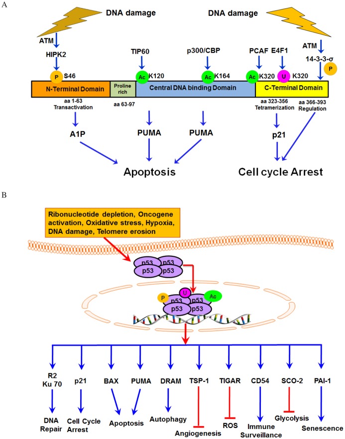
Fig. 2 illustrates the simplified structure and basic functions of p53 (Fig. 2A) and selected representative p53-interactive proteins (Fig. 2B). The p53 tumor suppressor plays a role in almost all types of DNA repair systems and is known to interact with Ape/ref-1, OGG1, and Polβ (base excision repair components). It also is involved in the ATM mediated induction of Ku70, a protein involved in non-homologous end joining; Ku70 interacts with BAX to inhibit its mitochondrial translocation and oligomerization leading to cell survival. The components of the mismatch repair system and the nucleotide excision repair are also upregulated by p53 in response to DNA damage[14]. The nature of the phenotypic responses to p53 activation is, at least partially, proportionate to the severity and nature of the activating signal. As shown in Fig. 2A, severe stresses induce more extreme and irreversible responses such as apoptosis and senescence, whereas milder stresses lead to a transient growth arrest coupled with an attempt to repair the damage caused.[12]
Fig. 2. p53, a tumor suppressor.
A: Selective Impact of p53 Modifications. Exemplary post-translational notifications via phosphorylation (P), acetylation (Ac), or ubiquitination (Ub) are depicted, which result in a specific cellular outcome in response to p53 activation and preferential activation of indicated target genes. E4F1 is an atypical ubiquitin ligase that modulates the p53 functions independently of degradation. E4F1-dependent Ub-p53 conjugates are associated with chromatin, and this induces a p53-dependent transcriptional program eliciting cell cycle arrest but not apoptosis. Following ATM activation, 14-3-3-σ is induced, and this causes dephosphorylation of p53 at S-376. HIPK2 induced S46 phosphorylation in p53 is essential for mediating its apoptotic functions. B: p53 contributes to multiple cellular processes in response to various cellular stresses via regulation of downstream targets and/or signaling pathways.
Additionally, p53 can also act as a transcriptional repressor, notably in the case of c-fos, myc, VEGF-A, and survivin gene expression-all of which modulate proliferation, survival, and angiogenesis pathways in a positive manner[14]–[16]. Many studies have also identified several microRNAs, most notably members of the miR-34 family, as being subject to transcriptional regulation by p53. Increased miR-34a activity due to induction or transactivation by p53 triggers enhanced apoptosis and changes in the expression of genes related to cell cycle, apoptosis, DNA repair, and angiogenesis[17]. Thus, we believe that p53 acts as a master regulator that functions as a node in numerous cellular signaling pathways and is involved in functions as diverse as embryo implantation, DNA metabolism, apoptosis, cell cycle regulation, senescence, energy metabolism, angiogenesis, immune response, cell differentiation, motility and migration, and cell-cell communication (Fig. 2B)[12]–[14].
Evidence suggests that proteins such as MDM2[9], PIRH2[18],[19], COP1[18], and ARF-BP1[18] can bind to p53 and act as p53 ubiquitin ligases, thus resulting in its degradation[9],[12]. However, the most important negative regulator of p53 is MDM2, which inhibits its biochemical activity through a negative feedback control. In the following sections, we further discuss MDM2 and the MDM2-p53 interaction.
MDM2 BIOIOGY
The mdm2 gene was first identified as the gene responsible for the spontaneous transformation of an immortalized murine cell line, BALB/c 3T3[20]–[22]. Early cell culture studies demonstrated that mdm2 overexpression rendered rodent fibroblasts tumorigenic in nude mice, thus establishing it as an oncogene[14]. The mdm2 gene was subsequently cloned and mapped to chromosome 12q13-14[23] and found to contain two transcriptional promoter elements termed P1 and P2 with the latter being p53-dependent. The mdm2 gene is expressed as different isoforms[24]–[26] with the full-length transcript of this gene encoding a protein of 491 amino acids[27]. Under normal conditions, MDM2 is expressed in the nucleus, but it translocates to the cytoplasm to mediate the degradation of some of its targets by the proteasome[11], [24].
Studies have shown that the mdm2 gene was amplified in over a third of 47 sarcomas, including common bone and soft tissue cancers[10]. A variety of mechanisms, such as amplification of the mdm2 gene[10], single nucleotide polymorphism at nucleotide 309 (SNP309) in its gene promoter[28]–[32], increased transcription and translation[33],[34], account for MDM2 overexpression. In human cancers, MDM2 has been associated with poor prognosis (especially in solid tumors of the breast, lung, stomach and esophagus; liposarcomas, glioblastomas, and leukemias)[10],[11],[31]. MDM2 overexpression also correlates with metastasis and advanced forms of the disease in osteosarcomas, and cancers of the colon, breast and prostate, and is often associated with more treatment resistant tumors[35].
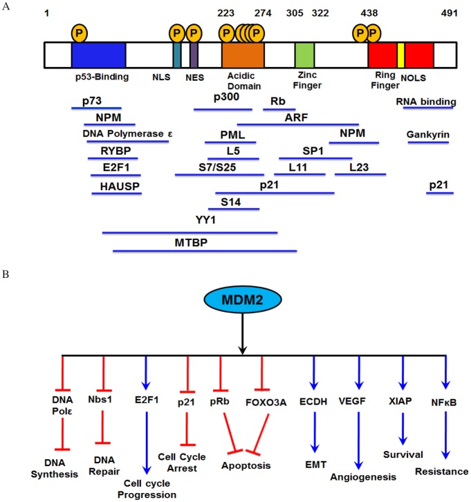
Fig. 3 depicts the basic structure and active domains (Fig. 3A) as well as its major cell functions and interactive partners (Fig. 3B). The activity and cellular localization of the evolutionarily conserved MDM2 oncoprotein is controlled by several known mechanisms[27]. The most widely studied mechanism being the p53-induced mdm2 transcription which is mediated via the P2 promoter, whereas basal transcription is initiated from the P1 promoter[36]. Additional transcription factors (such as NF-κB[37], Fli-ETS[38], IRF-8[27], SP1[27], and NFAT1[39]) as well as the Ras-Raf-MEK-MAPK[40] pathway can positively modulate the expression of MDM2 from either or both the P1 and the P2 promoters. On the other hand, the tumor suppressor PTEN decreases MDM2 expression, independent of p53[40]. Several microRNAs (miRNAs) such as miR-143, miR-145, miR-29 (through PI3K/Akt pathway) and miR-18b (upregulation of p53) have been proposed to block translation of MDM2 mRNA[41],[42].
Fig. 3. MDM2 as an oncogene.
A: MDM2 structure and binding sites for various interactive proteins. MDM2 protein domains and the cellular proteins interacting with different domains are listed. Blue region: p53 binding domain (aa 19-220); Teal blue region-Nuclear localization signal (NLS); Purple region: Nuclear export signal (NES); Orange region: Acidic domain (aa 223-274); Green region: Zinc finger domain (aa 305-322); Red region: RING finger domain (aa 438-478); Yellow region: Nucleolar localization signal (NOLS). B: MDM2 contributes to multiple processes leading to and promoting the development of cancer phenotype.
Another facet of MDM2 regulation involves post-translational modifications[43] including phosphorylation of the MDM2 protein by upstream molecules such as ATM (decreases MDM2 stability)[43]–[45] and Akt (increases MDM2 translocation from the cytoplasm into the nucleus, allowing p53 degradation)[46]–[49]. Other enzymes, such as CK2 and DNA-PK, as well as members of the Ras-Raf-MEK-MAPK pathway, also regulate MDM2 phosphorylation[27].
An increasing body of clinical and preclinical evidence suggests that MDM2 has important roles in the cell, independent of p53 (Fig. 3B). For example, MDM2 is able to affect processes such as DNA synthesis and repair by interaction with DNA polymerase ϵ[50],[51], DHFR[52], centrosome amplification[53] and the MRN DNA complex containing Nbs1[54],[55], etc. Similarly, MDM2 interacts with several proteins such as Rb/E2F-1 complex[55]–[57], the DNA methyltransferase DNMT3A[58], p107[59], MTBP[60],[61], the cyclin kinase inhibitor p21, independently of p53, and drives cell cycle progression (typically S-phase)[62],[63]. In an analogous fashion, the MDM2 oncoprotein interacts with the E2F1/Rb pathway to inhibit apoptosis[55]. MDM2's anti-apoptotic roles also include its interaction with well-known apoptosis mediators such as p73 (MDM2 mediates p73 NEDDylation and prevents p53 transactivation)[55],[64] and FOXO3a (MDM2 decreases FOXO3a protein stability)[65]. MDM2 upregulates the translation of anti-apoptotic XIAP, thus inactivating caspase-mediated apoptosis[66]. Therefore, MDM2 affects both pro-apoptotic as well as anti-apoptotic proteins.
Therefore, MDM2, in addition to being a negative regulator of p53, also affects the functions of other cellular proteins, which participate in pathways ranging from DNA repair to apoptosis to cell motility and invasion[27],[55],[67],[68]. However, most of the MDM2-protein interactions affect the steady-state levels of p53 in the cell, either directly or indirectly. Thus, it is evident that the MDM2-p53 interaction is at the heart of normal cell regulation, and has been studied minutely over the past few years.
MDM2-P53 AUTOREGULATORY FEED-BACK PATHWAY
As aforementioned, though MDM2 does have several p53-independent functions, the ability of MDM2 to act as an oncogene mainly stems from its capacity to bind the tumor suppressor p53 and to inhibit p53-mediated gene transactivation[11]–[13]. The proteasomal degradation of the p53 protein by MDM2 is essential to its repression of the tumor suppressor functions of p53, and many proteins intrude upon this activity, either enhancing or inhibiting it. Figure 4 shows the basic concept of the MDM2-p53 interaction, which was first established when the MDM2 protein was found to be physically associated with the tumor suppressor p53. Subsequent studies indicated that MDM2 overexpression decreased p53 levels in the cell, leading to the speculation that MDM2 is a negative regulator of p53. Furthermore, observations that MDM2 gene amplification is seen in several human sarcomas with wild-type p53 have established the validity of the hypothesis[10].
Fig. 4. The traditional MDM2-p53 regulatory pathway.
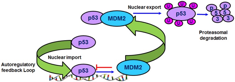
The feedback regulation involving the p53 and MDM2 is shown.
MDM2 targets p53 for ubiquitination and degradation by the proteasome[69]–[71], shuttles p53 out of the nucleus[69],[70], prevents p53 from interacting with transcriptional co-activators[72], and recruits transcriptional co-repressors to p53[73]–[75]. On the other hand, p53 regulates MDM2 oncoprotein expression by binding to its promoter[12],[13],[36]. The increased MDM2 levels cause it, in turn, to bind and inactivate p53 by directly blocking the p53 transactivational domain and by targeting the p53 protein for ubiquitin-dependent degradation by the proteasome[72],[73] (Fig. 4). This elegant autoregulatory loop helps to maintain low cellular levels of p53 in normal cells. The levels of p53 must be tightly controlled in unstressed cells since high levels of the anti-proliferative and pro-apoptotic p53 can be detrimental to normal cell growth and development[12].
The MDM2-p53 interaction was initially thought to result solely from the mutual binding of MDM2 and p53 via their N-terminal domains[75]. Recently, Poyurovsky et al. have discovered that alterations in the p53 C terminus (such as deletion, mutation or acetylation) can also affect the MDM2-p53 interaction[76]. In addition, the C-terminal RING finger domain MDM2 serves as an E3 ubiquitin ligase for p53 proteolysis and ubiquitinates p53 at several lysine residues[77]–[82]. Low levels of MDM2 activity induce the mono-ubiquitination and nuclear export of p53, whereas higher levels promote the poly-ubiquitination and nuclear degradation of p53[69],[70],[78]–[81]. MDM2's role in p53 regulation and in maintenance of life is further supported by the fact that targeted deletion of the mdm2 gene in mice is embryonically lethal[82]. These observations emphasize that the MDM2 interaction involves more than simple protein binding.
As can be envisaged, the MDM2-p53 interplay is a particularly attractive target for therapeutic intervention in cancer. Increasing the expression and activity of wild-type p53 is the ultimate goal in most treatment strategies, and therefore p53 gene therapy approaches have been enthusiastically pursued for several years. These include an adenovirus vector based p53 delivery system gaining approval in China in 2004 for the treatment of head and neck cancer[8]. Other strategies to restore wild-type p53 in the cell have been vaccines against mutant p53, small molecules that bind to mutant p53 to restore normal conformation and/or activity (e.g. ellipticine)[83]. Since MDM2 overexpression is seen in tumors containing wild-type p53, it has been postulated that attacking the MDM2-p53 interaction will help restore p53 levels and activity in the cancer cells, and an entire field of synthetic chemistry and pharmacology is dedicated to developing strategies to target this interaction for therapy[13].
MODULATORS OF THE MDM2-p53 PATHWAY
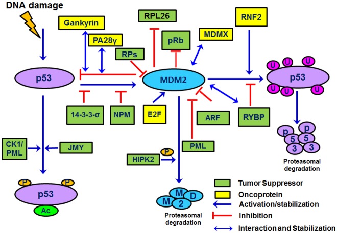
The MDM2-p53 feedback loop is crucial for restricting p53 levels and activity during normal cell physiology, and is tightly regulated by several other factors. These co-factors alter MDM2 or p53 conformation, binding, localization, expression, and modulate the E3 ligase activity of MDM2 towards itself, p53, and other substrates; consequently, regulating a variety of different cellular processes (Fig. 5). In the following section, we discuss exemplary cellular molecules that play a part in this interaction (Table 1).
Fig. 5. Several tumor suppressors and oncoproteins regulate the MDM2-p53 interaction.
Ribosomal proteins (RP-both the large subunit and small subunits) form a complex with p53 and MDM2 to inhibit MDM2-mediated p53 ubiquitination and stabilization of p53. ARF and PML sequester the MDM2 in the nucleolus, inhibiting MDM2 from binding and degrading p53. CK1 phosphorylates p53 at Thr18 in response to stress and DNA damage and, along with p53, localizes to the PML nuclear bodies. MDMX forms heteroligomers with MDM2 and induces p53 degradation. PA28γ protein interacts with both MDM2 and p53 proteins and promotes the MDM2-p53 interaction, leading to enhanced MDM2-mediated p53 ubiquitination and degradation. RYBP interacts with MDM2 to decrease MDM2-mediated p53 ubiquitination while RNF2 promotes p53 degradation. HIPK2, tumor suppressor (Ts) protein phosphorylates MDM2, promoting its proteasomal degradation while the Rb Ts forms a ternary complex with p53 and MDM2.
Table 1. MDM2-interactive proteins and the biological effects of the interaction.
| Protein name | Consequence of interaction on p53/MDM2 | Ubiquitination by MDM2 | Biological consequence of the interaction with MDM2 or p53 | Reference |
| 14-3-3-σ | MDM2 stability decreased, translocation to cytoplasm; p53 stability increased | None | P53 activation induces 14-3-3-σ causing G2/M phase arrest | [100] |
| p14 (ARF) | MDM2 activity decreased, MDM2 localized to the nucleolus; p53 stability increased. | Not reported | ARF localizes MDM2 to the nucleus preventing MDM2-p53 interactions while promoting rapid MDM2 degradation. | [94]–[96] |
| p73 | Increased stability and transcription of p53 | No; MDM2 promotes p73 NEDDylation | Increased apoptosis and cell cycle arrest due to increase in p53 stability | |
| Caspase-2 | Cleaves MDM2 at asp 367 leading to loss of C-terminal RING domain and increases p53 stability | None | Upon DNA damage, p53 induces the caspase-2-PIDDosome creating a positive feedback loop that inhibits MDM2 and reinforces p53 stability and activity, contributing to cell survival and drug resistance. | [126] |
| Gankyrin(PSM10) | E3 ligase activity of MDM2 increased; enhanced ubiquitination and degradation of p53 | None | Increased cell proliferation and decreased apoptosis due to decrease in p53 stability | [122] |
| HAUSP/USP7 | MDM2 stability increased due to de-ubiquitination p53. Stability decreased due to increased MDM2-mediated ubiquitination | MDM2 is deubiquitinatedby HAUSP | Increased cell proliferation and decreased apoptosis | [123],[124] |
| HIPK2 | HIPK2 and p53 co-localize with PML-3 into the nuclear bodies and cooperate in the activation of p53-dependent transcription and induction of apoptosis | Monoubiquitination at lysine 1182 | Increased apoptosis and cell cycle arrest due to p53 activation | [103]–[106] |
| IGF-1R | IGF-1R loss reduces translational synthesis of p53 and MDM2 protein. IGF-1R inhibition increases p53 protein stability by reducing p53 ubiquitination, decreases p53 synthesis, Z thus rendering p53 insensitive to stabilization after DNA damage | Polyubiquitination | Increased apoptosis and cell cycle arrest on IGF-1R overexpression | [127] |
| JMY | Augments p53 response to DNA damage. | Polyubiquitination | Induces p53 mediated cell cycle arrest and apoptosis; affects cell motility | [101],[102] |
| Merlin | Induces MDM2 degradation through its N-terminal and stabilizes p53 | Not reported | Decreased cell proliferation due to increase in p53 stability | [128] |
| MDMX | Hetero-oligomerization of MDM2 and MDMX via their RING domains suppresses p53 activity | Polyubiquitination | Increased cell proliferation and decreased apoptosis | [85]–[87] |
| NUMB | MDM2 increases its degradation and increases p53 activity | Monoubiquitination | Not well understood | [120] |
| Nucleosteo-min | Nucleoplasmic mobilization of nucleostemin stabilizes MDM2; decreases p53 transcriptional activity | Not reported | Decreased apoptosis and cell cycle arrest due to decreased p53 transcription | [129] |
| Nucleophos-min (NPM,B23) | NPM inhibits binding of p53 with MDM2 | Not reported | Increased apoptosis and cell cycle arrest due to p53 activation | [97] |
| PA28γ | Decreases stability of p53 | Increased ubiquitination of p53 | Enhanced the proteasomal degradation of various proteins involved in the cell cycle, leading to cell proliferation | [125] |
| PML | Decreases ubiquitinating ability. protects p53 from MDM2-mediated inhibition and degradation. | None | Increased apoptosis and cell cycle arrest due to increased accumulation of p53 in the cell | [99] |
| PCAF | Inhibits binding of MDM2 with p53; stimulates MDM2 auto-ubiquitination; Acetylates p53 in response to DNA damage; MDM2 increases its proteasomal degradation | Monoubiquitination | Increased apoptosis and cell cycle arrest due to activated p53 | [130] |
| Retinoblastoma protein (Rb) | Decreased expression and/or inhibition; P53-MDM2-Rb trimeric complex modulates pro-apoptotic function of p53 | None (some studies report poly-ubiquitination of Rb) | MDM2 overexpression inhibits Rb causing increased cell proliferation and decreased apoptosis; | [55]–[57] |
| Siva-1 | Increases MDM2-mediated p53 degradation. | None | Increased cell proliferation and decreased apoptosis due to decrease in p53 stability | [131] |
| Tip60 | Localization to PML bodies; decreased MDM2-mediated NEDDylation of p53; p53 acetylation promoted | Polyubiquitination | Increased apoptosis and cell cycle arrest due to activated p53 | [55],[132] |
| YY1 | YY1 promotes the assembly of the p53-Mdm2 complex. disrupts the interaction between p53 and the coactivator p300, blocks p300-dependent acetylation and stabilization of p53. | None | Increased cell proliferation and decreased apoptosis | [133] |
MDMX
MDMX, a splice variant of MDM2, possesses a high degree of homology to MDM2, especially in its N-terminal p53 binding domain and both proteins are believed to have non-redundant roles in maintaining low levels of p53 in the normal cell[84]–[88]. MDMX also directly binds to the transactivation domain of p53 and inhibits p53 activity, but does not induce p53 degradation. MDMX is overexpressed in several cancers and it heterodimerizes to MDM2 via its RING finger domain at its C-terminus[85],[89],[90], thereby modulating its E3 ligase activity. MDM2 and MDMX are proposed to form a complex that is more effective at inhibiting p53 transactivation or enhancing p53 turnover[84]–[87],[90],[91]. MDM2 can also directly ubiquitinate and degrade MDMX upon DNA-damage stimuli[86].
ARF
One of the first proteins discovered to interact with the MDM2-p53 loop was ARF, an alternate reading frame protein expressed from the INK4a locus. The ability of MDM2 to target p53 for proteolytic degradation is inhibited by ARF[92],[93]. This ARF-MDM2 interaction blocks MDM2 from shuttling between the nucleus and cytoplasm and sequesters MDM2 in the nucleolus; preventing it from degrading p53 resulting in the indirect activation of p53[92]–[96]. Conversely, ARF dysregulation may cause malignant transformation by increasing MDM2 levels[93]. The ARF (p14/p19) protein also increases MDM2 SUMOylation in a p53-independent manner[95]. While the SUMOylation of MDM2 by ARF does not appear to affect the MDM2-p53 loop, it may affect the p53-independent activities of MDM2[95].
Nucleophosmin (NPM)
The protein nucleophosmin (NPM) competes for the binding of MDM2 with p53 and can stabilize ARF, increasing its concentration in the nucleolus and resulting in decreased p53 degradation[97]. NPM and MDM2 have been shown to bind to the same region of p53, resulting in decreased p53 ubiquitination[97]. However, NPM also has several other p53 independent effects, and studies show that overexpression of NPM can enhance proliferation and oncogene-mediated transformation by c-Myc modulation; therefore, targeting NPM for cancer therapy may be controversial[27].
Promyelocytic Leukemia (PML)
The protein Promyelocytic leukemia protein (PML) mediates the localization of proteins to the nucleus. It is responsible for protecting p53 from MDM2-mediated ubiquitination by sequestering MDM2 in the nucleus[98],[99]. Casein kinase 1 (CK1) also plays a role in PML-mediated p53 protection by phosphorylating p53 at Thr18 in response to DNA damage and causing its localization to the PML nuclear bodies, thus protecting it from MDM2-mediated degradation[99].
14-3-3-σ
DNA damage activates several proteins, some of which are p53 downstream targets. The 14-3-3-σ protein is one such downstream target of p53 that is expressed following exposure to radiation[100]. It negatively regulates cell cycle progression through interactions with CDK2/4 and CDC2, preventing the cyclin-CDK interaction and causing G2 phase cell cycle arrest[100]. This protein can also decrease p53 degradation via an increase in MDM2 auto-ubiquitination and degradation, as well as by causing the translocation of MDM2 to the cytoplasm[100].
JMY
DNA damage also increases the accumulation of JMY, a p53 co-transcription factor. During DNA damage induced p53 response, JMY forms a DNA damage-dependent complex in the nucleus with the p300 co-activator and the MDM2 oncoprotein[101]. JMY and p300 are recruited to p53 in a protein complex subsequent to DNA damage and cooperate in boosting the p53 response. JMY is degraded following ubiquitination by the MDM2 RING domain[101]. Intriguingly, JMY has been recently reported to control cadherin expression and actin nucleation, thus influencing cell motility and invasion, finally integrating cytoskeletal events and cellular motility with the DNA damage response[102].
HIPK2
HIPK2 is a tumor suppressor that promotes apoptosis by modulating factors, directly or indirectly related to p53, such as the antiapoptotic transcriptional corepressor CtBP, the p53 inhibitor MDM2 and ΔNp63α[103]. HIPK2 phosphorylates MDM2 for proteasomal degradation, and may overcome the MDM2-induced p53 inactivation restoring p53 apoptotic activity. On the other hand, an interesting regulatory circuitry between MDM2 and HIPK2/p53 axis reveals that sub-lethal DNA damage leads to HIPK2 inhibition by a protein degradation mechanism which involves p53-induced MDM2 activity. These findings indicate a role for MDM2 to fine-tune the p53-mediated biological outcomes (that is, cell cycle arrest vs apoptosis), according to the requirements. This may also explain p53 inactivation in tumors overexpressing MDM2, regardless of the presence of wild-type p53[103]–[106].
Ribosomal Proteins
MDM2 is also prevented from targeting p53 for proteolytic degradation by a subset of ribosomal proteins. MDM2 is involved in the ribosome biogenesis occurring in both the cell cytoplasm and in the nucleolus of eukaryotic cells[107]. Several ribosomal proteins (both from the large as well as the small subunits), such as RPL5[108], RPL11[109], RPL23[110], RPS7[111],[112], RPS14[113], RPS25[114] and RPS 27/RPS27L[115],[116], have been shown to have a role in the regulation of the MDM2-p53 feedback loop in response to ribosomal stress. RPL5, RPL11, RPL23, and RPS14 have been shown to bind to the central acidic domain of MDM2 to inhibit its E3 ubiquitin ligase activity toward p53[116]. Furthermore, the S7 and S25 proteins bind MDM2 as well as p53, forming a ternary complex of MDM2-p53-ribosomal protein, which prevents p53 ubiquitination. Furthermore, the S7 protein has been demonstrated as a substrate for MDM2 E3 ligase in addition to it being a regulator of MDM2 mediated p53 degradation[111],[112]. Overexpression of these ribosomal proteins elevates p53 levels and transcriptional activity, leading to G1 or G2 arrest, reduced cell proliferation, and increased apoptosis.
Polycomb Proteins
Another tumor suppressor that activates p53 by destabilizing MDM2 is the pro-apoptotic polycomb group (PcG) RYBP (RING1-and YY1-binding protein), an ubiquitin-binding protein[117]. RYBP interacts with MDM2 to decrease MDM2-mediated p53 ubiquitination, leading to stabilization of p53 and an increase in p53 activity, leading to cell cycle arrest[117]. Contrastingly, another polycomb complex protein RNF2, also known as Ring1B/Ring2 is seen to bind with both p53 and MDM2 in colon cancer cell lines and promote MDM2-mediated p53 ubiquitination. RNF2 overexpression also increases the half-life of MDM2 and inhibits its ubiquitination[118],[119]. These observations indicate that polycomb proteins play important roles in p53/MDM2 regulation and may present novel targets for cancer therapy or prevention.
Proteasome-associated Proteins
From our earlier discussions, it is evident that MDM2's role as a p53 negative regulator stems from its ubiquitin ligase activity. MDM2 functions as an E3 ligase that ubiquitinates p53 at several lysine residues[70],[71],[77]–[79]. In addition to ubiquitinating p53, it also has the ability to ubiquitinate itself[81] and various other substrates, such as NUMB[120], pRb[55], and MDMX[85]. The protein CSN5, a part of the COP9 signalosome and a regulator of cell cycle proteins such as p27, has been shown to increase p53 proteasomal degradation by promoting p53 nuclear export and decreasing MDM2 auto-ubiquitination and degradation[121]. The property of MDM2 to ubiquitinate varied substrates as well as auto-ubiquitinate itself seems to be an attractive approach for developing targeted therapy.
Several proteasome-associated proteins other than MDM2 also associate with p53, affecting the MDM2-p53 interaction. For example, gankyrin, a seven-repeat protein associated with the 19S regulatory complex of the 26S proteasome and commonly overexpressed in early hepatocarcinogenesis facilitates the MDM2-p53 interaction by binding to MDM2, resulting in increased p53 ubiquitination and degradation[122]. Gankyrin also enhances the auto-ubiquitination of MDM2 in the absence of p53[122]. A de-ubiquitinating protein, HAUSP (herpes virus-associated ubiquitin-specific protease, also known as USP7; ubiquitin specific protease 7), cleaves ubiquitin from p53, thus stabilizing it[123]. Interestingly, it was later found to bind to MDM2 as well and increase MDM2 levels and stability by rescuing it from ubiquitination, resulting in p53 destabilization[124]. The dual control of p53 and MDM2 by HAUSP indicates a complex p53-MDM2-HAUSP regulatory pathway.
The proteasome activator PA28γ also regulates the MDM2-p53 interaction (independent of its proteasome-activator function) and serves as a co-factor for p53 degradation[125]. In addition, PA28γ binds p21 to regulate its degradation in an ubiquitin-independent manner. It also binds to the cell cycle control proteins p14/p19ARF and p16 (INK4A)[125]. These observations suggest that the MDM2-interactive proteins, such as PA28γ, p21, and p14ARF, may form a complex to enhance the proteasomal degradation of the various proteins involved in the cell cycle.
TARGETING THE MDM2-P53 PATHWAY FOR CANCER THERAPY: MORE THAN BINDING SITES
Our discussion, so far, has established that the tumor suppressor p53, in response to cellular stress, is activated and mediates responses such as cell cycle arrest, apoptosis, senescence and differentiation, thereby limiting malignant progression. The main regulator of p53 is the E3 ubiquitin ligase MDM2, which binds to p53's transactivation domain and functions by both preventing p53's transcriptional activity and targeting it for degradation. Activation of p53 in a tumor cell by antagonizing its negative regulator MDM2 or targeting the MDM2 oncogene itself offers a viable therapeutic strategy, and proof-of-concept experiments have already demonstrated the feasibility of this approach in vitro[134]–[136].
Strategies to target MDM2
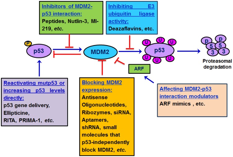
The major strategies (Fig. 6) that have been used for targeting the MDM2-p53 interaction are as follows:
Fig. 6. General strategies to inhibit the MDM2-p53 interaction.
RITA= Reactivation of p53 and induction of tumor apoptosis. Ellipticine binds to mutant p53 to restore normal conformation and/or activity; PRIMA-1 reactivates mutp53 by covalent binding to the core domain.
Blocking MDM2 expression. Inhibition of the MDM2 oncoprotein can limit its interaction with p53, thus preventing p53 degradation and resulting in higher levels of p53 in cells. Several gene silencing techniques (discussed later in this section) have already proved the effectiveness of such an approach.
Inhibiting MDM2-p53 binding. Inhibition of MDM2-p53 binding appears to be a desirable strategy for p53 stabilization and activation. However, targeting protein-protein interactions by small molecules is a challenging task. Protein-protein interactions usually involve large and flat surfaces that are difficult to disturb by low molecular weight compounds[13],[137]–[139]. However, in the case of the p53-MDM2 interaction, it has been demonstrated that only three amino acid residues, Phe19, Trp23 and Leu26 of p53, are crucial for the binding of the two proteins, and these are inserted into a deep hydrophobic pocket on the surface of the MDM2 molecule[13],[140],[141]. This protein architecture provides a framework to design small molecules that mimic this interaction. Several small molecule inhibitors such as nutlins[140]–[142], spiroxindoles[143], isolindones[144], and chalcone derivatives have been developed via combinatorial library screening, are based on this principle[144],[145].
Curtailing the E3 ubiquitin ligase activity of MDM2. MDM2 negatively regulates p53 by targeting the ubiquitin ligase activity of MDM2. A complementary approach to prevent p53 degradation by MDM2 is to develop agents designed to inhibit the E3 ligase activity of MDM2 directly so as to mimic the effects of ARF or the ribosomal protein L11. Recently, small-molecule inhibitors have been identified that specifically target the E3 ligase activity of MDM2[146]. The efficacy and molecular effects of these inhibitors on the biochemical functions of p53 still remain to be defined.
Gene Silencing Methods to Eliminate MDM2 Expression
Several early studies by our group using antisense oligonucleotides (ASOs) to inhibit MDM2 expression have established the proof-of-principle for the gene silencing approach for MDM2 inhibition in cells and mouse models of human cancer[134]–[136],[144]. These ASOs cause p53 stabilization and activation of the p53 pathway in cancer cells in tumor xenograft as well as cell culture in both p53 wild-type and mutant cells, possibly via the resulting p21 upregulation due to MDM2 inhibition[141]. Other gene targeting strategies include the use of MDM2 ribozymes, MDM2 aptamers, and RNA interference techniques[27],[147],[148]. All these techniques had antiproliferative and pro-apoptotic effects in the in vitro systems tested. However, none of the above approaches have subsequently progressed into preclinical or clinical development. Very recently in the past year, several groups have successfully used smart delivery approaches to deliver MDM2-siRNA for anticancer therapy. Reports from the Shizuoka University and Chinese Academy of Sciences indicate a successful delivery and accumulation of MDM2 siRNA into tumors by cationic liposomes and nanoparticles, respectively[149],[150]. These data suggest that targeted delivery of siRNAs by use of novel delivery approaches may have considerable potential for cancer treatment.
Small Molecule Inhibitors to Inhibit MDM2 Activity
Several different approaches have been taken to develop small molecule MDM2 inhibitors, with most efforts focused on the development of agents designed to inhibit the interaction between MDM2 and p53 (e.g. Nutlins, spiro-oxindoles, benzodiazepines and RITA-reactivation of p53 and induction of tumor cell apoptosis)[13],[27],[141],[143]–[145]. Most of these chemical entities possess the capability to displace p53 from MDM2 in vitro with nanomolar potency (IC50 = 90 nM for nutlin-3a). Crystal-structure studies demonstrate that nutlins bind to the p53 pocket of MDM2 in a way that remarkably mimics the molecular interactions of the three crucial amino acid residues from p53 (Phe19, Trp23 and Leu26)[13],[141].
Alternatively, the ubiquitin ligase activity inhibitors such as deazaflavins have been shown to inhibit the ubiquitination of p53 in vitro, with IC50 values in the 20-50 µM range. In cancer cells, they activate p53 signaling and induce apoptosis in a p53-dependent manner. They have no effect on the physical interaction between MDM2 and p53, suggesting that the mode of inhibition may be allosteric, perhaps by blocking a structural rearrangement of MDM2 necessary for p53 ubiquitination but not for MDM2 autoubiquitination[145],[146].
Additionally, several chemopreventive agents such as ginseng derived compounds, curcumin, and flavonoids such as genistein have been demonstrated to downregulate MDM2 oncoprotein expression. These compounds influence MDM2 levels in tumors with both wild-type p53 as well as mutant (non-functional) p53, thus indicating that their MDM2 blocking activities are independent of p53. Several compounds inhibit MDM2 interaction with other molecules, such as berberine which disrupts the MDM2-DAXX-HAUSP complex[151]. A comprehensive review on natural product inhibitors of MDM2 has appeared recently[151].
FUTURE DIRECTIONS
The p53-MDM2 interactions provide a focal point to improve cancer therapy. As MDM2 regulates p53 activity at the post-translational level, inhibition of the MDM2-p53 interaction permits an immediate p53-mediated response. Targeting the MDM2-p53 interaction directly and/or other cellular players that will ultimately increase functional p53 levels in the cell or decrease MDM2 levels are likely to offer viable approaches. The view that the MDM2-p53 interaction just constitutes binding of two proteins and the mutual regulation of one another is an extremely myopic view of the subject. The prime goal of p53 based cancer therapy has been to increase levels of functional p53 and/or inhibit MDM2 levels to prevent further p53 degradation. Compounds that mimic endogenous signaling components such as ARF, the ribosomal proteins which either sequester MDM2 preventing its interaction with p53 or that negatively affect its ubiquitinating capabilities present interesting strategies to overcome p53 attenuation in the cancer cell. In fact, the ubiquitin ligase modulators do exactly the same. Several transcription factors such as NFAT1 are known to upregulate MDM2 transcription; inhibition of these transcription factors may provide yet another strategy to inhibit MDM2 and increase p53 levels. A possible drawback of such an approach would be the undesired effects on other signaling pathways as transcription factors are known to regulate a broad spectrum of regulatory proteins. The interplay between MDM2 and MDMX presents yet another fascinating area of study. Emerging evidence indicates that MDM2 and MDMX possess both overlapping and non-overlapping roles in tumorigenesis, and that inactivation of only the MDM2-p53 interaction may not be able to protect the cell against the p53-inhibiting oncogenic activities of MDMX[152]–[155].
Disruption of the actual MDM2-p53 protein interaction with small molecule inhibitors is an attractive cancer therapeutic strategy but there still exist concerns as to how viable this concept would be therapeutically. For example, will it be possible to inhibit a protein-protein interaction with a drug selectively in human tumors? What are the consequences of increasing the p53 levels in normal tissues? Although clinical studies conducted on a nutlin series compound (RG7112) indicate good tolerance with dose escalation, long-term exposure to MDM2 inhibitors and toxicity upon repeated exposure need to be yet determined. Inhibitors of the MDM2-p53 interaction also present the risk for acquired resistance to p53 activation. Because expression of wild-type p53 is essential for the anti-cancer activity of MDM2 inhibitors, resistant clones of cancer cells may emerge from pre-existing microfoci of p53 mutant cells or through acquired p53 mutation. Endeavors to develop small-molecule inhibitors have addressed these issues and in so doing have increased our understanding of the MDM2-p53 protein-protein interaction and the effects of inhibiting the same.
Though a number of the MDM2 inhibitors have entered clinical trials, and have shown sufficient cancer selectivity, the ultimate proof of concept is yet to come. However, nutlins, in particular, have proved to be highly effective in the preclinical setup and may inhibit cancer growth by pathways other than MDM2 inhibition[156]–[163]. In order to critically evaluate the mechanism of action and therapeutic potential of a MDM2 inhibitor, the following properties are desirable: (a) a high binding affinity and specificity to MDM2, (b) potent cellular activity in cancer cells with wild-type p53, and (c) a an appropriate pharmacokinetic (PK) profile. Targeting individual interactive molecules or the interaction of MDM2 with specific co-factors or regulators (dependent or independent of p53) is also likely to provide effective therapeutic avenues.
There is a huge ongoing research effort in this field and medicinal chemists are actively generating novel synthetic scaffolds to target the MDM2-p53 interaction[145],[162],[163]. However, it is imperative to remember that, apart from the main actors, MDM2 and p53, there is a strong ‘supporting cast’ that encroach upon this apparently simplistic protein-protein interaction, subjecting it to multiple levels of regulation. More research is needed to elucidate the role(s) of each of these interactions, and to define the circumstances under which the interaction(s) can be successfully targeted. The use of modern combinatorial libraries and high throughput screening techniques, coupled with an increasingly in-depth understanding of the biochemistry and molecular biology of MDM2 and its regulators will, hopefully enable the development of new and effective inhibitors.
Acknowledgments
We thank Mr. Sukesh Voruganti and Dr. Wei Wang for helpful discussions in the preparation of the manuscript. We are grateful to all the current and former members of our laboratories for their excellent contributions to the research projects that were cited in this article. We apologize for not being able to cite all of the publications in the field due to the limitations of the length of the review.
Footnotes
This work was supported by the National Institutes of Health (NIH) grants R01 CA112029 and R01 CA121211 and a Susan G Komen Foundation grant BCTR0707731 (to R.Z.).
References
- 1.Croce CM. Oncogenes and cancer. N Engl J Med. 2008;358:502–11. doi: 10.1056/NEJMra072367. [DOI] [PubMed] [Google Scholar]
- 2.Zhang Z, Li M, Rayburn ER, Hill DL, Zhang R, Wang H. Oncogenes as novel targets for cancer therapy (part II): Intermediate signaling molecules. Am J Pharmacogenomics. 2005;5:247–57. doi: 10.2165/00129785-200505040-00005. [DOI] [PubMed] [Google Scholar]
- 3.Zhang Z, Li M, Rayburn ER, Hill DL, Zhang R, Wang H. Oncogenes as novel targets for cancer therapy (part III): transcription factors. Am J Pharmacogenomics. 2005;5:327–38. doi: 10.2165/00129785-200505050-00005. [DOI] [PubMed] [Google Scholar]
- 4.Hanahan D, Weinberg RA. Hallmarks of cancer: the next generation. Cell. 2011;144:646–74. doi: 10.1016/j.cell.2011.02.013. [DOI] [PubMed] [Google Scholar]
- 5.Perera S, Bapat B. Genetic Instability in Cancer. Atlas Genet CytogenetOncolHaematol. 2007 Jan; URL: http://AtlasGeneticsOncology.org/Deep/GenetInstabilityCancerID20056.html. [Google Scholar]
- 6.Weinstein IB, Joe A. Oncogene addiction. Cancer Res. 2008;68:3077–80. doi: 10.1158/0008-5472.CAN-07-3293. [DOI] [PubMed] [Google Scholar]
- 7.Letai AG. Diagnosing and exploiting cancer's addiction to blocks in apoptosis. Nature Rev Cancer. 2008;8:121–32. doi: 10.1038/nrc2297. [DOI] [PubMed] [Google Scholar]
- 8.Levine AJ, Oren M. The first 30 years of p53: growing ever more complex. Nat Rev Cancer. 2009;9:749–58. doi: 10.1038/nrc2723. [DOI] [PMC free article] [PubMed] [Google Scholar]
- 9.Moll UM, Petrenko O. The MDM2-p53 Interaction. Mol Cancer Res. 2003;1:1001–08. [PubMed] [Google Scholar]
- 10.Momand J, Jung D, Wilczynski S, Niland J. The MDM2 gene amplification database. Nucleic Acids Res. 1998;26:3453–9. doi: 10.1093/nar/26.15.3453. [DOI] [PMC free article] [PubMed] [Google Scholar]
- 11.Zhang, Wang H. MDM2 oncogene as a novel target for human cancer therapy. Curr Pharm Des. 2000;6:393–416. doi: 10.2174/1381612003400911. [DOI] [PubMed] [Google Scholar]
- 12.Vousden KH, Prives C. Blinded by the Light: The Growing Complexity of p53. Cell. 2009;13:413–31. doi: 10.1016/j.cell.2009.04.037. [DOI] [PubMed] [Google Scholar]
- 13.Shangary S, Wang S. Small-molecule inhibitors of the MDM2-p53 protein-protein interaction to reactivate p53 function: a novel approach for cancer therapy. Annu Rev Pharmacol Toxicol. 2009;49:223–41. doi: 10.1146/annurev.pharmtox.48.113006.094723. [DOI] [PMC free article] [PubMed] [Google Scholar]
- 14.Menendez D, Inga A, Resnick MA. The expanding universe of p53 targets. Nat Rev Cancer. 2009;9:724–37. doi: 10.1038/nrc2730. [DOI] [PubMed] [Google Scholar]
- 15.Ginsberg D, Mechta F, Yaniv M, Oren M. Wild-type p53 can down-modulate the activity of various promoters. Proc Natl Acad Sci U S A. 1991;88:9979–83. doi: 10.1073/pnas.88.22.9979. [DOI] [PMC free article] [PubMed] [Google Scholar]
- 16.Zhang L, Yu D, Hu M, Xiong S, Lang A, Ellis LM, Pollock RE. Wild-type p53 suppresses angiogenesis in human leiomyosarcoma and synovial sarcoma by transcriptional suppression of vascular endothelial growth factor expression. Cancer Res. 2000;60:3655–61. [PubMed] [Google Scholar]
- 17.Hünten S, Siemens H, Kaller M, Hermeking H. The p53/microRNA Network in Cancer: Experimental and Bioinformatics Approaches. Adv Exp Med Biol. 2013;774:77–101. doi: 10.1007/978-94-007-5590-1_5. [DOI] [PubMed] [Google Scholar]
- 18.Wang L, He G, Zhang P, Wang X, Jiang M, Yu L. Interplay between MDM2, MDMX, Pirh2 and COP1: the negative regulators of p53. Mol Biol Rep. 2011;38:229–36. doi: 10.1007/s11033-010-0099-x. [DOI] [PubMed] [Google Scholar]
- 19.Wang Z, Yang B, Dong L, Peng B, He X, Liu W. A novel oncoprotein Pirh2: rising from the shadow of MDM2. Cancer Sci. 2011;102:909–17. doi: 10.1111/j.1349-7006.2011.01899.x. [DOI] [PMC free article] [PubMed] [Google Scholar]
- 20.Momand J, Zambetti GP, Olson DC, George D, Levine AJ. The mdm2 oncogene product forms a complex with the p53 protein and inhibits p53-mediated transactivation. Cell. 1992;69:1237–45. doi: 10.1016/0092-8674(92)90644-r. [DOI] [PubMed] [Google Scholar]
- 21.Cahilly-Snyder L, Yang-Feng T, Francke U, George DL. Molecular analysis and chromosomal mapping of amplified genes isolated from a transformed mouse 3T3 cell line. Somat Cell Mol Genet. 1987;13:235–44. doi: 10.1007/BF01535205. [DOI] [PubMed] [Google Scholar]
- 22.Fakharzadeh SS, Trusko SP, George DL. Tumorigenic potential associated with enhanced expression of a gene that is amplified in a mouse tumor cell line. EMBO J. 1991;10:1565–9. doi: 10.1002/j.1460-2075.1991.tb07676.x. [DOI] [PMC free article] [PubMed] [Google Scholar]
- 23.Oliner JD, Kinzler KW, Meltzer PS, George DL, Vogelstein B. Amplification of a gene encoding a p53-associated protein in human sarcomas. Nature. 1992;358:80–3. doi: 10.1038/358080a0. [DOI] [PubMed] [Google Scholar]
- 24.Olson DC, Marechal V, Momand J, Chen J, Romocki C, Levine AJ. Identification and characterization of multiple mdm-2 proteins and mdm-2-p53 protein complexes. Oncogene. 1993;8:2353–60. [PubMed] [Google Scholar]
- 25.Evans SC, Viswanathan M, Grier JD, Narayana M, El-Naggar AK, Lozano G. An alternatively spliced HDM2 product increases p53 activity by inhibiting HDM2. Oncogene. 2001;20:4041–9. doi: 10.1038/sj.onc.1204533. [DOI] [PubMed] [Google Scholar]
- 26.Perry ME, Mendrysa SM, Saucedo LJ, Tannous P, Holubar M. p76(MDM2) inhibits the ability of p90(MDM2) to destabilize p53. J Biol Chem. 2000;275:5733–8. doi: 10.1074/jbc.275.8.5733. [DOI] [PubMed] [Google Scholar]
- 27.Rayburn ER, Ezell SJ, Zhang R. Recent advances in validating MDM2 as a cancer target. Anticancer Agents Med Chem. 2009;9:882–903. doi: 10.2174/187152009789124628. [DOI] [PMC free article] [PubMed] [Google Scholar]
- 28.Grochola LF, Zeron-Medina J, Mériaux S, Bond GL. Single nucleotide polymorphisms in the p53 signaling pathway. Cold Spring Harb Perspect Biol. 2010;2:a001032. doi: 10.1101/cshperspect.a001032. [DOI] [PMC free article] [PubMed] [Google Scholar]
- 29.Schmidt MK, Reincke S, Broeks A, Braaf LM, Hogervorst FB, Tollenaar RA, et al. Do MDM2 SNP309 and TP53 R72P interact in breast cancer susceptibility? A large pooled series from the breast cancer association consortium. Cancer Res. 2007;67:9584–90. doi: 10.1158/0008-5472.CAN-07-0738. [DOI] [PubMed] [Google Scholar]
- 30.Bond GL, Hirshfield KM, Kirchhoff T, Alexe G, Bond EE, Robins H, et al. MDM2 SNP309 accelerates tumor formation in a gender-specific and hormone-dependent manner. Cancer Res. 2006;66:5104–10. doi: 10.1158/0008-5472.CAN-06-0180. [DOI] [PubMed] [Google Scholar]
- 31.Wan Y, Wu W, Yin Z, Guan P, Zhou B. MDM2 SNP309, gene-gene interaction, and tumor susceptibility: an updated meta-analysis. BMC cancer. 2011;11:208. doi: 10.1186/1471-2407-11-208. [DOI] [PMC free article] [PubMed] [Google Scholar]
- 32.Wilkening S, Bermejo JL, Hemminki K. MDM2 SNP309 and cancer risk: a combined analysis. Carcinogenesis. 2007;28:2262–7. doi: 10.1093/carcin/bgm191. [DOI] [PubMed] [Google Scholar]
- 33.Watanabe T, Ichikawa A, Saito H, Hotta T. Overexpression of the MDM2 oncogene in leukemia and lymphoma. Leuk Lymphoma. 1996;21:391–7. doi: 10.3109/10428199609093436. [DOI] [PubMed] [Google Scholar]
- 34.Cordon-Cardo C, Latres E, Drobnjak M, Oliva MR, Pollack D, Woodruff JM, et al. Molecular abnormalities of mdm2 and p53 genes in adult soft tissue sarcomas. Cancer Res. 1994;54:794–9. [PubMed] [Google Scholar]
- 35.Rayburn E, Zhang R, He J, Wang H. MDM2 and human malignancies: expression, clinical pathology, prognostic markers, and implications for chemotherapy. Curr Cancer Drug Targets. 2005;5:27–41. doi: 10.2174/1568009053332636. [DOI] [PubMed] [Google Scholar]
- 36.Barak Y, Gottlieb E, Juven-Gershon T, Oren M. Regulation of mdm2 expression by p53: alternative promoters produce transcripts with non-identical translation potential. Genes Dev. 1994;8:1739–49. doi: 10.1101/gad.8.15.1739. [DOI] [PubMed] [Google Scholar]
- 37.Thomasova D, Mulay SR, Bruns H, Anders HJ. p53-independent roles of MDM2 in NF-κB signaling: implications for cancer therapy, wound healing, and autoimmune diseases. Neoplasia. 2012;14:1097–101. doi: 10.1593/neo.121534. [DOI] [PMC free article] [PubMed] [Google Scholar]
- 38.Truong AH, Cervi D, Lee J, Ben-David Y. Direct transcriptional regulation of MDM2 by Fli-1. Oncogene. 2005;24:962–9. doi: 10.1038/sj.onc.1208323. [DOI] [PubMed] [Google Scholar]
- 39.Zhang X, Zhang Z, Cheng J, Li M, Wang W, Xu W, et al. Transcription factor NFAT1 activates the mdm2 oncogene independent of p53. J Biol Chem. 2012;287:30468–76. doi: 10.1074/jbc.M112.373738. [DOI] [PMC free article] [PubMed] [Google Scholar]
- 40.Ries S, Biederer C, Woods D, Shifman O, Shirasawa S, Sasazuki T, et al. Opposing effects of Ras on p53: transcriptional activation of mdm2 and induction of p19ARF. Cell. 2000;103:321–30. doi: 10.1016/s0092-8674(00)00123-9. [DOI] [PubMed] [Google Scholar]
- 41.Zhang J, Sun Q, Zhang Z, Ge S, Han ZG, Chen WT. Loss of microRNA-143/145 disturbs cellular growth and apoptosis of human epithelial cancers by impairing the MDM2-p53 feedback loop. Oncogene. 2013;32:61–9. doi: 10.1038/onc.2012.28. [DOI] [PubMed] [Google Scholar]
- 42.Dar AA, Majid S, Rittsteuer C, de Semir D, Bezrookove V, Tong S, et al. The Role of miR-18b in MDM2-p53 Pathway Signaling and Melanoma Progression. J Natl Cancer Inst. 2013;105:433–42. doi: 10.1093/jnci/djt003. [DOI] [PMC free article] [PubMed] [Google Scholar]
- 43.de Toledo SM, Azzam EI, Dahlberg WK, Gooding TB, Little JB. ATM complexes with HDM2 and promotes its rapid phosphorylation in a p53-independent manner in normal and tumor human cells exposed to ionizing radiation. Oncogene. 2000;19:6185–93. doi: 10.1038/sj.onc.1204020. [DOI] [PubMed] [Google Scholar]
- 44.Meulmeester E, Pereg Y, Shiloh Y, Jochemsen AG. ATM-mediated phosphorylations inhibit Mdmx/Mdm2 stabilization by HAUSP in favor of p53 activation. Cell Cycle. 2005;4:1166–70. doi: 10.4161/cc.4.9.1981. [DOI] [PubMed] [Google Scholar]
- 45.Maya R, Balass M, Kim ST, Shkedy D, Leal JF, Shifman O, et al. ATM-dependent phosphorylation of Mdm2 on serine 395: role in p53 activation by DNA damage. Genes Dev. 2001;15:1067–77. doi: 10.1101/gad.886901. [DOI] [PMC free article] [PubMed] [Google Scholar]
- 46.Mayo LD, Donner DB. A phosphatidylinositol 3-kinase/Akt pathway promotes translocation of Mdm2 from the cytoplasm to the nucleus. Proc Natl Acad Sci U S A. 2001;98:11598–603. doi: 10.1073/pnas.181181198. [DOI] [PMC free article] [PubMed] [Google Scholar]
- 47.Gama V, Gomez JA, Mayo LD, Jackson MW, Danielpour D, Song K, et al. Hdm2 is a ubiquitin ligase of Ku70-Akt promotes cell survival by inhibiting Hdm2-dependent Ku70 destabilization. Cell Death Differ. 2009;16:758–69. doi: 10.1038/cdd.2009.6. [DOI] [PMC free article] [PubMed] [Google Scholar]
- 48.Ogawara Y, Kishishita S, Obata T, Isazawa Y, Suzuki T, Tanaka K, et al. Akt enhances Mdm2-mediated ubiquitination and degradation of p53. J Biol Chem. 2002;277:21843–50. doi: 10.1074/jbc.M109745200. [DOI] [PubMed] [Google Scholar]
- 49.Zhou BP, Liao Y, Xia W, Zou Y, Spohn B, Hung MC. HER-2/neu induces p53 ubiquitination via Akt-mediated MDM2 phosphorylation. Nat Cell Biol. 2001;3:973–82. doi: 10.1038/ncb1101-973. [DOI] [PubMed] [Google Scholar]
- 50.Asahara H, Li Y, Fuss J, Haines DS, Vlatkovic N, Boyd MT, et al. Stimulation of human DNA polymerase epsilon by MDM2. Nucleic Acids Res. 2003;31:2451–9. doi: 10.1093/nar/gkg342. [DOI] [PMC free article] [PubMed] [Google Scholar]
- 51.Vlatkovic N, Guerrera S, Li Y, Linn S, Haines DS, Boyd MT. MDM2 interacts with the C-terminus of the catalytic subunit of DNA polymerase epsilon. Nucleic Acids Res. 2000;28:3581–6. doi: 10.1093/nar/28.18.3581. [DOI] [PMC free article] [PubMed] [Google Scholar]
- 52.Maguire M, Nield PC, Devling T, Jenkins RE, Park BK, Polański R, et al. MDM2 regulates dihydrofolatereductase activity through monoubiquitination. Cancer Res. 2008;68:3232–42. doi: 10.1158/0008-5472.CAN-07-5271. [DOI] [PMC free article] [PubMed] [Google Scholar]
- 53.Carroll PE, Okuda M, Horn HF, Biddinger P, Stambrook PJ, Gleich LL, et al. Centrosome hyperamplification in human cancer: chromosome instability induced by p53 mutation and/or Mdm2 overexpression. Oncogene. 1999;18:1935–44. doi: 10.1038/sj.onc.1202515. [DOI] [PubMed] [Google Scholar]
- 54.Alt JR, Bouska A, Fernandez MR, Cerny RL, Xiao H, Eischen CM. Mdm2 binds to Nbs1 at sites of DNA damage and regulates double strand break repair. J Biol Chem. 2005;280:18771–81. doi: 10.1074/jbc.M413387200. [DOI] [PubMed] [Google Scholar]
- 55.Bouska A, Lushnikova T, Plaza S, Eischen CM. Mdm2 promotes genetic instability and transformation independent of p53. Mol Cell Biol. 2008;28:4862–74. doi: 10.1128/MCB.01584-07. [DOI] [PMC free article] [PubMed] [Google Scholar]
- 56.Hsieh JK, Chan FS, O'Connor DJ, Mittnacht S, Zhong S, Lu X. RB regulates the stability and the apoptotic function of p53 via MDM2. Mol Cell. 1999;3:181–93. doi: 10.1016/s1097-2765(00)80309-3. [DOI] [PubMed] [Google Scholar]
- 57.Uchida C, Miwa S, Kitagawa K, Hattori T, Isobe T, Otani S, et al. Enhanced Mdm2 activity inhibits pRB function via ubiquitin-dependent degradation. EMBO J. 2005;24:160–9. doi: 10.1038/sj.emboj.7600486. [DOI] [PMC free article] [PubMed] [Google Scholar]
- 58.Tang YA, Lin RK, Tsai YT, Hsu HS, Yang YC, Chen CY, et al. MDM2 overexpression deregulates the transcriptional control of RB/E2F leading to DNA methyltransferase 3A overexpression in lung cancer. Clin Cancer Res. 2012;18:4325–33. doi: 10.1158/1078-0432.CCR-11-2617. [DOI] [PubMed] [Google Scholar]
- 59.Dubs-Poterszman MC, Tocque B, Wasylyk B. MDM2 transformation in the absence of p53 and abrogation of the p107 G1 cell-cycle arrest. Oncogene. 1995;11:2445–9. [PubMed] [Google Scholar]
- 60.Boyd MT, Vlatkovic N, Haines DS. A novel cellular protein (MTBP) binds to MDM2 and induces a G1 arrest that is suppressed by MDM2. J Biol Chem. 2000;275:31883–90. doi: 10.1074/jbc.M004252200. [DOI] [PubMed] [Google Scholar]
- 61.Brady M, Vlatkovic N, Boyd MT. Regulation of p53 and MDM2 activity by MTBP. Mol Cell Biol. 2005;25:545–53. doi: 10.1128/MCB.25.2.545-553.2005. [DOI] [PMC free article] [PubMed] [Google Scholar]
- 62.Zhang Z, Wang H, Li M, Agrawal S, Chen X, Zhang R. MDM2 is a negative regulator of p21WAF1/CIP1, independent of p53. J Biol Chem. 2004;279:16000–6. doi: 10.1074/jbc.M312264200. [DOI] [PubMed] [Google Scholar]
- 63.Xu H, Zhang Z, Li M, Zhang R. MDM2 promotes proteasomal degradation of p21Waf1 via a conformation change. J Biol Chem. 2010;285:18407–14. doi: 10.1074/jbc.M109.059568. [DOI] [PMC free article] [PubMed] [Google Scholar]
- 64.Malaguarnera R, Vella V, Pandini G, Sanfilippo M, Pezzino V, Vigneri R, et al. TAp73 alpha increases p53 tumor suppressor activity in thyroid cancer cells via the inhibition of Mdm2-mediated degradation. Mol Cancer Res. 2008;6:64–77. doi: 10.1158/1541-7786.MCR-07-0005. [DOI] [PubMed] [Google Scholar]
- 65.Fu W, Ma Q, Chen L, Li P, Zhang M, Ramamoorthy S, et al. MDM2 acts downstream of p53 as an E3 ligase to promote FOXO ubiquitination and degradation. J Biol Chem. 2009;284:13987–4000. doi: 10.1074/jbc.M901758200. [DOI] [PMC free article] [PubMed] [Google Scholar]
- 66.Gu L, Zhu N, Zhang H, Durden DL, Feng Y, Zhou M. Regulation of XIAP translation and induction by MDM2 following irradiation. Cancer Cell. 2009;15:363–75. doi: 10.1016/j.ccr.2009.03.002. [DOI] [PMC free article] [PubMed] [Google Scholar]
- 67.Manfredi JJ. The Mdm2-p53 relationship evolves: Mdm2 swings both ways as an oncogene and a tumor suppressor. Genes Dev. 2010;24:1580–9. doi: 10.1101/gad.1941710. [DOI] [PMC free article] [PubMed] [Google Scholar]
- 68.Li Q, Lozano G. Molecular pathways: targeting Mdm2 and Mdm4 in cancer therapy. Clin Cancer Res. 2013;19:34–41. doi: 10.1158/1078-0432.CCR-12-0053. [DOI] [PMC free article] [PubMed] [Google Scholar]
- 69.Haupt Y, Maya R, Kazaz A, Oren M. Mdm2 promotes the rapid degradation of p53. Nature. 1997;387:296–9. doi: 10.1038/387296a0. [DOI] [PubMed] [Google Scholar]
- 70.Honda R, Tanaka H, Yasuda H. Oncoprotein MDM2 is a ubiquitin ligase E3 for tumor suppressor p53. FEBS Lett. 1997;420:25–27. doi: 10.1016/s0014-5793(97)01480-4. [DOI] [PubMed] [Google Scholar]
- 71.Kubbutat MH, Jones SN, Vousden KH. Regulation of p53 stability by Mdm2. Nature. 1997;387:299–303. doi: 10.1038/387299a0. [DOI] [PubMed] [Google Scholar]
- 72.Oliner JD, Pietenpol JA, Thiagalingam S, Gyuris J, Kinzler KW, Vogelstein B. Oncoprotein MDM2 conceals the activation domain of tumour suppressor p53. Nature. 1993;362:857–860. doi: 10.1038/362857a0. [DOI] [PubMed] [Google Scholar]
- 73.Wu X, Bayle JH, Olson D, Levine AJ. The p53-mdm-2 autoregulatory feedback loop. Genes Dev. 1993;7:1126–1132. doi: 10.1101/gad.7.7a.1126. [DOI] [PubMed] [Google Scholar]
- 74.Thut CJ, Goodrich JA, Tjian R. Repression of p53-mediated transcription by MDM2: a dual mechanism. Genes Dev. 1997;11:1974–1986. doi: 10.1101/gad.11.15.1974. [DOI] [PMC free article] [PubMed] [Google Scholar]
- 75.Chi SW, Lee SH, Kim DH, Ahn MJ, Kim JS, Woo JY, et al. Structural details on mdm2-p53 interaction. J Biol Chem. 2005;280:38795–02. doi: 10.1074/jbc.M508578200. [DOI] [PubMed] [Google Scholar]
- 76.Poyurovsky MV, Katz C, Laptenko O, Beckerman R, Lokshin M, Ahn J, et al. The C terminus of p53 binds the N-terminal domain of MDM2. Nat Struct Mol Biol. 2010;17:982–9. doi: 10.1038/nsmb.1872. [DOI] [PMC free article] [PubMed] [Google Scholar]
- 77.Nakamura S, Roth JA, Mukhopadhyay T. Multiple lysine mutations in the C-terminal domain of p53 interfere with MDM2-dependent protein degradation and ubiquitination. Mol Cell Biol. 2000;20:9391–98. doi: 10.1128/mcb.20.24.9391-9398.2000. [DOI] [PMC free article] [PubMed] [Google Scholar]
- 78.Rodriguez MS, Desterro JM, Lain S, Lane DP, Hay RT. Multiple C-terminal lysine residues target p53 for ubiquitin-proteasome-mediated degradation. Mol Cell Biol. 2000;20:8458–67. doi: 10.1128/mcb.20.22.8458-8467.2000. [DOI] [PMC free article] [PubMed] [Google Scholar]
- 79.Fang S, Jensen JP, Ludwig RL, Vousden KH, Weissman AM. Mdm2 is a RING finger-dependent ubiquitin protein ligase for itself and p53. J Biol Chem. 2000;275:8945–51. doi: 10.1074/jbc.275.12.8945. [DOI] [PubMed] [Google Scholar]
- 80.Lee JT, Gu W. The multiple levels of regulation by p53 ubiquitination. Cell Death Differ. 2010;17:86–92. doi: 10.1038/cdd.2009.77. [DOI] [PMC free article] [PubMed] [Google Scholar]
- 81.Lai Z, Ferry KV, Diamond M, Wee K, Kim YB, Ma J, et al. Human Mdm2 mediates multiple mono-ubiquitination of p53 by a mechanism requiring enzyme isomerization. J Biol Chem. 2001;276:31357–67. doi: 10.1074/jbc.M011517200. [DOI] [PubMed] [Google Scholar]
- 82.Montes de Oca Luna R, Wagner DS, Lozano G. Rescue of early embryonic lethality in mdm2-deficient mice by deletion of p53. Nature. 1995;378:203–6. doi: 10.1038/378203a0. [DOI] [PubMed] [Google Scholar]
- 83.Xu GW, Mawji IA, Macrae CJ, Koch CA, Datti A, Wrana JL, et al. A high-content chemical screen identifies ellipticine as a modulator of p53 nuclear localization. Apoptosis. 2008;13:413–22. doi: 10.1007/s10495-007-0175-4. [DOI] [PubMed] [Google Scholar]
- 84.Wang X, Jiang X. Mdm2 and MdmX partner to regulate p53. FEBS Lett. 2012;586:1390–6. doi: 10.1016/j.febslet.2012.02.049. [DOI] [PubMed] [Google Scholar]
- 85.Marine JC, Jochemsen AG. Mdmx and Mdm2: brothers in arms? Cell Cycle. 2004;3:900–4. [PubMed] [Google Scholar]
- 86.Marine JC, Jochemsen AG. Mdmx as an essential regulator of p53 activity. Biochem Biophys Res Commun. 2005;331:750–60. doi: 10.1016/j.bbrc.2005.03.151. [DOI] [PubMed] [Google Scholar]
- 87.Zdzalik M, Pustelny K, Kedracka-Krok S, Huben K, Pecak A, Wladyka B, et al. Interaction of regulators Mdm2 and Mdmx with transcription factors p53, p63 and p73. Cell Cycle. 2010;9:4584–91. doi: 10.4161/cc.9.22.13871. [DOI] [PubMed] [Google Scholar]
- 88.Danovi D, Meulmeester E, Pasini D, Migliorini D, Capra M, Frenk R, et al. Amplification of Mdmx (or Mdm4) directly contributes to tumor formation by inhibiting p53 tumor suppressor activity. Mol Cell Biol. 2004;24:5835–43. doi: 10.1128/MCB.24.13.5835-5843.2004. [DOI] [PMC free article] [PubMed] [Google Scholar]
- 89.Prodosmo A, Giglio S, Moretti S, Mancini F, Barbi F, Avenia N, et al. Analysis of human MDM4 variants in papillary thyroid carcinomas reveals new potential markers of cancer properties. J Mol Med (Berl.) 2008;86:585–96. doi: 10.1007/s00109-008-0322-6. [DOI] [PMC free article] [PubMed] [Google Scholar]
- 90.Huang L, Yan Z, Liao X, Li Y, Yang J, Wang ZG, et al. The p53 inhibitors MDM2/MDMX complex is required for control of p53 activity in vivo. Proc Natl Acad Sci U S A. 2011;108:12001–6. doi: 10.1073/pnas.1102309108. [DOI] [PMC free article] [PubMed] [Google Scholar]
- 91.Popowicz GM, Czarna A, Holak TA. Structure of the human Mdmx protein bound to the p53 tumor suppressor transactivation domain. Cell Cycle. 2008;7:2441–3. doi: 10.4161/cc.6365. [DOI] [PubMed] [Google Scholar]
- 92.denBesten W, Kuo ML, Tago K, Williams RT, Sherr CJ. Ubiquitination of, and sumoylation by, the Arf tumor suppressor. Isr Med Assoc J. 2006;8:249–51. [PubMed] [Google Scholar]
- 93.Honda R, Yasuda H. Association of p19 (ARF) with Mdm2 inhibits ubiquitin ligase activity of Mdm2 for tumor suppressor p53. EMBO J. 1999;18:22–7. doi: 10.1093/emboj/18.1.22. [DOI] [PMC free article] [PubMed] [Google Scholar]
- 94.Kamijo T, Weber JD, Zambetti G, Zindy F, Roussel MF, Sherr CJ. Functional and physical interactions of the ARF tumor suppressor with p53 and Mdm2. Proc Natl Acad Sci U S A. 1998;95:8292–7. doi: 10.1073/pnas.95.14.8292. [DOI] [PMC free article] [PubMed] [Google Scholar]
- 95.Sherr CJ, Bertwistle D, DEN Besten W, Kuo ML, Sugimoto M, Tago K, et al. p53-dependent and -independent functions of the Arf tumor suppressor. Cold Spring Harb Symp Quant Biol. 2005;70:129–37. doi: 10.1101/sqb.2005.70.004. [DOI] [PubMed] [Google Scholar]
- 96.Weber JD, Kuo ML, Bothner B, DiGiammarino EL, Kriwacki RW, Roussel MF, et al. Cooperative signals governing ARF-mdm2 interaction and nucleolar localization of the complex. Mol Cell Biol. 2000;20:2517–28. doi: 10.1128/mcb.20.7.2517-2528.2000. [DOI] [PMC free article] [PubMed] [Google Scholar]
- 97.Korgaonkar C, Hagen J, Tompkins V, Frazier AA, Allamargot C, Quelle FW, et al. Nucleophosmin (B23) targets ARF to nucleoli and inhibits its function. Mol Cell Biol. 2005;25:1258–71. doi: 10.1128/MCB.25.4.1258-1271.2005. [DOI] [PMC free article] [PubMed] [Google Scholar]
- 98.Louria-Hayon I, Grossman T, Sionov RV, Alsheich O, Pandolfi PP, Haupt Y. The promyelocytic leukemia protein protects p53 from Mdm2-mediated inhibition and degradation. J Biol Chem. 2003;278:33134–41. doi: 10.1074/jbc.M301264200. [DOI] [PubMed] [Google Scholar]
- 99.Alsheich-Bartok O, Haupt S, Alkalay-Snir I, Saito S, Appella E, Haupt Y. PML enhances the regulation of p53 by CK1 in response to DNA damage. Oncogene. 2008;27:3653–61. doi: 10.1038/sj.onc.1211036. [DOI] [PubMed] [Google Scholar]
- 100.Yang HY, Wen YY, Chen CH, Lozano G, Lee MH. 14-3-3 sigma positively regulates p53 and suppresses tumor growth. Mol Cell Biol. 2003;23:7096–107. doi: 10.1128/MCB.23.20.7096-7107.2003. [DOI] [PMC free article] [PubMed] [Google Scholar]
- 101.Coutts AS, Boulahbel H, Graham A, La Thangue NB. Mdm2 targets the p53 transcription cofactor JMY for degradation. EMBO Rep. 2007;8:84–90. doi: 10.1038/sj.embor.7400855. [DOI] [PMC free article] [PubMed] [Google Scholar]
- 102.Coutts AS, Weston L, La Thangue NB. Actin nucleation by a transcription co-factor that links cytoskeletal events with the p53 response. Cell Cycle. 2010;9:1511–5. doi: 10.4161/cc.9.8.11258. [DOI] [PubMed] [Google Scholar]
- 103.Bitomsky N, Hofmann TG. Apoptosis and autophagy: Regulation of apoptosis by DNA damage signaling- roles of p53, p73 and HIPK2. FEBS J. 2009;276:6074–83. doi: 10.1111/j.1742-4658.2009.07331.x. [DOI] [PubMed] [Google Scholar]
- 104.D'Orazi G, Rinaldo C, Soddu S. Updates on HIPK2: a resourceful oncosuppressor for clearing cancer. J Exp Clin Cancer Res. 2012;31:63. doi: 10.1186/1756-9966-31-63. [DOI] [PMC free article] [PubMed] [Google Scholar]
- 105.Rinaldo C, Prodosmo A, Mancini F, Iacovelli S, Sacchi A, Moretti F, Soddu S. MDM2 -regulated degradation of HIPK2 prevents p53Ser46 phosphorylation and DNA damage-induced apoptosis. Mol Cell. 2007;25:739–50. doi: 10.1016/j.molcel.2007.02.008. [DOI] [PubMed] [Google Scholar]
- 106.Puca R, Nardinocchi L, Givol D, D'Orazi G. Regulation of p53 activity by HIPK2: molecular mechanisms and therapeutical implications in human cancer cells. Oncogene. 2010;29:4378–87. doi: 10.1038/onc.2010.183. [DOI] [PubMed] [Google Scholar]
- 107.Zhang Y, Lu H. Signaling to p53: ribosomal proteins find their way. Cancer Cell. 2009;16:369–77. doi: 10.1016/j.ccr.2009.09.024. [DOI] [PMC free article] [PubMed] [Google Scholar]
- 108.Dai MS, Lu H. Inhibition of MDM2-mediated p53 ubiquitination and degradation by ribosomal protein L5. J Biol Chem. 2004;279:44475–82. doi: 10.1074/jbc.M403722200. [DOI] [PubMed] [Google Scholar]
- 109.Zhang Y, Wolf GW, Bhat K, Jin A, Allio T, Burkhart WA, et al. Ribosomal protein L11 negatively regulates oncoprotein MDM2 and mediates a p53-dependent ribosomal-stress checkpoint pathway. Mol Cell Biol. 2003;23:8902–12. doi: 10.1128/MCB.23.23.8902-8912.2003. [DOI] [PMC free article] [PubMed] [Google Scholar]
- 110.Dai MS, Zeng SX, Jin Y, Sun XX, David L, Lu H. Ribosomal protein L23 activates p53 by inhibiting MDM2 function in response to ribosomal perturbation but not to translation inhibition. Mol Cell Biol. 2004;24:7654–68. doi: 10.1128/MCB.24.17.7654-7668.2004. [DOI] [PMC free article] [PubMed] [Google Scholar]
- 111.Zhu Y, Poyurovsky MV, Li Y, Biderman L, Stahl J, Jacq X, et al. Ribosomal protein S7 is both a regulator and a substrate of MDM2. Mol Cell. 2009;35:316–26. doi: 10.1016/j.molcel.2009.07.014. [DOI] [PMC free article] [PubMed] [Google Scholar]
- 112.Chen D, Zhang Z, Li M, Wang W, Li Y, Rayburn ER, et al. Ribosomal protein S7 as a novel modulator of p53-MDM2 interaction: binding to MDM2, stabilization of p53 protein, and activation of p53 function. Oncogene. 2007;26:5029–37. doi: 10.1038/sj.onc.1210327. [DOI] [PubMed] [Google Scholar]
- 113.Zhou X, Hao Q, Liao J, Zhang Q, Lu H. Ribosomal protein S14 unties the MDM2-p53 loop upon ribosomal stress. Oncogene. 2013;32:388–96. doi: 10.1038/onc.2012.63. [DOI] [PMC free article] [PubMed] [Google Scholar]
- 114.Zhang X, Wang W, Wang H, Wang MH, Xu W, Zhang R. Identification of ribosomal protein S25 (RPS25)-MDM2-p53 regulatory feedback loop. Oncogene. 2012 doi: 10.1038/onc.2012.289. [DOI] [PMC free article] [PubMed] [Google Scholar]
- 115.He H, Sun Y. Ribosomal protein S27L is a direct p53 target that regulates apoptosis. Oncogene. 2007;26:2707–16. doi: 10.1038/sj.onc.1210073. [DOI] [PubMed] [Google Scholar]
- 116.Xiong X, Zhao Y, He H, Sun Y. Ribosomal protein S27-like and S27 interplay with p53-MDM2 axis as a target, a substrate and a regulator. Oncogene. 2011;30:1798–811. doi: 10.1038/onc.2010.569. [DOI] [PMC free article] [PubMed] [Google Scholar]
- 117.Chen D, Zhang J, Li M, Rayburn ER, Wang H, Zhang R. RYBP stabilizes p53 by modulating MDM2. EMBO Rep. 2009;10:166–72. doi: 10.1038/embor.2008.231. [DOI] [PMC free article] [PubMed] [Google Scholar]
- 118.Su WJ, Fang JS, Cheng F, Liu C, Zhou F, Zhang J. RNF2/Ring1b negatively regulates p53 expression in selective cancer cell types to promote tumor development. Proc Natl Acad Sci U S A. 2013;110:1720–5. doi: 10.1073/pnas.1211604110. [DOI] [PMC free article] [PubMed] [Google Scholar]
- 119.Wen W, Peng C, Kim MO, Ho Jeong C, Zhu F, Yao K, et al. Knockdown of RNF2 induces apoptosis by regulating MDM2 and p53 stability. Oncogene. 2013 doi: 10.1038/onc.2012.605. [Epub ahead of print] [DOI] [PMC free article] [PubMed] [Google Scholar]
- 120.Yogosawa S, Miyauchi Y, Honda R, Tanaka H, Yasuda H. Mammalian Numb is a target protein of Mdm2, ubiquitin ligase. Biochem Biophys Res Commun. 2003;302:869–72. doi: 10.1016/s0006-291x(03)00282-1. [DOI] [PubMed] [Google Scholar]
- 121.Zhang XC, Chen J, Su CH, Yang HY, Lee MH. Roles for CSN5 in control of p53/MDM2 activities. J Cell Biochem. 2008;103:1219–30. doi: 10.1002/jcb.21504. [DOI] [PubMed] [Google Scholar]
- 122.Higashitsuji H, Itoh K, Sakurai T, Nagao T, Sumitomo Y, Sumitomo H, et al. The oncoprotein gankyrin binds to MDM2/HDM2, enhancing ubiquitylation and degradation of p53. Cancer Cell. 2005;8:75–87. doi: 10.1016/j.ccr.2005.06.006. [DOI] [PubMed] [Google Scholar]
- 123.Li M, Chen D, Shiloh A, Luo J, Nikolaev AY, Qin J, et al. Deubiquitination of p53 by HAUSP is an important pathway for p53 stabilization. Nature. 2002;416:648–53. doi: 10.1038/nature737. [DOI] [PubMed] [Google Scholar]
- 124.Cummins JM, Vogelstein B. HAUSP is required for p53 destabilization. Cell Cycle. 2004;3:689–92. [PubMed] [Google Scholar]
- 125.Zhang Z, Zhang R. Proteasome activator PA28 gamma regulates p53 by enhancing its MDM2-mediated degradation. EMBO J. 2008;27:852–64. doi: 10.1038/emboj.2008.25. [DOI] [PMC free article] [PubMed] [Google Scholar]
- 126.Oliver TG, Meylan E, Chang G.P, Xue W, Burke JR, Humpton TJ, et al. Caspase-2-mediated cleavage of Mdm2 creates a p53-induced positive feedback loop. Mol Cell. 2011;43:57–71. doi: 10.1016/j.molcel.2011.06.012. [DOI] [PMC free article] [PubMed] [Google Scholar]
- 127.Xiong L, Kou F, Yang Y, Wu J. A novel role for IGF-1R in p53-mediated apoptosis through translational modulation of the p53-Mdm2 feedback loop. J Cell Biol. 2007;178:995–1007. doi: 10.1083/jcb.200703044. [DOI] [PMC free article] [PubMed] [Google Scholar]
- 128.Kim H, Kwak NJ, Lee JY, Choi BH, Lim Y, Ko YJ, et al. Merlin neutralizes the inhibitory effect of Mdm2 on p53. J Biol Chem. 2004;279:7812–7818. doi: 10.1074/jbc.M305526200. [DOI] [PubMed] [Google Scholar]
- 129.Lo D, Lu H. Nucleostemin: Another nucleolar “Twister” of the p53-MDM2 loop. Cell Cycle. 2010;9:3227–32. doi: 10.4161/cc.9.16.12605. [DOI] [PMC free article] [PubMed] [Google Scholar]
- 130.Linares LK, Kiernan R, Triboulet R, Chable-Bessia C, Latreille D, Cuvier O, et al. Intrinsic ubiquitination activity of PCAF controls the stability of the oncoprotein Hdm2. Nat Cell Biol. 2007;9:331–38. doi: 10.1038/ncb1545. [DOI] [PubMed] [Google Scholar]
- 131.Ray RM, Bhattacharya S, Johnson LR. Mdm2 inhibition induces apoptosis in p53 deficient human colon cancer cells by activating p73- and E2F1-mediated expression of PUMA and Siva-1. Apoptosis. 2011;16:35–44. doi: 10.1007/s10495-010-0538-0. [DOI] [PubMed] [Google Scholar]
- 132.Dohmesen C, Koeppel M, Dobbelstein M. Specific inhibition of Mdm2-mediated neddylation by Tip60. Cell Cycle. 2008;7:222–31. doi: 10.4161/cc.7.2.5185. [DOI] [PubMed] [Google Scholar]
- 133.Sui G, Affar EIB, Shi Y, Brignone C, Wall NR, Yin P, et al. Yin Yang 1 is a negative regulator of p53. Cell. 2004;117:859–72. doi: 10.1016/j.cell.2004.06.004. [DOI] [PubMed] [Google Scholar]
- 134.Zhang Z, Wang H, Li M, Rayburn E, Agrawal S, Zhang R. Novel MDM2 p53-independent functions identified through RNA silencing technologies. Ann N Y AcadSci. 2005;1058:205–14. doi: 10.1196/annals.1359.030. [DOI] [PubMed] [Google Scholar]
- 135.Zhang Z, Wang H, Prasad G, Li M, Yu D, Bonner JA, et al. Radiosensitization by antisense anti-MDM2 mixed-backbone oligonucleotide in in vitro and in vivo human cancer models. Clin Cancer Res. 2004;10:1263–73. doi: 10.1158/1078-0432.ccr-0245-03. [DOI] [PubMed] [Google Scholar]
- 136.Zhang R, Wang H, Agrawal S. Novel antisense anti-MDM2 mixed-backbone oligonucleotides: proof of principle, in vitro and in vivo activities, and mechanisms. Curr Cancer Drug Targets. 2005;5:43–9. doi: 10.2174/1568009053332663. [DOI] [PubMed] [Google Scholar]
- 137.Lauria A, Tutone M, Ippolito M, Pantano L, Almerico AM. Molecular modeling approaches in the discovery of new drugs for anti-cancer therapy: the investigation of p53-MDM2 interaction and its inhibition by small molecules. Curr Med Chem. 2010;17:3142–54. doi: 10.2174/092986710792232021. [DOI] [PubMed] [Google Scholar]
- 138.Chène P. Inhibiting the p53-MDM2 interaction: an important target for cancer therapy. Nat Rev Cancer. 2003;3:102–9. doi: 10.1038/nrc991. [DOI] [PubMed] [Google Scholar]
- 139.Zhao J, Wang M, Chen J, Luo A, Wang X, Wu M, et al. The initial evaluation of non-peptidic small-molecule HDM2 inhibitors based on p53-HDM2 complex structure. Cancer Lett. 2002;183:69–77. doi: 10.1016/s0304-3835(02)00084-8. [DOI] [PubMed] [Google Scholar]
- 140.Klein C, Vassilev LT. Targeting the p53-MDM2 interaction to treat cancer. Br J Cancer. 2004;91:1415–9. doi: 10.1038/sj.bjc.6602164. [DOI] [PMC free article] [PubMed] [Google Scholar]
- 141.Vassilev LT. Small-molecule antagonists of p53-MDM2 binding: research tools and potential therapeutics. Cell Cycle. 2004;3:419–21. [PubMed] [Google Scholar]
- 142.Elsawy KM, Verma CS, Joseph TL, Lane DP, Twarock R, Caves LS. On the interaction mechanisms of a p53 peptide and nutlin with the MDM2 and MDMX proteins: A Brownian dynamics study. Cell Cycle. 2013;12:394–404. doi: 10.4161/cc.23511. [DOI] [PMC free article] [PubMed] [Google Scholar]
- 143.Shangary S, Qin D, McEachern D, Liu M, Miller RS, Qiu S, et al. Temporal activation of p53 by a specific MDM2 inhibitor is selectively toxic to tumors and leads to complete tumor growth inhibition. Proc Natl Acad Sci U S A. 2008;105:3933–8. doi: 10.1073/pnas.0708917105. [DOI] [PMC free article] [PubMed] [Google Scholar]
- 144.Buolamwini JK, Addo J, Kamath S, Patil S, Mason D, Ores M. Small molecule antagonists of the MDM2 oncoprotein as anticancer agents. Curr Cancer Drug Targets. 2005;5:57–68. doi: 10.2174/1568009053332672. [DOI] [PubMed] [Google Scholar]
- 145.Kamal A, Mohammed AA, Shaik TB. p53-Mdm2 inhibitors: patent review (2009-2010) Expert Opin Ther Pat. 2012;22:95–105. doi: 10.1517/13543776.2012.656593. [DOI] [PubMed] [Google Scholar]
- 146.Yang Y, Ludwig RL, Jensen JP, Pierre SA, Medaglia MV, Davydov IV, et al. Small molecule inhibitors of HDM2 ubiquitin ligase activity stabilize and activate p53 in cells. Cancer Cell. 2005;7:547–59. doi: 10.1016/j.ccr.2005.04.029. [DOI] [PubMed] [Google Scholar]
- 147.Brummelkamp TR, Fabius AW, Mullenders J, Madiredjo M, Velds A, Kerkhoven RM, et al. An shRNA barcode screen provides insight into cancer cell vulnerability to MDM2 inhibitors. Nat Chem Biol. 2006;2:202–6. doi: 10.1038/nchembio774. [DOI] [PubMed] [Google Scholar]
- 148.Brown CJ, Dastidar SG, See HY, Coomber DW, Ortiz-Lombardía M, Verma C, et al. Rational design and biophysical characterization of thioredoxin-based aptamers: insights into peptide grafting. J Mol Biol. 2010;395:871–83. doi: 10.1016/j.jmb.2009.10.069. [DOI] [PubMed] [Google Scholar]
- 149.Kenjo E, Asai T, Yonenaga N, Ando H, Ishii T, Hatanaka K, et al. Systemic delivery of small interfering RNA by use of targeted polycation liposomes for cancer therapy. Biol Pharm Bull. 2013;36:287–91. doi: 10.1248/bpb.b12-00817. [DOI] [PubMed] [Google Scholar]
- 150.Yu H, Zou Y, Jiang L, Yin Q, He X, Chen L, et al. Induction of apoptosis in non-small cell lung cancer by downregulation of MDM2 using pH-responsive PMPC-b-PDPA/siRNA complex nanoparticles. Biomaterials. 2013;34:2738–47. doi: 10.1016/j.biomaterials.2012.12.042. [DOI] [PubMed] [Google Scholar]
- 151.Qin JJ, Nag S, Voruganti S, Wang W, Zhang R. Natural product MDM2 inhibitors: anticancer activity and mechanisms of action. Curr Med Chem. 2012;19:5705–25. doi: 10.2174/092986712803988910. [DOI] [PMC free article] [PubMed] [Google Scholar]
- 152.Wade M, Li YC, Wahl GM. MDM2, MDMX and p53 in oncogenesis and cancer therapy. Nat Rev Cancer. 2013;13:83–96. doi: 10.1038/nrc3430. [DOI] [PMC free article] [PubMed] [Google Scholar]
- 153.Wang X. p53 regulation: teamwork between RING domains of Mdm2 and MdmX. Cell Cycle. 2011;10:4225–9. doi: 10.4161/cc.10.24.18662. [DOI] [PubMed] [Google Scholar]
- 154.Zak K, Pecak A, Rys B, Wladyka B, Dömling A, Weber L, et al. Mdm2 and MdmX inhibitors for the treatment of cancer: a patent review (2011-present) Expert OpinTher Pat. 2013 doi: 10.1517/13543776.2013.765405. [DOI] [PubMed] [Google Scholar]
- 155.Pazgier M, Liu M, Zou G, Yuan W, Li C, Li J, et al. Structural basis for high-affinity peptide inhibition of p53 interactions with MDM2 and MDMX. Proc Natl Acad Sci U S A. 2009;106:4665–70. doi: 10.1073/pnas.0900947106. [DOI] [PMC free article] [PubMed] [Google Scholar]
- 156.Chappell WH, Lehmann BD, Terrian DM, Abrams SL, Steelman LS, McCubrey JA. p53 expression controls prostate cancer sensitivity to chemotherapy and the MDM2 inhibitor Nutlin-3. Cell Cycle. 2012;11:4579–88. doi: 10.4161/cc.22852. [DOI] [PMC free article] [PubMed] [Google Scholar]
- 157.Michaelis M, Rothweiler F, Barth S, Cinatl J, van Rikxoort M, Löschmann N, et al. Adaptation of cancer cells from different entities to the MDM2 inhibitor nutlin-3 results in the emergence of p53-mutated multi-drug-resistant cancer cells. Cell Death Dis. 2011;2:e243. doi: 10.1038/cddis.2011.129. [DOI] [PMC free article] [PubMed] [Google Scholar]
- 158.Park EJ, Choi KS, Yoo YH, Kwon TK. Nutlin-3, a small-molecule MDM2 inhibitor, sensitizes Caki cells to TRAIL-induced apoptosis through p53-mediated PUMA upregulation and ROS-mediated DR5 upregulation. Anticancer Drugs. 2013;24:260–9. doi: 10.1097/CAD.0b013e32835c0311. [DOI] [PubMed] [Google Scholar]
- 159.Schilling D, Düwel M, Molls M, Multhoff G. Radiosensitization of wildtype p53 cancer cells by the MDM2-inhibitor PXN727 is associated with altered heat shock protein 70 (Hsp70) levels. Cell Stress Chaperones. 2013;18:183–91. doi: 10.1007/s12192-012-0369-8. [DOI] [PMC free article] [PubMed] [Google Scholar]
- 160.Vatsyayan R, Singhal J, Nagaprashantha LD, Awasthi S, Singhal SS. Nutlin-3 enhances sorafenib efficacy in renal cell carcinoma. Mol Carcinog. 2013;52:39–48. doi: 10.1002/mc.20875. [DOI] [PMC free article] [PubMed] [Google Scholar]
- 161.Wang B, Fang L, Zhao H, Xiang T, Wang D. MDM2 inhibitor Nutlin-3a suppresses proliferation and promotes apoptosis in osteosarcoma cells. ActaBiochimBiophys Sin (Shanghai) 2012;44:685–91. doi: 10.1093/abbs/gms053. [DOI] [PubMed] [Google Scholar]
- 162.Zhuang C, Miao Z, Zhu L, Dong G, Guo Z, Wang S, et al. Discovery, synthesis, and biological evaluation of orally active pyrrolidone derivatives as novel inhibitors of p53-MDM2 protein-protein interaction. J Med Chem. 2012;55:9630–42. doi: 10.1021/jm300969t. [DOI] [PubMed] [Google Scholar]
- 163.Rothweiler U, Czarna A, Krajewski M, Ciombor J, Kalinski C, Khazak V, Ross G, Skobeleva N, Weber L, Holak TA. Isoquinolin-1-one inhibitors of the MDM2-p53 interaction. Chem Med Chem. 2008;3:1118–1128. doi: 10.1002/cmdc.200800025. [DOI] [PubMed] [Google Scholar]


