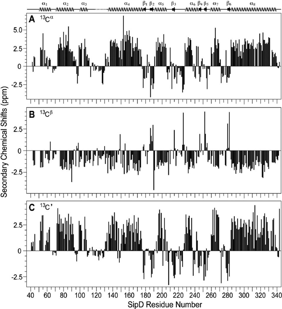
Fig. 2.
Secondary structures of SipD39–343 based on the (A) Cα, (B) Cβ and (C) C’ secondary NMR chemical shifts. The secondary structures of the SipD39–343 crystal are also shown and are denoted by arrow (beta strand), wavy line (helix); solid line (loop), and broken line (disordered loop). Residues 118–133 lacked electron density in the SipD39–343 crystal.

