Abstract
Magnetic resonance imaging (MRI) has become a common and important life-saving diagnostic tool in recent times, for diseases of the head and neck region. Dentists should be aware of the interactions of various restorative dental materials and different technical factors put to use by an MRI scanning machine. Specific knowledge about these impacts, at the dentist level and at the level of the personnel at the MRI centers can save valuable time for the patient and prevent errors in MRI images. Artifacts from metal restorations are a major hindrance at such times, as they result in disappearance or distortion of the image and loss of important information.
Keywords: Magnetic, magnetic resonance imaging, metal artifacts, magnetic resonance image
In addition to palpation and inspection, the imaging modalities used to evaluate the oral cavity include panoramic radiography, ultrasound, computed tomography (CT), Magnetic resonance imaging (MRI), and positron emission tomography (PET). CT and MRI remain the primary methods for evaluation of oral cavity cancers. However, CT and MRI often do not accurately.
Evaluate the primary tumors in patients with dental metallic implants or dentures.[1] MRI is thought to be more useful than CT in diagnosis of soft tissue and blood flow within the bone in implants insertion and osteomyelitis.[2] MR imaging is considered contraindicated in patients with ferromagnetic metallic implants or other ferromagnetic materials, primarily because of the potential risks associated with movement or dislodgment of these objects.[3–6] Artifacts caused by metallic objects, such as dental crowns, dental implants and metallic orthodontic appliances, are a common problem in head and neck MRI.[7–21]
MRI
It’s a diagnostic tool that uses powerful magnetic field, radio waves and a computer, to create images of tissues and organs throughout the body.[7] The powerful magnetic field aligns atomic particles called protons that are present in most of body tissues especially the soft tissues. The applied radio waves then cause these particles to produce signals that are picked up by the receiver within MR scanner.
Magnetic force
It is measured in tesla (T). MRI uses 1.0-1.5 T, more powerful MR scanner uses 3.0 T. Compared to earth′s magnetic force (50 μT); it is 10,000 times more powerful.
Types of MRI
Based on the magnetic field strength:
Low-field MRI scanners (0.23 T-0.3 T): They are typically identified as open MRI scanners. Low-field MRI scanners have decreased image quality and require a longer scan time compared to high-field MRI scanners.
High-field MRI scanners (1.5 T to 3.0 T): These are typically identified as closed MRI scanners. A 1.5 T MRI scanner provides great image quality, fast scan times, and the ability to evaluate how certain structures in the body function. The 3.0 T MRI scanner is great for visualizing very fine detail, such the vessels of the brain or heart.
Ultra-high field MRI scanners (7.0 T to 10 T): It is not widely available and is typically used for research.
Materials Used in Dentistry
Dental materials may be classified in many ways. Here it is classified based on magnetic susceptibility as:[7,8,11]
Ferromagnetic
These are those types of materials which are strongly attracted to a magnet. Their permeability is very high in the range of hundreds and thousands. Examples include chromium oxide, cobalt, ferrite (iron), cadolinium, nickel, rare earth magnet, magnetite, yttrium, etc.
Paramagnetic
These are those materials which are not very strongly attracted to the magnet. They are slightly magnetized when placed in a strong magnetic field and act in the direction of the magnetic field. Their relative permeability is slightly more than one. Examples of such materials are magnesium, tin, platinum, lithium, tantalum, aluminum, molybdenum, etc.
Diamagnetic
These are those materials which are repelled by a magnet. They are slightly magnetized when placed in a strong magnetic field and act in the direction opposite to that of the magnetic field. Their permeability is slightly less than one. For example wood, zinc, copper, bismuth, silver, gold, etc., are diamagnetic materials.
Impact of Magnetic Force on Materials used in Dentistry
Projectile accidents
The powerful magnetic field of MR system will attract iron containing (or ferromagnetic) objects and may cause them to move suddenly and with a great force like a “missile”. This can cause possible risk to patient or anyone in an objects “flight path”. It can pull any ferromagnetic object in the body too.
Thermal heating
Tissue injury can be caused due to heating of prosthesis.[17,18,22] Radio frequency (RF) heating was confirmed to take place at both ends of the implants in spite of their different shapes.[22] The maximum temperature rise was observed at the tip where there is large curvature.[22]
Failure of prosthesis
It also causes failure of prosthesis like crown or FPD dislodgement. Failure of magnets used in overdentures, magnetic keepers, etc., and deflection of orthodontic wires are possible.
Dental restorations/appliances which may cause safety problem
Impact of Dental Materials in MRI
Artifact
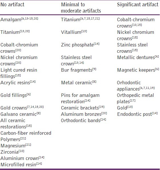
An artifact may be defined as a distortion of signal intensity or void that does not have any anatomic basis in the plane being imaged.[11] It is so defined as the pixels that do not faithfully represent the tissue components being studied.[8] Even a dental bur fragments can cause artifact [Table 1].
Table 1.
Dental materials and artifacts

Factors Affecting the Size of Artifact
Size, shape and position of the material
Larger the size of the material, greater will be the artifact. Maximum area of signal loss will be, when the material is within the radius of 10 cm inside the region of interest. Artifacts are generated even with paramagnetic metals, and that the causative factor is related to the shape of the material.[2] Artifact in clinical case is due to ring-shaped attachments inside the oral cavity, even if they are of the non-magnetic metal, and they may produce artifacts depending on the orientation during MR imaging.
Region that requires MRI
Significant artifact will be produced in orofacial imaging. Importance for neck and brain imaging depends on plane of section.
Factors that influence the risk of using MRI in a patient with implant or ferromagnetic material:[4] (1) The strength of the gradient and static magnetic fields, (2) the degree of ferromagnetism of the implant or material, (3) the geometry of the implant or material, (4) the location and orientation of the implant or material in situ, and (5) the amount of time the implant or material has been in place.
Steps/guidelines to prevent these interactions
Knowledge about the interaction of dental materials and MRI is essential for dentists and radiologists. MRI centers can be notified whether the restoration/appliance is MR friendly or not.
Also we can prefer nonmetals like all ceramic restorations over metal ceramic.
Even in metal ceramic it is better to choose a noble metal alloy.
Removable appliances/prosthesis is not a problem, since patient can remove it.
Treat all material as MR unsafe, if the dentist is not sure about the type of prosthesis/appliance. It is advisable to remove the prosthesis/appliances prior to MRI.
Materials for prosthetic restoration should be selected based not only on their biological compatibility and functional and esthetic qualities, but also on whether they generate minimum artifacts in MRI. Thus ALL ceramic restorations will be a valuable alternative in dentistry.
Conclusions
Further study about the interactions is needed. Undergraduate curriculum should include these interactions and safety measures, especially the magnetic property of various dental materials.
The US Food and Drug Administration reported in 2011 that MRI accidents in the US have risen over 500% from 2000 to 2009. The overwhelming majority of reported injuries fell into one of three categories: Burns, projectiles and hearing damage. However, these injuries are not accidents: They are directly related to the failure to assure proper safety standards.
Most of these accidents were not reported. No records or data is maintained about these accidents. Proper survey in future will reveal exactly the amount of hazards that have happened in the past and will help us to take preventive steps.
Footnotes
Source of Support: Nil.
Conflict of Interest: None declared.
References
- 1.Baek CH, Chung MK, Son YI, Choi JY, Kim HJ, Yim YJ, et al. Tumor volume assessment by 18F-FDG PET/CT in patients with oral cavity cancer with dental artifacts on CT or MR images. J Nucl Med. 2008;49:1422–8. doi: 10.2967/jnumed.108.051649. [DOI] [PubMed] [Google Scholar]
- 2.Taniyama T, Sohmura T, Etoh T, Aoki M, Sugiyama E, Takahashi J. Metal artifacts in MRI from non-magnetic dental alloy and its FEM analysis. Dent Mater J. 2010;29:297–302. doi: 10.4012/dmj.2009-116. [DOI] [PubMed] [Google Scholar]
- 3.Shellock FG, Crues JV. High-field-strength MR imaging and metallic biomedical implants: An ex vivo evaluation of deflection forces. AJR Am J Roentgenol. 1988;151:389–92. doi: 10.2214/ajr.151.2.389. [DOI] [PubMed] [Google Scholar]
- 4.Shellock FG. MR imaging of metallic implants and materials: A compilation of the literature. AJR Am J Roentgenol. 1988;151:811–4. doi: 10.2214/ajr.151.4.811. [DOI] [PubMed] [Google Scholar]
- 5.Gegauff AG, Laurell KA, Thavendrarajah A, Rosenstiel SF. A potential MRI hazard: Forces on dental magnet keepers. J Oral Rehabil. 1990;17:403–10. doi: 10.1111/j.1365-2842.1990.tb01411.x. [DOI] [PubMed] [Google Scholar]
- 6.New PF, Rosen BR, Brady TJ, Buonanno FS, Kistler JP, Burt CT, et al. Potential hazards and artifacts of ferromagnetic and nonferromagnetic surgical and dental materials and devices in nuclear magnetic resonance imaging. Radiology. 1983;147:139–48. doi: 10.1148/radiology.147.1.6828719. [DOI] [PubMed] [Google Scholar]
- 7.Costa AL, Appenzeller S, Yasuda CL, Pereira FR, Zanardi VA, Cendes F. Artifacts in brain magnetic resonance imaging due to metallic dental objects. Med Oral Patol Oral Cir Bucal. 2009;14:E278–82. [PubMed] [Google Scholar]
- 8.Chen DP, Wu GY, Wang YN. Influence of galvano-ceramic and metal-ceramic crowns on magnetic resonance imaging. Chin Med J (Engl) 2010;123:208–11. [PubMed] [Google Scholar]
- 9.Kaneda T, Minami M, Curtin HD, Utsunomiya T, Shirouzu I, Yamashiro M, et al. Dental bur fragments causing metal artifacts on MR images. AJNR Am J Neuroradiol. 1998;19:317–9. [PMC free article] [PubMed] [Google Scholar]
- 10.Raphael B, Haims AH, Wu JS, Katz LD, White LM, Lynch K. MRI comparison of periprosthetic structures around zirconium knee prostheses and cobalt chrome prostheses. AJR Am J Roentgenol. 2006;186:1771–7. doi: 10.2214/AJR.05.1077. [DOI] [PubMed] [Google Scholar]
- 11.Harris TM, Faridrad MR, Dickson JA. The benefits of aesthetic orthodontic brackets in patients requiring multiple MRI scanning. J Orthod. 2006;33:90–4. doi: 10.1179/146531205225021465. [DOI] [PubMed] [Google Scholar]
- 12.Ernstberger T, Heidrich G, Bruening T, Krefft S, Buchhorn G, Klinger HM. The interobserver-validated relevance of intervertebral spacer materials in MRI artifacting. Eur Spine J. 2007;16:179–85. doi: 10.1007/s00586-006-0064-5. [DOI] [PMC free article] [PubMed] [Google Scholar]
- 13.Abbaszadeh K, Heffez LB, Mafee MF. Effect of interference of metallic objects on interpretation of T1-weighted magnetic resonance images in the maxillofacial region. Oral Surg Oral Med Oral Pathol Oral Radiol Endod. 2000;89:759–65. doi: 10.1067/moe.2000.105942. [DOI] [PubMed] [Google Scholar]
- 14.Hinshaw DB, Jr, Holshouser BA, Engstrom HI, Tjan AH, Christiansen EL, Catelli WF. Dental material artifacts on MR images. Radiology. 1988;166:777–9. doi: 10.1148/radiology.166.3.3340777. [DOI] [PubMed] [Google Scholar]
- 15.Lissac M, Metrop D, Brugirard J, Coudert JL, Pimmel P, Briguet A, et al. Dental materials and magnetic resonance imaging. Invest Radiol. 1991;26:40–5. doi: 10.1097/00004424-199101000-00008. [DOI] [PubMed] [Google Scholar]
- 16.Guermazi A, Miaux Y, Zaim S, Peterfy CG, White D, Genant HK. Metallic artefacts in MR imaging: Effects of main field orientation and strength. Clin Radiol. 2003;58:322–8. doi: 10.1016/s0009-9260(02)00540-8. [DOI] [PubMed] [Google Scholar]
- 17.Cunningham AS, Harding S, Chatfield DA, Hutchinson P, Carpenter TA, Pickard JD, et al. Metallic neurosurgical implants for cranial reconstruction and fixation: Assessment of magnetic field interactions, heating and artefacts at 3.0 Tesla. Br J Neurosurg. 2005;19:167–72. doi: 10.1080/02688690500145720. [DOI] [PubMed] [Google Scholar]
- 18.Schueler BA, Parrish TB, Lin JC, Hammer BE, Pangrle BJ, Ritenour ER, et al. MRI compatibility and visibility assessment of implantable medical devices. J Magn Reson Imaging. 1999;9:596–603. doi: 10.1002/(sici)1522-2586(199904)9:4<596::aid-jmri14>3.0.co;2-t. [DOI] [PubMed] [Google Scholar]
- 19.Okano Y, Yamashiro M, Kaneda T, Kasai K. Magnetic resonance imaging diagnosis of the temporomandibular joint in patients with orthodontic appliances. Oral Surg Oral Med Oral Pathol Oral Radiol Endod. 2003;95:255–63. doi: 10.1067/moe.2003.37. [DOI] [PubMed] [Google Scholar]
- 20.Starcuková J, Starcuk Z, Jr, Hubálková H, Linetskiy I. Magnetic susceptibility and electrical conductivity of metallic dental materials and their impact on MR imaging artifacts. Dent Mater. 2008;24:715–23. doi: 10.1016/j.dental.2007.07.002. [DOI] [PubMed] [Google Scholar]
- 21.Ernstberger T, Buchhorn G, Heidrich G. Artifacts in spine magnetic resonance imaging due to different intervertebral test spacers: An in vitro evaluation of magnesium versus titanium and carbon-fiber-reinforced polymers as biomaterials. Neuroradiology. 2009;51:525–9. doi: 10.1007/s00234-009-0537-4. [DOI] [PMC free article] [PubMed] [Google Scholar]
- 22.Muranaka H, Horiguchi T, Ueda Y, Usui S, Tanki N, Nakamura O. Evaluation of RF heating on hip joint implant in phantom during MRI examinations. Nihon Hoshasen Gijutsu Gakkai Zasshi. 2010;66:725–33. doi: 10.6009/jjrt.66.725. [DOI] [PubMed] [Google Scholar]


