

Abstract
Introduction:
“The fibers running anteroposteriorly within the core and in concentric curves across the base of each ruga” determine their orientation and forms. The varying shapes of palatal rugae can be attributed to the fact that rugae develop as localized regions of epithelial proliferation and thickening. Fibroblasts and collagen fibers then accumulate in the connective tissue beneath the thickened epithelium and assume distinct orientation.
Aim:
The aim of the present study was to assess the morphology, gender difference of rugae pattern in 5-15 year old children.
Materials and Methods:
The various diagnostic dental stone cast available in Department of Pedodontics were analyzed by the method based on Thomas and Kotze classification in 1983.
Results:
There was a female prediction in the total count and primary rugae pattern. Comparing the shapes of rugae both in male and female study models showed a predominance in wave shape followed by cure. No circular pattern was observed in the study population. No statistical difference in the direction and unification of rugae among males and females.
Conclusion:
The fingerprint-like uniqueness of rugae to each individual has become accepted as a possible aid to person identification. This may help narrow the field for identification and give results in conjunction with the other methods such as visual, fingerprints, and dental characteristics in forensic sciences.
Keywords: Forensic dentistry, identification, palatal rugae
Palatal rugae, also called plicae palatinae transversae and rugae palatina, refer to the ridges on the anterior part of the palatal mucosa, each side of the median palatal raph′e and behind the incisive papilla.[1,2] In human embryos, rugae are relatively prominent and occupy most of the length of the palatal shelves at the time of their elevation.[3] At the 550 mm stage of embryonic development, there are five to seven rather symmetrically disposed ridges, with the anterior ones beginning at the raph′e, the others more laterally, towards the end of intra-uterine life, the pattern of rugae becomes less regular, posterior ones disappearing and those anterior become considerably more pronounced and compressed.[1]
The palatine rugae are permanent and unique to each person and can establish identity through discrimination (via casts, tracings or digitized rugae patterns).[4,5] The anatomical position of the rugae inside the mouth-surrounded by cheeks, lips, tongue, buccal pad of fat, teeth and bone-keeps them well-protected from trauma and high temperatures.[6]
Materials and Methods
The study was conducted at the Department of Pedodontics, study sample consists of 100 diagnostic study models of 50 male and 50 female, which was available in the department which were free from palatal defects was used to determine the number, pattern and also to assess the predominant pattern of rugae in children from 5 to 15 years and differences in rugae pattern among two genders in the selected groups.
The materials used to assess the rugae pattern are [Figure 1]:
Figure 1.

The materials used to assess the rugae pattern
Magnifying glass
Study models
Marking pencil
Divider
Metal scale
Figure 2 shows the study models used in the study. The method of identification was based on Thomas and Kotze classification in 1983 [Figure 3].
Figure 2.
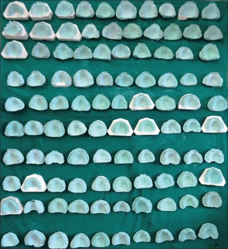
Study models used in the study
Figure 3.

Thomas and Kotze classification
The rugae were classified based on their length as:
Primary: > 5 mm, Secondary: 3-5 mm, Fragmentary: < 3 mm, rugae less than 2 mm were ignored [Figure 4].
Figure 4.

Cast showing tracing of palatal rugae patterns
The rugae were divided into 4 types based on their shape.
Curved: They had a crescent shape and curved gently.
Wavy: If there was a slight curve at the origin or termination of a curved rugae.
Straight: They run directly from their origin to termination.
Circular: Rugae that form a definite continuous ring.
The direction of the rugae was determined by measuring the angle formed by the line joining its origin and termination and the line perpendicular to the median raphe. Based on the direction rugae were classified as:
Forwardly directed rugae: Associated with positive angles.
Backwardly directed rugae: Associated with negative angles.
Perpendicular rugae: Associated with zero angles.
Unification was said to have occurred when two rugae joined at their origin or termination:
Diverging: If two rugae had the same origin from the midline but immediately branched.
Converging: Rugae with different origins from midline, but which joined on their lateral portions.
All the details from each dental models were documented. Association between rugae forms and gender were tested by using unpaired t-test.
Results
The present study was an attempt to determine the number, pattern and also to assess the predominant pattern of rugae in children from 5 to 15 years.
An attempt was also made to determine the differences in rugae pattern among two genders in the selected groups.
The total number of rugae was more in female study models when compared to male [Table 1].
Table 1.
Total no. of subjects and the mean value of rugae in males and females

Total number of primary rugae was also more in female study models when compared to male [Table 2].
Table 2.
Total no. of subjects and the mean value of primary rugae in males and females

Comparing the shapes of rugae both in male and female study models showed a predominance in wave shape followed by cure [Table 3].
Table 3.
Number of the various shapes of rugae among males and females

Straight ruga was seen in 1 male study model only.
None of the study models showed circular shape rugae.
In the present study direction and unification of rugae did not show any statistically difference among males and females [Table 4].
Table 4.
Total no of subjects and the mean value of various unification of rugae in males and females

Discussion
Palatal rugoscopy was first proposed in 1932, by Spanish investigator Troban Hermaso. Since then, various classifications had been given. Most studies are based on systems devised by Lysell and Thomas and Kotze, although they may differ in detail.
Hauser et al. 1989 Controversy still exists about the stability of quantitative and qualitative characteristics of rugae during growth, and the extent of differences between ethnic groups and sexes, the mean ruga count changes moderately in adolescence, then increases markedly from the age of 35-40 years.[3] In contrast, Lysell 1955 the number of ruga decreased from 23 years of age onwards.[1] The characteristic pattern of the palatal rugae did not change as a result of growth, remaining stable from time of development until the oral mucosa degenerated at death.[4,7] Events such as trauma, extreme finger sucking in infancy, and persistent pressure with orthodontic treatment and dentures can contribute the change in rugae patterns.[1,8] The changes in the length of rugae with age result from underlying palatal growth.[1,3,9] The anterior rugae do not increase in length after 10 years of age according to Van der Linden. Other qualitative characteristics such as shape, direction and unification remain stable throughout life.[10]
Lysell[1] there is a tendency for the backward direction of rugae to decrease with age, due to an increase in the width of the palate and forward movement of the teeth in relation to the rugae, resulting in rugae being located in a wider and shallower part of the palate than originally. Another possible explanation given for this tendency is the forward movement of lateral parts of the rugae in connection with forward growth of the dental arch. In contrast, 53% of the rugae that changed direction actually moved backwards. This indicates a high percentage of rugae moving in a posterior direction in Aborigines. Could this be attributed to the different ethnicity of the subjects in each study? With a difference in ethnicity come differences in the pattern and extent of growth of the palate, genetic variation, and different patterns of movement of teeth due to crowding and wear pattern Dohke and Osato[8] the direction of rugae is influenced by the formation of the dental arch associated with replacement of teeth and by growth and developmental changes in the palate.
The palatine rugae are unique to each patient[1,11] and are reasonably stable during the patient′s growth;[12] thus, they may serve as suitable reference points from which the clinician can derive the reference planes necessary for longitudinal cast analysis.[10] It is a well-established fact that the rugae pattern is as unique to a human as are his or her fingerprints,[13–19] and it retains its shape throughout the life.[5,18,20]
Songnnaes[21] use of casts made from jaws rather than from dentures for a more reliable. Jacob and shalla[22] dental stone casts derived from maxillary tissues and from internal aspect of maxillary dentures for postmortem identification of edentulous people showed 79% accuracy when tracing the rugae. Indira AP 2011 at post orthodontic treatment, rugae pattern remain unchanged during an individual′s lifetime and also following action of the force.[23] This was in consistent with Sabet and Abdel.[4,18,24] The core within the palatal rugae of humans contains elements that are believed to contribute to the maintenance of its shape. The main structural element contains glycosaminoglycans which by its hydrophilic nature causes the tissues to swell and contributes to the maintenance of the shape of rugae throughout life. Fibroblasts and collagen fibers beneath the thickened epithelium contribute to the stability of palatal rugae.[23]
Sassuouni no two palates are alike in their configuration and that the palato print did not change during growth. Ritter the rugae of twins and found that the pattern was similar but not identical. Hauser[3] the characteristic picture of the palate does not change as a result of growth in children from birth to 9 years. Leontsinis ascertained that rugae do not change from the time they develop until the oral mucosa degenerates at death.[4]
Park et al.[25] the pattern of palatine rugae in submucosal clefts had a unique feature in 87.5% of the submucosal clefts and in 100% of the isolated clefts: one or more of the palatine rugae curved toward the region of the bony notch in the posterior border of the hard palate.
Kratzsch and Opitz[26] the number and type of rugae before and after surgical repair of the cleft palate, each segment had four or five rugae, similar to the number in people without a cleft palate. After palatal cleft repair, the rugae counts per segment decreased significantly, but the third ruga was never lost after surgery. The primary rugae in unilateral and bilateral cleft lip and palate were the same as those in isolated cleft palates, and they did not differ from those in people who did not have cleft lip or palate. The linear distance from the tuberosity line to the rugal zone increased in the unilateral and bilateral cleft segments before palatal cleft repair, indicating sagittal maxillary development in the posterior area of the palate.
Kratzsch and Opitz[27] investigated the relationship of palatine rugae to points (landmarks) and distances on the cleft palate during the period from birth to the time of early mixed dentition and identified changes in the distances from the lateral palatine rugae points of the first and third rugae to the incisal point, the canine point and the tuberosity line. They indicated that a comparison of distances from the palatine rugae with distances between equivalent points revealed the changes that occurred in the anterior palate during various stages of orthodontic therapy and growth.
Palatal rugae patter did not sustain any changes after thermal effects and decomposition of pan facial third - degree burn victims and the ability to resist decomposition changes for up to 7 days after death was also noted.[14] Thus, palatal rugae appear to possess the features of an ideal forensic identification parameter-uniqueness, postmortem resistance, and stability. Hence, they can be used in postmortem identification provided an ante mortem record exists.[6]
Thomas and Kotze[28,29] postulated that an evolutionary trend exists in which the rugae of primates, including man, are becoming attenuated. Paul Jibi[30] environmental influences such as food habits along with genetic factor over the decades would have influenced the rugae pattern, thus would give a difference in the pattern between the two communities and sexes. The most difficult aspect of observing rugae is the application of the classification while its characteristics have been defined as fully as possible; interpretation of features is sometimes difficult.[31] There was a female prediction in the total count and primary rugae pattern. Comparing the shapes of rugae both in male and female study models showed a predominance in wave shape followed by cure. No circular pattern was observed in the study population. No statistical difference in the direction and unification of rugae among males and females.
It has been suggested that a classification system that is simple and reliable be used in rugae studies. The classification method used in this study was found to be more practical and easiest to apply compared with other methods such as those of with this classification, difference could be appreciated between population groups with certain patterns being more significant, thus indicating the applicability of rugae pattern in population differentiation.
Conclusions
Located in the anterior half of the roof of the mouth, they serve as a reference landmark in various dental treatment modalities and can be used in the identification of submucosal clefts.
The fingerprint-like uniqueness of rugae to each individual has become accepted as a possible aid to person identification. This may help narrow the field for identification and give results in conjunction with the other methods such as visual, fingerprints, and dental characteristics in forensic sciences. The use of palatal rugae in forensic identification is preferred because of their low utilization cost, simplicity and reliability. The authors recognize that the above-mentioned interpretations are precluded by limited sample size and therefore, the preceding analysis should only be considered as preliminary. Within the limitations of the present study, it may be concluded that the rugae pattern may be an additional method of differentiation between the Indian male and female children.
Footnotes
Source of Support: Nil.
Conflict of Interest: None declared.
References
- 1.Lysell L. Plicae palatinae transversae and papilla incisiva in man: A morphologic and genetic study. Acta Odontol Scand. 1955;13(Suppl 18):5–137. [PubMed] [Google Scholar]
- 2.Thomas CJ, Kotze TJ. The palatal rugae pattern: A new classification. J Dent Assoc South Afr. 1983;38:153–7. [PubMed] [Google Scholar]
- 3.Hauser G, Daponte A, Roberts MJ. Palatal rugae. J Anat. 1989;165:237–49. [PMC free article] [PubMed] [Google Scholar]
- 4.English WR, Robison SF, Summitt JB, Oesterle LJ, Brannon RB, Morlang WM. Individuality of human palatal rugae. J Forensic Sci. 1988;33:718–26. [PubMed] [Google Scholar]
- 5.Harrison A. The palatal rugae in man. Proc Acad Nat Soc. 1889;6:245. [Google Scholar]
- 6.Patil MS, Patil SB, Acharya AB. Palatine rugae and their significance in clinical dentistry: A review of the literature. J Am Dent Assoc. 2008;139:1471–8. doi: 10.14219/jada.archive.2008.0072. [DOI] [PubMed] [Google Scholar]
- 7.Peavy DC, Jr, Kendrick GS. The effects of tooth movement on the palatine rugae. J Prosthet Dent. 1967;18:536–42. doi: 10.1016/0022-3913(67)90219-3. [DOI] [PubMed] [Google Scholar]
- 8.Dohke M, Osato S. Morphological study of the palatal rugae in Japanese. 1 Bilateral differences in the regressive evolution of the palatal rugae. Jpn J Oral Biol. 1994;36:126–40. [Google Scholar]
- 9.Simmons JD, Moore RN, Erickson LC. A longitudinal study of anteroposterior growth changes in the palatine rugae. J Dent Res. 1987;66:1512–5. doi: 10.1177/00220345870660092001. [DOI] [PubMed] [Google Scholar]
- 10.van der Linden FP. Changes in the position of posterior teeth in relation to ruga points. Am J Orthod. 1978;74:142–61. doi: 10.1016/0002-9416(78)90081-7. [DOI] [PubMed] [Google Scholar]
- 11.Thomas CJ, Van Wyk CW. Elastic fibre and hyaluronic acid in the core of human palatal rugae. J Biol Buccale. 1987;15:171–4. [PubMed] [Google Scholar]
- 12.Moyers RE, Van der Linden FP, Riolo ML, McNamara JA. Standards of human occlusal development: Monograph no. 5 craniofacial growth series. Ann Arbor, Mich: Center for human growth and development, University of Michigan; 1976. p. 187. [Google Scholar]
- 13.Carrea JU. La Identificacion humana por las rugosidades palatinas. Rev Orthodont (Buenos Aires) 1937;1:3–23. [Google Scholar]
- 14.Caldas IM, Magalhães T, Afonso A. Establishing identity using cheiloscopy and palatoscopy. Forensic Sci Int. 2007;165:1–9. doi: 10.1016/j.forsciint.2006.04.010. [DOI] [PubMed] [Google Scholar]
- 15.Antonopoulos AN. The palatal rugae as a guide in the correct arrangement of upper anterior teeth in complete denture construction. Odontostomatol Proodos. 1972;26:44–50. [PubMed] [Google Scholar]
- 16.Comoy J. La rugoscopie. Chir Dent Fr. 1973;43:59–60. [Google Scholar]
- 17.Kogon SL, Ling SC. A new technique for palatal rugae comparison in forensic odontology. Can Soc Forensic Sci J. 1973;63:3–10. [Google Scholar]
- 18.Abou EF, Mona M, Gamal ZH. A study of palatal rugae pattern (rugoscopy) in Egyptian population. Egypt Dent J. 1998;44:3177–84. [Google Scholar]
- 19.Buchner A. The identification of human remains. Int Dent J. 1985;35:307–11. [PubMed] [Google Scholar]
- 20.Limson KS, Julian R. Computerized recording of the palatal rugae pattern and an evaluation of its application in forensic identification. J Forensic Odontostomatol. 2004;22:1–4. [PubMed] [Google Scholar]
- 21.Sognnaes RD. Forensic stomatology (third of three parts) N Engl J Med. 1977;296:197–203. doi: 10.1056/NEJM197701272960405. [DOI] [PubMed] [Google Scholar]
- 22.Jacob RF, Shalla CL. Postmortem identification of the edentulous deceased: Denture tissue surface anatomy. J Forensic Sci. 1987;32:698–702. [PubMed] [Google Scholar]
- 23.Indira AP, Manish Gupta, David MP. Rugoscopy for establishing individuality. Indian J Dent Adv. 2011;3:427–32. [Google Scholar]
- 24.Abdel-Aziz HM, Sabet NE. Palatal rugae area: A landmark for analysis of pre- and post-orthodontically treated adult Egyptian patients. East Mediterr Health J. 2001;7:60–6. [PubMed] [Google Scholar]
- 25.Park S, Eguti T, Kato K, Nitta N, Kitano I. The pattern of palatal rugae in submucous cleft palates and isolated cleft palates. Br J Plast Surg. 1994;47:395–9. doi: 10.1016/0007-1226(94)90066-3. [DOI] [PubMed] [Google Scholar]
- 26.Kratzsch H, Opitz C. Investigations on the palatal rugae pattern in cleft patients. Part I: A morphological analysis. J Orofac Orthop. 2000;61:305–17. doi: 10.1007/pl00001901. [DOI] [PubMed] [Google Scholar]
- 27.Kratzsch H, Opitz C. Investigations on the palatal rugae pattern in cleft patients. Part II: Changes in the distances from the palatal rugae to maxillary points. J Orofac Orthop. 2000;61:421–31. doi: 10.1007/pl00001910. [DOI] [PubMed] [Google Scholar]
- 28.Thomas CJ, Kotze TJ. The palatal ruga pattern in six Southern African human populations. Part III: An evolutionary perspective. J Dent Assoc S Afr. 1983;38:173–6. [PubMed] [Google Scholar]
- 29.Thomas CJ, Kotze TJ, Van der Merwe CA. An improved statistical method for the racial classification of man by means of palatal rugae. Arch Oral Biol. 1987;32:315–7. doi: 10.1016/0003-9969(87)90028-8. [DOI] [PubMed] [Google Scholar]
- 30.Jibi PM, Gautam KK, Basappa N, Raju OS. Morphological pattern of palatal rugae in children of Davangere. J Forensic Sci. 2011;56:1192–7. doi: 10.1111/j.1556-4029.2011.01831.x. [DOI] [PubMed] [Google Scholar]
- 31.Thomas CJ, Kotze TJ. The palatal ruga pattern in six Southern African human populations. Part I: A description of the populations an a method for its investigation. J Dent Assoc S Afr. 1983;38:158–65. [PubMed] [Google Scholar]