Abstract
Introduction:
Supernumerary teeth refers to an excessive number of teeth and are relatively common in the oral cavity. The supernumerary tooth located in the midline of the maxilla between the central incisors is referred to as “mesiodens”. The present study emphasizes the importance of multidisciplinary management and long-term follow-up of patients with mesiodens.
Aim of the Study:
This study investigates the characteristics of mesiodens among children in patients reported to department of pediatric and preventive dentistry in an institution in the state of Tamil Nadu, India.
Materials and Methods:
The study population consisted of 55 children below 14 years. The characteristics of mesiodens were obtained from radiographs and clinical examination. And all these were confirmed as mesiodens by surgical intervention.
Results:
Results showed that males were affected approximately 2 times as frequently as females. Predominant shape of the mesiodens was conical (79.7%). 27 (49.09%) of 55 patients had multiple mesiodens. 25 (45.45%) of 55 patients required orthodontic correction after surgery.
Conclusions:
Mesiodens can cause many complications. So routine radiographic examination of all pediatric patients is advised. And when identified, they should be extracted as early as possible in children and adolescents in order to avoid possible complications.
Keywords: Mesiodens, supernumerary teeth, radiograph, clinical
The term mesiodens refers to a supernumerary tooth present in the midline of the maxilla between central incisors. It has overall prevalence of 0.15-1.9%. Mesiodens can occur individually or as multiples, may appear unilaterally or bilaterally, and often do not erupt.[1–4] Mesiodens can significantly alter both occlusion and appearance by altering the eruption path and the position of the permanent incisors. The literature report 3 theories in the etiology of mesiodens, but this subject remains controversial. (a) Phylogenic reversion or atavism[5] (b) Dichotomy theory[1] and (c) Hyperactivity of the dental lamina.[6] Autosomal dominant inheritance with incomplete penetration has been the proposed genetic theory. A sex-linked pattern has also been proposed as males are affected twice as frequently as females.[7] Mesiodens can be classified on the basis of their occurrence in the permanent dentition (rudimentary mesiodens) or the primary dentition (supplementary mesiodens) and according to their morphology (conical, tuberculate or molariform).[3,8]
Materials and Methods
The population studied consisted of 55 patients treated for mesiodens at the Department of Pedodontics and Preventive Dentistry, Vinayaka Missions Sankarachariyar Dental College, Salem, Tamil Nadu. 82 mesiodens from 55 children were identified by studying radiographs. In addition to gender and age, the following information was recorded about the mesiodens: (1) Number; (2) Shape; (3) Position;(4) whether requiring ortho treatment or not and (5) Number of mesiodens per patient and their types. Either a periapical or panoramic radiograph, or both were taken in all 55 patients.
Results
Age and sex distribution
Of the 82 mesiodens were diagnosed in 55 patients. The predominant age group was from 7 to 14 years old. There were 37 male and 18 female patients with a gender ratio of 2.05:1 [Table 1, Graph 1].
Table 1.
Sex wise distribution of mesiodens

Graph 1.
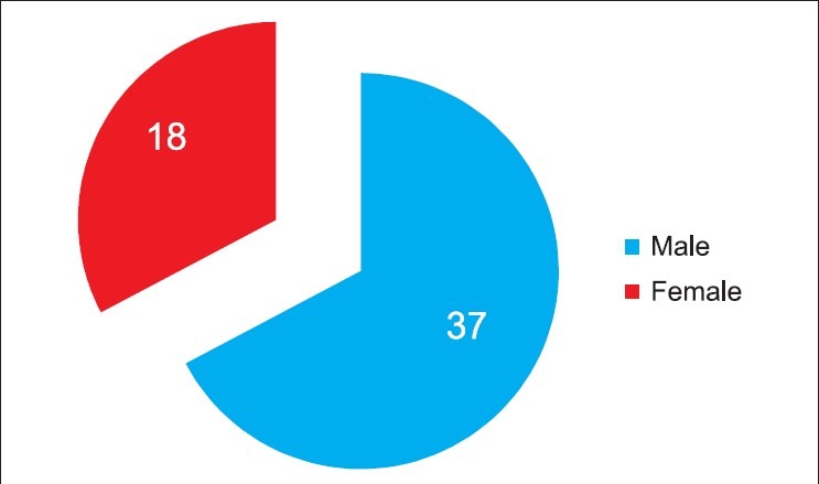
Sex wise distribution of mesiodens
Shapes
The shapes of the extracted mesiodens were classified as conical, tuberculate, eumorphic and molariform. The most common shape was conical (79.74%) [Table 2, Graph 2].
Table 2.
Shape wise distribution of mesiodens

Graph 2.
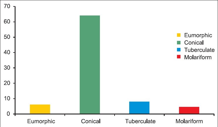
Table 2 shape wise distribution of mesiodens
Position
Of the 82 mesiodens, 55 were erupted into the oral cavity while 27 (32.92%) were fully impacted [Table 3, Graph 3].
Table 3.
Distribution of mesiodens based on whether erupted or impacted

Graph 3.
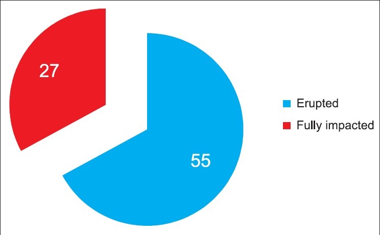
Distribution of mesiodens based on wether erupted or impacted
Requiring orthodontic correction
In 25 (45.45%) patients orthodontic treatment was done following surgical removal of mesiodens, while remaining 30 patients did not require any orthodontic correction [Table 4, Graph 4].
Table 4.
Distribution of mesiodens based on the requirement of orthodontic treatment

Graph 4.
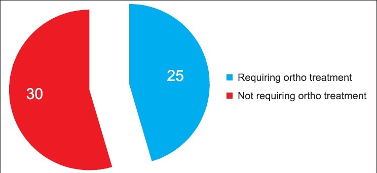
Distribution of mesiodens based on the requirement of orthodontic treatment
Number of mesiodens per patient and types
Approximately 28 patients (50.90%) had only 1 mesiodens and 27 patients (49.09%) had multiple mesiodens, out of 55 patients. Out of the 82 mesiodens, 24 were erupted multiple, 31 were erupted single, 15 were unerupted multiple and 12 were unerupted single [Table 5, Graph 5].
Table 5.
Distribution based on the number of mesiodens present

Graph 5.
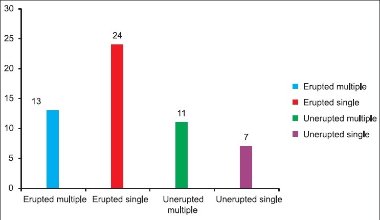
Distribution of mesiodens based on the number present
Discussion
Etiology
Phylogenic reversion or atavism postulated that mesiodens represented a phylogenetic relic of extinct ancestors who had 3 central incisors.[5] This theory is largely discarded by embryologists. Dichotomy theory suggests that the tooth bud is split to create 2 teeth, one of which is the mesiodens.[1] The third theory, involving hyperactivity of the dental lamina, is the most widely supported. According to this theory, remnants of the dental lamina or palatal offshoots of active dental lamina are induced to develop into an extra tooth bud, which results in a supernumerary tooth. Genetics is also thought to contribute to the development of mesiodens as such teeth have been diagnosed in twins, siblings and sequential generations of a single family.[6] In twins, unilateral mesiodens may present as mirror images, and the same number of supernumerary teeth are located in similar regions of the mouth.[9]
The incidence of mesiodens has been estimated at 0.15% to 1% of the population. It occurs more frequently in boys than in girls, with the ratio being approximately 2:1.[4] In my study, a male:female ratio of 2.05:1 for the prevalence for mesiodens was found. Mesiodens range in appearance from a simple conical shape to those that have a complicated crown shape with a number of tubercles.[5] Tuberculate mesiodens tend to develop later and manifest as incompletely developed roots. In the present study, the shape obtained was mainly conical (79.74%) which was similar to many other studies.
The clinical complications of mesiodens are as follows:
Impaction of maxillary central incisor;
Tooth retention or delayed eruption of permanent incisors;
Axial rotation or inclination of erupted permanent incisors;
Eruption within the nasal cavity; formation of diastema;
Intraoral infection, pulpitis of mesiodens;
Root anomaly;
Root resorption of adjacent teeth;
As a consequence of these clinical implications, it is recommended that mesiodens be removed surgically. Early diagnosis and treatment of patients with mesiodens is important to prevent complications. In this present study, 25 patients (45.45%) required orthodontic correction following removal of mesiodens.
Conclusion
Mesiodens may cause pathological conditions; so early diagnosis with regular radiographic examination is advised. Mesiodens should be extracted in children and adolescents in order to avoid possible adverse effects on adjacent teeth as well as cyst formation. In the recent years dental pulp stem cells derived from the mesiodens were studied and were identified to be capable of differentiating into adipogenic and osteogenic lineages. Therefore, the supernumerary teeth usually discarded after extraction might represent a valuable source of human dental pulp stem cells.[10]
Footnotes
Source of Support: Nil.
Conflict of Interest: None declared.
References
- 1.Sedano HO, Gorlin RJ. Familial occurrence of mesiodens. Oral Surg Oral Med Oral Pathol. 1969;27:360–1. doi: 10.1016/0030-4220(69)90366-1. [DOI] [PubMed] [Google Scholar]
- 2.Sykaras SN. Mesiodens in primary and permanent dentitions. Report of a case. Oral Surg Oral Med Oral Pathol. 1975;39:870–4. doi: 10.1016/0030-4220(75)90107-3. [DOI] [PubMed] [Google Scholar]
- 3.Primosch RE. Anterior supernumerary teeth: Assessment and surgical intervention in children. Pediatr Dent. 1981;3:204–15. [PubMed] [Google Scholar]
- 4.Russell KA, Folwarczna MA. Mesiodens: Diagnosis and management of a common supernumerary tooth. J Can Dent Assoc. 2003;69:362–6. [PubMed] [Google Scholar]
- 5.von Arx T. Anterior maxillary supernumerary teeth: A clinical and radiographic study. Aust Dent J. 1992;37:189–95. doi: 10.1111/j.1834-7819.1992.tb00741.x. [DOI] [PubMed] [Google Scholar]
- 6.McKibben DR, Brearley LJ. Radiographic determination of the prevalence of selected dental anomalies in children. ASDC J Dent Child. 1971;28:390–8. [PubMed] [Google Scholar]
- 7.Hattab FN, Yassin OM, Rawashdeh MA. Supernumerary teeth: Report of three cases and review of the literature. ASDC J Dent Child. 1994;61:382–93. [PubMed] [Google Scholar]
- 8.Foster TD, Taylor GS. Characteristics of supernumerery teeth in the upper central incisor region. Dent Pract Dent Rec. 1969;20:8–12. [PubMed] [Google Scholar]
- 9.Chol KL, Chang CH, Samuel TH, Chuang Bilateral mesiodentes in identical twins: A case report. Chin Dent J. 1990;9:116–22. [PubMed] [Google Scholar]
- 10.Huang AH, Chen YK, Lin LM, Shieh TY, Chan AW. Isolation and characterization of dental pulp stem cells from a supernumerary tooth. J Oral Pathol Med. 2008;37:571–4. doi: 10.1111/j.1600-0714.2008.00654.x. [DOI] [PubMed] [Google Scholar]


