

Abstract
Kawasaki Disease (KD) is an acute systemic vasculitis of unknown etiology. In many developed countries, KD has replaced rheumatic heart disease as the leading cause of acquired heart disease in children. Among the classical criteria for a diagnosis of KD are oral manifestations such as strawberry tongue, erythematous cracked lip, and oropharyngeal mucositis. We report the case of a 24-year-old Saudi female with a history of Kawasaki disease who presented to our oral medicine clinic with recurrent painless swelling of the upper lip. As lip swelling has not previously been reported as an oral manifestation of KD, this case represents a novel presentation of recurrent Kawasaki disease in an adult female.
Keywords: Kawasaki disease, Oral manifestation, Recurrence, Lip swelling, Vasculitis, IVIG
1. Introduction
Kawasaki Disease (KD) was named after the Japanese pediatrician Tomisaku Kawasaki, who in 1967 described 50 cases in which a rash and fever in early childhood were accompanied by edema, conjunctival infection, redness and cracking of the lips, “strawberry tongue,” convalescent desquamation, and occasionally lymphadenopathy (Kawasaki, 1967; Kawasaki et al., 1974).
Also known as Mucocutaneous Lymph Node Syndrome, KD is an idiopathic infantile multi-organ vasculitis of medium and small-sized arteries. It typically affects children of Asian ethnicity younger than 4 years of age, although it may also occur in adults, where it is often misdiagnosed due to its nonspecific clinical presentation (Kanno et al., 2011; Sève et al., 2005). KD occurs more often in boys than in girls (1.5:1 male:female ratio) (Cox and Sallis, 2009; Pemberton et al., 1999),and is considered to be the most common cause of acquired pediatric cardiac disease in developed countries (Bhatnagar et al., 2003) .
Kawasaki disease has three phases. Most signs and symptoms appear during the initial acute febrile phase, which lasts from 1 to 2 weeks. This is followed by a subacute phase, characterized by desquamation, arthralgia, and elevated platelet counts. This phase lasts from the end of the fever until approximately day 25. Finally, during the convalescent phase, the clinical signs disappear and acute-phase reactants such as erythrocyte sedimentation rate (ESR) return to normal. The average duration of Kawasaki disease is 6–8 weeks.
A diagnosis of KD requires the presence of at least five of the six classic clinical features (Table 1) (Bhatnagar et al., 2003; Scardina, 2007). Patients who exhibit fewer than this number are considered to have Incomplete Kawasaki Disease (IKD) or Atypical Kawasaki Disease (AKD) (Bhatnagar et al., 2003; Cox and Sallis, 2009; Wolff et al., 2007). Although there are no diagnostic laboratory tests available to rule the disease in or out, some test results can support the clinical diagnosis. For example; ESR, C-reactive protein, and platelets are usually elevated, while antinuclear antibodies (ANAs) and rheumatoid factors (RFs) are typically negative ((Bhatnagar et al., 2003; Cox and Sallis, 2009). These tests can also be used to rule out infectious diseases with a similar presentation, such as scarlet fever caused by streptococcal infection, staphylococcal infection, erythema multiforme, and measles. Where the diagnosis is uncertain, failure of the fever to respond to antibiotics and steroids should lead the clinician to consider KD (Sève et al., 2005; Bhatnagar et al., 2003).
Table 1.
CDC diagnostic criteria for Kawasaki disease.
| Fever lasting for 5 or more days without another, more reasonable, explanation and at least four of the following criteria | |
|---|---|
| 1 | Bilateral nonexudative conjunctival infection |
| 2 | At least one of the following oral mucous membranes changes: |
| – Red or dry fissured lip | |
| – Pharyngeal erythema | |
| – Strawberry tongue | |
| 3 | At least one of the following extremities changes |
| – Erythema of the palms and soles | |
| – Edema of the hands and feet | |
| – Generalized or periungual desquamation | |
| 4 | Polymorphous rash |
| 5 | Cervical lymphadenopathy, usually unilateral (at least one lymph node > 1 cm in diameter) |
Echocardiography should be obtained at the time of examination for all patients with an unresponsive fever where KD or IKD is suspected (Pemberton et al., 1999).
Management of KD is aimed primarily at reducing inflammation in the myocardium and the coronary arterial walls during the acute phase of the disease, with the goal of preventing coronary thrombosis. Aspirin and intravenous immunoglobulin (IVIG) are the cornerstones of therapy (Bhatnagar et al., 2003; Wolff et al., 2007).
The Office of Rare Diseases (ORD) of the National Institutes of Health (NIH) lists Kawasaki disease as a rare disease, with fewer than 200,000 people in the US affected by KD or one of its subtypes “NIH Office of Rare Diseases Research (ORDR)”. The prevalence of Kawasaki disease in the Middle East region is not well documented, although isolated cases have been reported from countries in the Arabian Gulf (Bhatnagar et al., 2003; Ghazal et al., 1998; Al-Harbi, 2010; Muzaffer and Al-Mayouf, 2002; Al-Mosawi et al., 2006; Owa et al., 1995).
The recurrence rate of KD has been reported to be 0.8% in the United States and 3% in Japan (Parmar et al., 2003; Pemberton et al., 1999).
Dentists should be familiar with the oral and facial manifestations of KD, as they might be involved in the diagnosis and management of these patients (Bhatnagar et al., 2003; Scardina, 2007). In particular, they should be alert to the features of the acute disease, especially in patients with a history of KD, and be aware of the possibility of recurrence and cardiac valvular defects requiring antibiotic prophylaxis prior to relevant dental treatment (Bhatnagar et al., 2003).
2. Case report
A 24-year-old Saudi female presented to the oral medicine clinic at the College of Dentistry, King Saud University in Saudi Arabia with a chief complaint of recurrent unilateral swelling of primarily the upper lip which usually resolved spontaneously.
Informed consent was taken from the patient for publishing her photographs. The ethical approval to carry out the investigations was obtained from the College of Dentistry Research Center, King Saud University with project registration no. NF 2345.
She reported that the swelling was accompanied by a mild skin rash as well as mild pain in the extremities, particularly the hands and fingers. She recently began to notice severe bruising, which did not seem to be the result of trauma and which persisted up to 3 weeks. She had also developed a number of new food allergies. She has been particularly concerned with the recent onset of unilateral lip swellings, which typically begin with itching, followed by swelling that lasted up to several hours before subsiding spontaneously. One month after the first episode of lip swelling (Fig. 1), she became aware of accompanying swelling of the ipsilateral eyelid (Fig. 2) and cheek. She began to develop skin rashes which appeared to alternate with the episodes of lip swelling. Usually occurring on the extremities, the rashes began as areas of itching followed by mild swelling with no formation of vesicle or crusts. She additionally developed an allergy to a wide variety of dental materials used in the dental school she was attending. Most of these symptoms developed while regularly taking antihistamines.
Figure 1.
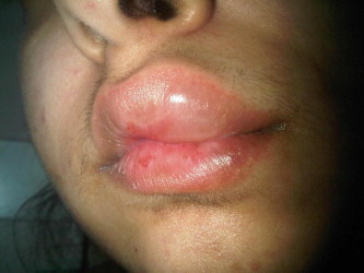
Unilateral lip swelling.
Figure 2.
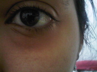
Edema of the eye lid.
The patient had a history of Kawasaki disease. She was first admitted to the hospital at the age of 4, after having had a fever for more than 2 days, which had initially been diagnosed as an upper respiratory tract infection. The fever persisted and 5 days later she was readmitted for further investigation. On physical examination she was found to have congested eyes, a red tongue, and a congested throat. ESR was elevated at 109 mm/h. The patient’s symptoms did not respond to treatment with an antibiotic. Three days later, she developed right upper quadrant abdominal swelling and was found to have hepatomegaly. Echocardiogram and electrocardiogram (ECG) results were normal and the patient appeared at the time to have no cardiovascular complications. She remained unresponsive to antibiotics and subsequently developed a truncal maculopapular rash and was found to have mild left cervical lymphadenopathy. One day after she commenced a high dose of aspirin (100 mg/kg/d orally in 4 doses) the patient became afebrile. However, after 2 days of aspirin therapy she developed vomiting, epigastric pain, and occult blood in the stool. Aspirin was stopped and then restarted at a lower dose (80 mg/kg p.o. daily) with Maalox suspension. Her ESR subsequently dropped to 72 mm/h.
Five days later her mycoplasma titer was 1:640 and her Salmonella typhi titer was 1:640, suggesting an immunoregulatory abnormality that was likely secondary to vasculitis. Due to the unavailability of IVIG in the hospital at that time, the patient was discharged and then readmitted 5 days later when she was given IVIG at a dose of 400 mg/kg/d over 4 successive days. After treatment, the patient was found to be afebrile with an ESR of 69 mm/h. She was then discharged on a low dose of aspirin (50 mg p.o daily). One month later the patient was readmitted complaining of fever, cough, and chest pain. Her platelet count was 1,026,000 mm3 and ESR was elevated at 100 mm/h. Two-dimensional echocardiography revealed a mild degree of long segment ectasia of both the left and right coronary arteries. The patient was clinically diagnosed with Kawasaki disease complicated by non-giant coronary aneurysms and was started on a brief regimen of a high-dose aspirin therapy (100 mg/kg/day) then followed by a low dose (4 mg/kg/day) and IVIG in a dose of 400 mg/kg/day by infusion in 4 successive days. The treatment plan included follow-up by ECG and 2-D echocardiography to monitor cardiac involvement. The patient was also to be continued on a low dose of aspirin (2–5 mg/kg once daily) as well as dipyridamole (3–5 mg/kg/d in 3 divided doses) to inhibit platelet aggregation as long as the coronary aneurysms persisted.
Four years ago the patient complained of multiple episodes of nausea, numbness in the extremities and sweating. A diagnosis of vasovagal syncope was made and the physicians advised the patient to avoid any stress or physical exertion. She was referred to a cardiologist who performed echocardiography which showed mild mitral regurgitation with a good ejection fraction of 65% and no wall motion abnormality. A 24-h ECG Holter Monitor investigation was normal except for two short runs of sinus tachycardia while walking with a heart rate of 170 associated with central chest pressure-like pain. The most likely diagnosis was positional vertigo. However, there was a need to rule out any sequelae of KD associated with cardiac arrhythmias which could be responsible for her complaint. The cardiologist recommended a cardiac CT scan which was highly suggestive of KD with involvement of the proximal segment of the coronary arteries mainly the left main, left anterior descending and the right coronary arteries. The affected arteries demonstrated multiple areas of thickening and aneurysmal formation with mild to moderate stenosis and a small aneurysmal formation ranging from 3 to 5 mm in diameter. The patient was strongly advised to continue aspirin therapy as well as regular follow-up visits for observation. She was informed that if the coronary arterial diameters were to grow to more than 70% increase over normal values, she would require a coronary bypass operation.
The patient continued to take Aspirin (80 mg/day), although she has expressed some resistance toward continuing this therapy as per the advice of her physicians.
She was advised to see an immunologist for skin patch testing. In 2010 she was diagnosed as having mild mitral regurgitation and as a result has also been advised to see a cardiologist for follow-up of this condition.
3. Discussion
Kawasaki disease is predominantly a disease of childhood, although cases have been reported in adults (Gomard-Mennesson et al., 2010; Wolff et al., 2007). It occurs primarily in the Asian population. The mean annual incidence is 90–112 per 100,000 people. In the United States the mean annual incidence is 6–9 per 100,000 children under 5 years of age, with a peak incidence in children between 1 and 2 years of age (Gedalia, 2002; Rozo et al., 2004). The mean annual incidence in Britain and Australia is 3.6 and 3.7 per 100,000 people, respectively.
In the Arab Gulf countries(Oman, Bahrain and Saudi Arabia), cases of KD are infrequently reported, although the disease may be under diagnosed because of resemblance of the clinical signs and symptoms with other diseases (Al-Harbi, 2010; Al-Mosawi et al., 2006; Bhatnagar et al., 2003; Muzaffer and Al-Mayouf, 2002; Owa et al., 1995). In one retrospective study conducted in the Madinah region of Saudi Arabia, investigators reviewed all pediatric cases over a 3-year period in which a diagnosis of KD was suspected and in which the patient underwent cardiac evaluation. Of 51 cases, only 24 patients were confirmed to have Kawasaki disease. Of the confirmed cases, most were of patients younger than 5 years of age, with a male to female ratio of 1.7:1 (Al-Harbi, 2010;Bhatnagar et al., 2003; Ghazal et al., 1998; Muzaffer and Al-Mayouf, 2002).
The likelihood of recurrence of Kawasaki disease increases with age. Recurrence within 2 years of the initial onset of the disease is unusual. Multiple recurrences have been reported in rare cases having recurrences in adulthood (Parmar et al., 2003; Pemberton et al., 1999). A high index of suspicion is appropriate in patients with a confirmed history of Kawasaki disease who present with clinical signs consistent with the disease (Parmar et al., 2003; Pemberton et al., 1999).
Untreated disease usually resolves spontaneously after several weeks (Pemberton et al., 1999; Rozo et al., 2004) even though the major pathologic feature of KD is acute systemic vasculitis. Coronary artery lesions usually develop early in the acute phase, although they can infrequently develop more than 4 weeks after the onset of the disease. These early lesions are characterized by endothelial cell edema, proliferation, necrosis, and adhesion of polymorphonuclear leukocytes to the endothelium (Cox and Sallis, 2009; Thabet et al., 2011). Because one of the classical criteria for Kawasaki disease is oral involvement, including lip erythema, dry cracked lips, and involvement of the oropharyngeal mucosa (Bhatnagar et al., 2003; Kanno et al., 2011),these signs may also be present in the setting of recurrent KD (Parmar et al., 2003).
At the time of her last admission, the patient we report here presented with five of the six principal criteria for Kawasaki disease. These included: (1) a remittent fever that was unresponsive to antibiotics; (2) bilateral conjunctival injection; (3) a polymorphous truncal rash without vesicles or crusts; (4) unilateral cervical lymphadenopathy; and (5) thrombocytosis. She also exhibited oral manifestations including erythema of the lips and oropharyngeal mucosa as well as a strawberry tongue.
The patient’s chief complaint when she was presented to the oral medicine clinic was lip swelling involving the cheek and eye lids on the ipsilateral side. She was on an antihistamine to exclude any allergic reaction to cats and foods, however, she did not respond to the medication. Also, histories of physical or surgical traumas causing any lip swellings were excluded as stated by the patient. No sign of any insect bite was observed since the swelling repeated almost on the same site with no response to antihistamine drugs. No relevance with any medical syndromes was observed which might indicate the involvement of any of the signs and symptoms e.g. Crohns’ Disease or Melkersson–Rosenthal Disease. Any other possible cause for lip swellings was investigated with no direct causative relevance emerged. All symptoms subsided spontaneously. She also experienced a single episode of laryngoedema which also resolved without treatment. Symptoms that the patient had experienced were not related to any environmental changes. The most recent laboratory results showed an ESR rate of 22 mm/h. Some of the clinical signs and symptoms including lip involvement and the spontaneous resolution of the allergic-like reaction may suggest the possibility of a recurrence of KD in this case.
Early diagnosis of KD is essential, because administration of IVIG within the first 10 days of illness has been shown to reduce the incidence of coronary artery aneurysms by a factor of five (Bhatnagar et al., 2003; Cox and Sallis, 2009). Cardiac complications occur in 20–25% of untreated patients, but in only 4% of treated patients (Bhatnagar et al., 2003; Cox and Sallis, 2009). The most common cardiac complication is coronary artery aneurysm, although other sequelae have been reported, including decreased myocardial contractility, congestive heart failure, arrhythmias, pancarditis, pericardial effusion, and myocardial ischemia (Cox and Sallis, 2009). Coronary artery aneurysms usually become apparent 1–3 weeks after the onset of fever, and resolve in 50% of patients within 5–18 months (Cox and Sallis, 2009; Rozo et al., 2004). Estimates of the incidence of under diagnosed conditions attempt to justify a larger incidence rate than is reported by doctors or medical authorities; whereas other rates may use only the official reported rates. In view of these potentially avoidable outcomes and because KD is under diagnosed, it is important to have a high degree of suspicion for the disease in any child with prolonged unexplained fevers (Bhatnagar et al., 2003).
The standard of care for children with acute Kawasaki disease is a single, 2 mg/kg infusion of IVIG along with aspirin at a dose of 80–100 mg/kg/day in 4 divided doses (Wolff et al., 2007). Aspirin can be decreased to 3–5 mg/kg/d once the fever has resolved doses (Burns et al., 2000; Ghazal et al., 1998; Wolff et al., 2007). In patients with coronary artery aneurysms, aspirin should be continued until 2 years after the aneurysms resolve. If aneurysms do not resolve, then aspirin therapy should be continued indefinitely to prevent coronary artery thrombosis (Wolff et al., 2007).
Although the patient reported here was admitted during the initial episode of her disease and presented with all of the criteria of the acute febrile phase, the delayed diagnosis led to the delayed administration of IVIG. This may ultimately have been responsible for the observed late recurrence of the oral manifestations (Parmar et al., 2003; Pemberton et al., 1999).
Dentists and oral medicine specialists are not usually involved in the diagnostic process of Kawasaki disease in children since the persistent fever tends to be the parents’ main concern. The oral manifestations of the disease are important to recognize since they may be overlooked by the general physician. Dentists have the opportunity to play an important role in assisting other health professionals in the early diagnosis of this rare disease.
Footnotes
The manuscript was presented as a Poster in the American Academy of Oral Medicine (AAOM) 66th Annual Meeting at Charleston/South Carolina, 17–21 April 2012.
Peer review under responsibility of King Saud University.

References
- Al-Harbi K.M. Kawasaki disease in Western Saudi Arabia. Saudi Med. J. 2010;31(11):1217. [PubMed] [Google Scholar]
- Al-Mosawi Z., Mohammad A., Al-Saif A., Al Madhoob A. Kawasaki disease: a retrospective study. Bahrain Med. Bull. 2006;28(2):1–8. [Google Scholar]
- Bhatnagar S.K., Paul G., Subramanian R., Al Hosni M.S., Al Khusaiby S.M. Kawasaki disease in Oman – a clinical study. J. Trop. Pediatr. 2003;49(6):361–366. doi: 10.1093/tropej/49.6.361. [DOI] [PubMed] [Google Scholar]
- Burns J.C., Kushner H.I., Bastian J.F., Shike H., Shimizu C., Matsubara T. Kawasaki disease: a brief history. Pediatrics. 2000;106(2) doi: 10.1542/peds.106.2.e27. e27. [DOI] [PubMed] [Google Scholar]
- Center for Disease Control and Prevention. Kawasaki Syndrome-CDC Diagnostic Criteria for Kawasaki Disease. <http://www.cdc.gov/kawasaki/> (cited 28.03.12).
- Cox J.R., Sallis R.E. Recognition of Kawasaki disease. Perm. J. 2009;13(1):57–61. doi: 10.7812/tpp/08-042. [DOI] [PMC free article] [PubMed] [Google Scholar]
- Gedalia A. Kawasaki disease: an update. Curr. Rheumatol. Rep. 2002;4(1):25–29. doi: 10.1007/s11926-002-0020-2. [DOI] [PubMed] [Google Scholar]
- Ghazal S.S., Alhowasi M., el Samady M.M. Kawasaki disease in a paediatric hospital in Riyadh. Ann. Trop. Paediatr. 1998;18(4):295–299. doi: 10.1080/02724936.1998.11747963. [DOI] [PubMed] [Google Scholar]
- Gomard-Mennesson E., Landron C., Dauphin C., Epaulard O., Petit C., Green L. Kawasaki disease in adults. Medicine. 2010;89(3):149–158. doi: 10.1097/MD.0b013e3181df193c. [DOI] [PubMed] [Google Scholar]
- Kanno K., Sakai H., Nakajima M., Satake A., Konishi T., Iizuka H. An adult case of kawasaki disease in a pregnant Japanese woman: a case report. Case Rep. Dermatol. 2011;3(1):98–102. doi: 10.1159/000327937. [DOI] [PMC free article] [PubMed] [Google Scholar]
- Kawasaki T. Acute febrile mucocutaneous syndrome with lymphoid involvement with specific desquamation of the fingers and toes in children. Clinical observation of 50 patients. Jpn. J. Allergy. 1967;16:178–222. [PubMed] [Google Scholar]
- Kawasaki T., Kosaki F., Okawa S., Shigematsu I., Yanagawa H. A new infantile acute febrile mucocutaneous lymph node syndrome (MLNS) prevailing in Japan. Pediatrics. 1974;54(3):271–276. [PubMed] [Google Scholar]
- Muzaffer M.A., Al-Mayouf S.M. Pattern of clinical features of Kawasaki disease. Saudi Med. J. 2002;23(4):409–412. [PubMed] [Google Scholar]
- NIH.NIH Office of Rare Diseases Research (ORDR). <http://www.rarediseases.info.nih.gov/Wrapper.aspx?src=asp/resources/ord_brochure.html> (cited 28.03.12).
- Owa J., Ahmad N., Abusrair H., Shawfey S. Kawasaki disease: report of three cases. Bahrain Med. Bull. [Internet] 1995;17(2) (cited 28.03.12) [Google Scholar]
- Parmar R., Somale A., Bavdekar S., Muranjan M. Incomplete Kawasaki disease with recurrent skin peeling: a case report with the review of literature. J. Postgrad. Med. 2003;49:72. doi: 10.4103/0022-3859.924. [DOI] [PubMed] [Google Scholar]
- Pemberton M.N., Doughty I.M., Middlehurst R.J., Thornhill M.H. Recurrent Kawasaki disease. Br. Dent. J. 1999;186(6):270–271. doi: 10.1038/sj.bdj.4800085. [DOI] [PubMed] [Google Scholar]
- Rozo J.C., Jefferies J.L., Eidem B.W., Cook P.J. Kawasaki disease in the adult. Tex. Heart Inst. J. 2004;31(2):160–164. [PMC free article] [PubMed] [Google Scholar]
- Scardina G.A. Oral manifestation of Kawasaki disease. Res. J. Biol. Sci. 2007;2(4):431–433. [Google Scholar]
- Sève P., Stankovic K., Smail A., Durand D.V., Marchand G., Broussolle C. Adult Kawasaki disease: report of two cases and literature review. Semin. Arthritis Rheum. 2005;34(6):785–792. doi: 10.1016/j.semarthrit.2005.01.012. [DOI] [PubMed] [Google Scholar]
- Thabet F., Bafaqih H., Al-Mohaimeed S., Al-Hilali M., Al-Sewairi W., Chehab M. Shock: an unusual presentation of Kawasaki disease. Eur. J. Pediatr. 2011;170(7):941–943. doi: 10.1007/s00431-011-1426-5. [DOI] [PubMed] [Google Scholar]
- Wolff A.E., Hansen K.E., Zakowski L. Acute Kawasaki disease: not just for kids. J. Gen. Intern. Med. 2007;22(5):681–684. doi: 10.1007/s11606-006-0100-5. [DOI] [PMC free article] [PubMed] [Google Scholar]