Background: Notch regulates bone mass and induces Hairy and Enhancer of Split-related with YRPW motif (HEY) in osteoblasts, but the skeletal function of individual HEY proteins is unclear.
Results: In male mice, Hey2 inactivation increases bone mass, whereas HEY2 overexpression enhances bone resorption, reducing bone mass.
Conclusion: HEY2 regulates skeletal remodeling in male mice, decreasing bone mass.
Significance: HEY2 mediates selected skeletal effects of Notch.
Keywords: Bone, Interleukin, Notch, Osteoblasts, Osteoclast, HEY2, Bone Remodeling
Abstract
Notch induces Hairy and Enhancer of Split-related with YRPW motif (Hey)1, Hey2, and HeyL expression in osteoblasts, but the contributions of these genes to the skeletal effects of Notch are not fully understood. HEY1 misexpression has limited skeletal impact, female HeyL null mice display increased bone mass, and Hey2 inactivation is developmentally lethal. To inactivate Hey2 in immature or mature osteoblasts, Hey2loxP/loxP mice were crossed with transgenics expressing CRE under the control of the osterix (Osx-Cre) or osteocalcin (Oc-Cre) promoters to generate Osx-Cre+/−;Hey2Δ/Δ or Oc-Cre+/−;Hey2Δ/Δ mice. Trabecular bone volume increased in 3-month-old Osx-Cre+/−;Hey2Δ/Δ and Oc-Cre+/−;Hey2Δ/Δ male mice and in 1-month-old Oc-Cre+/−;Hey2Δ/Δ female mice, although 3-month-old Oc-Cre+/−;Hey2Δ/Δ females developed osteopenia. Alkaline phosphatase liver/bone/kidney (ALPL) expression and activity were suppressed in osteoblasts from Oc-Cre+/−;Hey2Δ/Δ mice of both sexes. To overexpress HEY2 in osteoblasts, transgenic mice where a 3.6-kb fragment of the rat collagen type-I α1 promoter directs HEY2 expression were created. Three-month-old Hey2 transgenic males exhibited decreased osteoblast activity and increased bone resorption and developed osteopenia at 6 months of age. Hey2 transgenic females exhibited reduced osteoblast number and function, but no changes in bone resorption. HEY2 overexpression in osteoblasts from mice of both sexes inhibited ALPL expression and activity and suppressed osteocalcin transcripts in cells from male mice only. HEY2 overexpression in osteoblasts from male mice enhanced bone resorption by co-cultured splenocytes and induced interleukin-6, a molecule that promotes osteoclastogenesis. In conclusion, HEY2 decreases skeletal mass and regulates bone remodeling in male mice.
Introduction
Hairy and Enhancer of Split (HES)2-related with YRPW motif (HEY) genes encode 3 helix-loop-helix transcription factors termed HEY1, HEY2, and HEY-Like (HEYL) (1–3). HEY proteins display structural similarities with HES transcription factors and are targets of canonical Notch signaling, a critical regulator of cell differentiation during development and postnatal life (4). HEY proteins play an important role in cardiovascular development. Inactivation of Hey2 results in embryonic lethality due to cardiovascular defects, and the dual inactivation of Hey1 and Hey2 phenocopies the global deletion of Notch1. Similarly, the deletion of Hey1 and HeyL impairs cardiovascular development in mice (5–9).
The continuous renewal of skeletal tissue is carried out in basic multicellular units. There, osteoclasts resorb bone, and following a reversal phase, new bone matrix is deposited by osteoblasts. Osteoblasts are derived from multipotent mesenchymal stem cells that can differentiate toward the osteoblastic, chondrocytic, or adipocytic lineages (10). The commitment of mesenchymal cells to the osteoblastic fate is controlled by a signaling network that includes bone morphogenetic proteins, Wnt, and Notch (11–16). Osteoclasts are multinucleated cells formed by fusion and osteoclastic differentiation of mononuclear cell precursors, a process that requires macrophage-colony stimulating factor (M-CSF) and the receptor activator of nuclear factor-κb ligand (RANKL). RANK is expressed by osteoclast precursors and is activated following contact with cells expressing the membrane-bound RANKL. The activity of RANKL is opposed by osteoprotegerin (OPG), a soluble RANKL decoy receptor, and the ratio of RANKL over OPG regulates osteoclastogenesis (17, 18).
Notch is a transmembrane receptor activated by direct contact with Notch ligands. In the canonical signaling pathway, the Notch intracellular domain is released following a series of cleavages and forms a complex with Epstein-Barr virus latency C promoter binding factor 1, Suppressor of Hairless and Lag-1 (CSL), also termed Rbpjκ in mice, and with mastermind-like (MAML). The effects of Notch in the skeleton appear to be mediated by the canonical signaling pathway, but the genes responsible for the biology of Notch in bone have not been defined (19, 20). Hes1, Hes5, Hey1, Hey2, and HeyL are targets of the canonical Notch signaling pathway in skeletal cells, and as such should account, singly or in combination, for the effects of Notch in the skeleton (21). Previously, we reported that HES1 causes osteopenia by inhibiting bone formation and inducing bone resorption in mice (22). These effects of HES1 phenocopied only partially the skeletal phenotype of mice misexpressing Notch in the skeleton, suggesting that HEY proteins may be downstream effectors of Notch signaling in the skeleton (22). Transgenic expression of HEY1 under the control of the ubiquitously expressed β-actin promoter as well as the global inactivation of Hey1 caused mild to modest osteopenia, and global HeyL null mice display increased bone mass (23, 24).
Notch signaling controls osteoclastic differentiation of bone marrow mononuclear precursors and the expression of RANKL and OPG in osteoblastic cells (15, 25, 26). Activation of the Notch signaling pathway in osteoclast precursors regulates osteoclastogenesis, and the cellular context or experimental conditions determine whether Notch suppresses or stimulates osteoclast differentiation (25–29).
In this study, we examined the function of HEY2 in the postnatal skeleton. For this purpose, the skeletal phenotype of mice misexpressing HEY2 was investigated by microcomputed tomography (μCT) and by histomorphometric analysis. To understand the cellular mechanisms involved, the differentiation and function of osteoblasts misexpressing HEY2 were examined in vitro.
EXPERIMENTAL PROCEDURES
Hey2 Conditional Null Mice
To study the skeletal consequences of Hey2 inactivation in cells of the osteoblastic lineage, mice where the Hey2 sequence comprised between exon 1 and exon 4 was flanked by loxP sites in a 129Sv/C57BL/6 background, were provided by E. N. Olson (University of Texas Southwestern Medical Center, Dallas, TX) (6). To express CRE recombinase at early stages of osteoblastic differentiation, we obtained C57BL/6 mice where the CRE coding sequence is cloned downstream of an osterix (Osx) promoter (Osx-Cre) (The Jackson Laboratory) (30). In these mice, tetracycline suppresses the Osx promoter activity by virtue of a Tet-Off cassette (31). To express CRE preferentially in mature osteoblasts, mice where a 3.9-kb fragment of the human osteocalcin promoter directs CRE expression (Oc-Cre) were obtained from T. Clemens (Johns Hopkins Medicine, Baltimore, MD) and backcrossed for seven generations into a C57BL/6 genetic background. Osx-Cre or Oc-Cre transgenics were crossed with Hey2loxP/loxP mice to create Osx-Cre+/− or Oc-Cre+/−;Hey2loxP/wt mice, which were mated with Hey2loxP/loxP to obtain Osx-Cre+/− or Oc-Cre+/−;Hey2loxP/loxP mice. The latter were crossed with Hey2loxP/loxP to generate an experimental cohort, in which CRE excises the loxP-flanked sequences from the Hey2loxP allele (Osx-Cre+/− or Oc-Cre+/−;Hey2Δ/Δ) and littermate controls (Hey2loxP/loxP). To prevent Osx-Cre expression during embryonic development, pregnant mothers were administered chow containing 625 mg/kg of doxycycline (Harlan Laboratories, Indianapolis, IN) from the time of conception to delivery. The presence of the Osx-Cre and Oc-Cre transgenes and of the Hey2loxP allele was determined by PCR in tail DNA extracts in newborns and adult mice, and primers specific for fatty acid-binding protein 1 (Fabp1) were used as positive controls in the PCR reactions. Recombination of sequences flanked by loxP sites was assessed by PCR in DNA extracts from parietal bone, using primers specific for the Hey2Δ allele. All animal experiments were approved by the Animal Care and Use Committee of Saint Francis Hospital and Medical Center.
Col3.6-Hey2 Transgenic Mice
For preferential expression of HEY2 in osteoblasts, a 1047-bp DNA fragment coding for murine HEY2 (American Type Culture Collection; ATCC, Manassas, VA), preceded by a Kozak consensus sequence, was cloned downstream of a 3.6-kb fragment of the rat Col1a1 (collagen type I α1) promoter and upstream of the bovine growth hormone polyadenylation signal (32). Microinjection of linearized DNA into pronuclei of fertilized oocytes from Friend leukemia virus strain B (FVB) mice (Charles River Laboratories, Wilmington, MA) and transfer of microinjected embryos into pseudopregnant mice were carried out at the Gene Targeting and Transgenic Facility of the University of Connecticut Health Center (Farmington, CT). Positive founders were identified by Southern blot analysis of tail DNA and bred to wild type FVB mice to create Col3.6-Hey2 transgenic lines (33). To assess the effects of HEY2 overexpression, heterozygous Col3.6-Hey2 mice were mated to wild type FVB mice to generate heterozygous Col3.6-Hey2 transgenic mice and wild type littermate controls. The presence of the Col3.6-Hey2 transgene was documented by PCR in tail DNA. To assess mRNA expression in skeletal cells, calvariae were frozen in liquid nitrogen at the time of harvest and transferred to −80 °C for storage before RNA extraction. To determine levels of bone resorption, fasting serum concentration of collagen type I C-terminal telopeptide (CTX-I), was measured by using the RatLaps enzyme-linked immunosorbent assay in accordance with the manufacturer's instructions (Immuno Diagnostic Systems, Scottsdale, AZ) (34).
Microcomputed Tomography
Femurs were scanned in 70% ethanol at an energy level of 55 kVp, an intensity of 145 μA, and an integration time of 200 ms on a μCT 40 scanner (Scanco Medical AG, Bassersdorf, Switzerland). Trabecular bone volume fraction and microarchitecture were evaluated starting ∼1.0 mm proximal to the femoral condyles. A total of 160 consecutive slices acquired at an isotropic voxel size of 216 μm3 and a slice thickness of 6 μm were chosen for analysis. Contours were manually drawn every 10 slices a few voxels away from the endocortical boundary to define the region of interest for analysis. The contours of the remaining slices were iterated automatically. Trabecular regions were assessed for bone volume fraction, trabecular thickness, number and separation, connectivity density, and structure model index (SMI), using a Gaussian filter (σ = 0.8) and a user-defined threshold (35). A total of 100 slices for the cortical region were measured at the mid-diaphysis of each femur with an isotropic voxel size of 216 μm3 and a slice thickness of 6 μm. For mid-diaphysis analysis, contours were iterated across the 100 slices along the cortical shell, excluding the bone marrow cavity. Analysis for cortical thickness was performed using a Gaussian filter (σ = 0.8) and a user-defined threshold (35).
Bone Histomorphometric Analysis
Static and dynamic histomorphometry of femurs was carried out after injection with 20 mg/kg calcein and 50 mg/kg demeclocycline, at an interval of 2 days for 1-month-old mice and 7 days for 3- and 6-month-old mice. Animals were sacrificed by CO2 inhalation 2 days after the demeclocycline injection. Femurs were sectioned on a microtome at a thickness of 5 μm (Microm, Richards-Allan Scientific, Kalamazoo, MI) and stained with 0.1% toluidine blue. Static parameters of bone formation and resorption were measured in a defined area between 360 and 2160 μm from the growth plate, using an OsteoMeasure morphometry system (Osteometrics, Atlanta, GA) (36). For dynamic histomorphometry, mineralizing surface per bone surface and mineral apposition rate were measured on unstained sections under ultraviolet light, using a triple diamino-2-phenylindole fluorescein set long pass filter, and bone formation rate was calculated. The terminology and units used are those recommended by the Histomorphometry Nomenclature Committee of the American Society for Bone and Mineral Research (37).
Osteoblast Cultures
Osteoblast-enriched cells were isolated from parietal bones of 3–5-day-old male or female Oc-Cre+/−;Hey2Δ/Δ or Col3.6-Hey2 transgenic mice and littermate controls of the same sex by sequential collagenase digestion, as described (38). The sex of newborn mice was determined by PCR analysis of tail DNA with 5′-GAGAGCATGGAGGGCAT-3′ forward and 5′-GAGTACAGGTGTGCAGCTC-3′ reverse primers amplifying a 400-bp fragment of sex-determining region Y, located on chromosome Y, and with 5′-TGGACAGGACTGGACCTCTGCTTTCC-3′ forward and 5′-TAGAGCTTTGCCACATCACAGGTCAT-3′ reverse primers amplifying a 200-bp fragment of the autosomal gene fatty acid-binding protein 1. Cells were cultured in Dulbecco's modified Eagle's medium (DMEM, Life Technologies) supplemented with nonessential amino acids (Life Technologies), 20 mm HEPES, 100 μg/ml ascorbic acid (both from Sigma-Aldrich), and 10% fetal bovine serum (FBS; Atlanta Biologicals, Norcross, GA) at 37 °C in a humidified 5% CO2 incubator.
Quantitative Reverse Transcription-PCR (qRT-PCR)
Total RNA was extracted from cells with the RNeasy mini kit, according to the manufacturer's instructions (Qiagen, Valencia, CA), and from frozen calvariae by phenol/chloroform extraction (Sigma-Aldrich), and changes in mRNA levels were determined by qRT-PCR. 0.5–1 μg of total RNA was reverse-transcribed using the iScript cDNA synthesis kit (Bio-Rad Laboratories) and amplified in the presence of 5′-TGGTATGGGCGTCTCCACAGTAACC-3′ forward and 5′-CTTGGAGAGGGCCACAAAGG-3′ reverse primers for alkaline phosphatase liver/bone/kidney (Alpl; NM_007431), 5′-CCCCTCTGGAAAGCTGTGGCGT-3′ forward and 5′-AGCTTCCCGTTCAGCTCTGG-3′ reverse primers for glyceraldehyde 3-phosphate dehydrogenase (Gapdh; NM_008084), 5′-AGCGAGAACAATTACCCTGGGCAC-3′ forward and 5′-ATTCTTGCCCTTCGCCTCTT-3′ reverse primers for Hey2 (NM_013904), 5′-CGGCCTTCCCTACTTCACAAGTCCG-3′ forward and 5′-CAGGTCTGTTGGGAGTGGTATCC-3′ reverse primers for interleukin-6 (Il6; NM_031168), 5′-GACTCCGGCGCTACCTTGGGTAAG-3′ forward and 5′-CCCAGCACAACTCCTCCCTA-3′ reverse primers for osteocalcin (NM_001037939), 5′-CAGAAAGGAAATGCAACACATGACAAC-3′ forward and 5′-GCCTCTTCACACAGGGTGACATC-3′ reverse primers for osteoprotegerin (Opg; NM_008764), 5′-TATAGAATCCTGAGACTCCATGAAAAC-3′ forward and 5′-CCCTGAAAGGCTTGTTTCATCC-3′ reverse primers for Rankl (NM_011613), and 5′-AGAACAAGGATAATGTGAAGTTCAAGGTTC-3′ forward and 5′-CTGCTTCAGCTTCTCTGCCTTT-3′ reverse primers for ribosomal protein L38 (Rpl38; NM_001048057, NM_001048058, and NM_023372) and iQ SYBR Green supermix (Bio-Rad Laboratories) at 60 °C for 45 cycles, according to the manufacturer's instructions. Transcript copy number was estimated by comparison with a dilution series of Alpl and Rpl38 (both from ATCC), Gapdh (R. Wu, Cornell University, Ithaca, NY), Hey2 (T. Iso, University of Southern California, Los Angeles, CA), Il6 and Opg (both from Open Biosystems, Huntsville, AL), osteocalcin (J. B. Lian, University of Massachusetts, Worcester, MA), and Rankl (Source BioScience, Nottingham, UK) cDNA (39–43). Reactions were conducted in a CFX96 qRT-PCR detection system (Bio-Rad Laboratories), and fluorescence was monitored at the annealing step of every PCR cycle. Specificity of the reaction was confirmed by the presence of a single peak in the melt curve analysis of PCR products.
Alkaline Phosphatase Activity
Alkaline phosphatase activity was determined in cell extracts by the hydrolysis of p-nitrophenyl phosphate to p-nitrophenol, measured by spectroscopy at 405 nm according to manufacturer's instructions (Sigma-Aldrich). Data are expressed as nanomoles of p-nitrophenol released per minute per μg of protein measured by the DC protein assay (Bio-Rad Laboratories).
Osteoblast-Splenocyte Co-Cultures and Pit Formation Assay
Primary osteoblasts from male Col3.6-Hey2 transgenic mice and male littermate controls were seeded on BioCoat discs (BD Biosciences), and after reaching confluence, cultured in the presence of 100 μg/ml ascorbic acid and 5 mm β-glycerophosphate. Primary splenocytes were harvested from spleens aseptically removed from 1-month-old wild type FVB male mice, and 1 × 10⁶ cells/cm2 were seeded on the layer of primary osteoblasts in the presence of 10 nm 1,25 dihydroxyvitamin D3 (BioMol International, Plymouth Meeting, PA) or phosphate-buffered saline, as control (44–46). Splenocytes and primary osteoblasts were cultured for 7 days, cells were removed with bleach for 5 min, and BioCoat discs were stained with von Kossa. Stained discs were photographed on a white light background and a digital grayscale image analyzed with Adobe Photoshop (Adobe Systems, Inc., San Jose, CA). The negative image was obtained with the invert feature of Adobe Photoshop, brightness and contrast were maximized, and the area of resorption was calculated as the percentage of pixels contained in half of the grayscale, determined by using the histogram function of Adobe Photoshop (22).
Statistical Analysis
Data are expressed as means ± S.E. Statistical differences were determined by Student's t test or analysis of variance with Schaeffe's post hoc analysis for pairwise or multiple comparisons, respectively (47).
RESULTS
Hey2 Inactivation in Cells of the Osteoblastic Lineage Causes a Transient Increase in Bone Volume
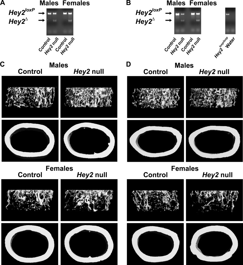
To investigate the function of Hey2 at early stages of osteoblast maturation and in differentiated osteoblasts, the skeletal phenotype of Osx-Cre+/−;Hey2Δ/Δ or Oc-Cre+/−;Hey2Δ/Δ mice was compared with the phenotype of littermate Hey2loxP/loxP controls of the same sex. Recombination of Hey2 sequences flanked by loxP sites was detected in DNA extracts from calvariae of 1- and 3-month-old Osx-Cre+/−;Hey2Δ/Δ and Oc-Cre+/−;Hey2Δ/Δ mice, whereas recombination was not observed in littermate controls (Fig. 1, A and B, and data not shown). Osx-Cre+/−;Hey2Δ/Δ and Oc-Cre+/−;Hey2Δ/Δ mice were viable, appeared normal, and did not exhibit differences in mortality when compared with littermate controls.
FIGURE 1.
Hey2 inactivation in osteoblastic cells transiently increases trabecular bone volume in male mice. In A and B, DNA was extracted from parietal bones of 3-month-old male or female Osx-Cre+/− (A) or Oc-Cre+/− (B); Hey2Δ/Δ mice (Hey2 null), or littermate Hey2loxP/loxP controls of the same sex (Control). Recombination of the Hey2loxP allele was determined by PCR. Control reactions conducted in tail DNA from female Hey2loxP/loxP mouse or water were performed to confirm specificity of the primers. All reaction products were separated by electrophoresis on the same gel, and results from representative individuals are shown. C and D display microcomputed tomography images of proximal trabecular bone and cortical bone at the midshaft of femurs from representative 3-month-old male or female Osx-Cre+/− (C) or Oc-Cre+/− (D); Hey2Δ/Δ mice; (Hey2 null) or littermate Hey2loxP/loxP controls of the same sex (Control).
In initial experiments, we demonstrated that Oc-Cre, Osx-Cre, and Hey2loxP/loxP did not exhibit an appreciable skeletal phenotype as determined by μCT at 1 month of age (48). One-month-old Osx-Cre+/−;Hey2Δ/Δ male mice did not display a skeletal phenotype. μCT analysis of femurs from 3-month-old Osx-Cre+/−;Hey2Δ/Δ male mice revealed increased trabecular bone volume and connectivity, secondary to increased trabecular number (Table 1, Fig. 1C). However, no changes in parameters of bone formation or bone resorption were observed by histomorphometric analysis (data not shown). No skeletal phenotype was observed in Osx-Cre+/−;Hey2Δ/Δ female mice by μCT analysis (Table 1, Fig. 1C), indicating that Hey2 inactivation at early stages of osteoblast differentiation increases bone mass only in male mice.
TABLE 1.
Microcomputed tomography (μCT) of the femur of 1- and 3-month-old Osx-Cre+/−;Hey2Δ/Δ (Hey2 null) male or female mice or littermate Hey2loxP/loxP controls (Control) of the same sex
Values are means ± S.E.; n = 5–9. *, significantly different between Hey2 null and control, p < 0.05; +, p < 0.07.
| 1 month |
3 month |
|||
|---|---|---|---|---|
| Control | Hey2 null | Control | Hey2 null | |
| μCT (males) | ||||
| Bone volume fraction (%) | 4.9 ± 0.8 | 4.9 ± 0.6 | 4.1 ± 0.5 | 6.6 ± 0.8* |
| Trabecular separation (μm) | 243 ± 19 | 222 ± 7 | 236 ± 5 | 203 ± 5* |
| Trabecular number (mm−1) | 4.3 ± 0.3 | 4.6 ± 0.1 | 4.2 ± 0.1 | 4.9 ± 0.1* |
| Trabecular thickness (μm) | 24.2 ± 1.0 | 23.4 ± 0.8 | 28.5 ± 0.5 | 30.0 ± 1.8 |
| Connectivity density (mm−3) | 161 ± 38 | 192 ± 58 | 96 ± 18 | 218 ± 34* |
| Structure model index (SMI) | 2.97 ± 0.10 | 2.93 ± 0.10 | 2.98 ± 0.08 | 2.72 ± 0.09+ |
| Cortical thickness (μm) | 102 ± 3 | 103 ± 2 | 165 ± 6 | 167 ± 6 |
| μCT (females) | ||||
| Bone volume fraction (%) | 4.8 ± 0.6 | 4.0 ± 0.4 | 2.9 ± 0.4 | 3.3 ± 0.4 |
| Trabecular separation (μm) | 234 ± 16 | 244 ± 8 | 330 ± 12 | 294 ± 14 |
| Trabecular number (mm−1) | 4.4 ± 0.3 | 4.2 ± 0.2 | 3.1 ± 0.1 | 3. 5 ± 0.1 |
| Trabecular thickness (μm) | 24.0 ± 0.6 | 22.7 ± 0.6 | 33.3 ± 0.5 | 32.4 ± 0.5 |
| Connectivity density (mm−3) | 135 ± 25 | 110 ± 23 | 63 ± 11 | 92 ± 14 |
| Structure model index (SMI) | 2.85 ± 0.07 | 3.04 ± 0.07 | 3.04 ± 0.07 | 2.96 ± 0.10 |
| Cortical thickness (μm) | 100 ± 2 | 104 ± 1 | 178 ± 3 | 164 ± 5 |
Oc-Cre+/−;Hey2Δ/Δ male mice at 1 month of age did not exhibit changes in trabecular bone volume, although these mice displayed a limited reduction in mineral apposition rate and bone formation rate (Fig. 1D, Table 2). Three-month-old Oc-Cre+/−;Hey2Δ/Δ male mice developed increased bone volume/tissue volume, indicating that the suppressed osteoblast function at 1 month of age does not translate into osteopenia in older mice (Table 2, Fig. 1D). In addition, inactivation of Hey2 in 3-month-old male mice resulted in lower SMI (Table 2, Fig. 1D), indicative of a higher ratio of plate-like over rod-like trabeculae (49). A modest increase in cortical thickness was observed (Table 2, Fig. 1D). Female Oc-Cre+/−;Hey2Δ/Δ mice did not exhibit a skeletal phenotype at 1 month of age except for a modest reduction in mineral apposition rate, and at 3 months of age, they exhibited modest osteopenia by μCT, but not by histomorphometry (Table 2, Fig. 1D). The skeletal phenotypes of Osx-Cre+/−;Hey2Δ/Δ and Oc-Cre+/−;Hey2Δ/Δ mice were transient, and at 6 months of age, these mice were not different from littermate Hey2loxP/loxP controls, as determined by μCT (not shown).
TABLE 2.
Microcomputed tomography (μCT) and histomorphometry of the femur of 1, and 3 month old Oc-Cre+/−;Hey2Δ/Δ (Hey2 null) male or female mice, or littermate Hey2loxP/loxP (Control) of the same sex
Values are means ± S.E.; n = 4–9. * Significantly between Hey2 null and Control, p < 0.05; +p < 0.07.
| 1 month |
3 month |
|||
|---|---|---|---|---|
| Control | Hey2 null | Control | Hey2 null | |
| μCT (males) | ||||
| Bone volume fraction (%) | 2.9 ± 0.6 | 2.8 ± 0.2 | 5.1 ± 0.6 | 6.9 ± 0.2* |
| Trabecular separation (μm) | 342 ± 27 | 321 ± 26 | 241 ± 12 | 221 ± 3 |
| Trabecular number (mm−1) | 3.05 ± 0.28 | 3.22 ± 0.26 | 4.19 ± 0.18 | 4.49 ± 0.06 |
| Trabecular thickness (μm) | 22.3 ± 0.6 | 22.2 ± 0.6 | 30.9 ± 1.6 | 33.2 ± 0.8 |
| Connectivity density (mm−3) | 103 ± 46 | 49 ± 12 | 127 ± 29 | 176 ± 10 |
| Structure model index (SMI) | 3.07 ± 0.15 | 3.16 ± 0.26 | 2.80 ± 0.07 | 2.60 ± 0.06* |
| Cortical thickness (μm) | 89 ± 2 | 87 ± 5 | 162 ± 1 | 170 ± 2* |
| Histomorphometry (males) | ||||
| Bone volume/tissue volume (%) | 7.3 ± 1.0 | 4.8 ± 0.9 | 8.5 ± 1.2 | 9.9 ± 1.6 |
| Osteoblast surface/bone surface (%) | 23.2 ± 1.2 | 21.7 ± 1.8 | 12.8 ± 1.2 | 16.3 ± 1.0 |
| Osteoblasts/bone perimeter (mm−1) | 20.4 ± 1.0 | 20.0 ± 1.1 | 14.2 ± 1.3 | 17.2 ± 1.9 |
| Osteoid surface/bone surface (%) | 3.9 ± 0.7 | 3.9 ± 0.5 | 1.1 ± .5 | 1.8 ± 0.5 |
| Osteoclast surface/bone surface (%) | 19.2 ± 1.3 | 19.3 ± 1.4 | 5.8 ± 0.7 | 5.2 ± 0.5 |
| Osteoclasts/bone perimeter (mm−1) | 8.6 ± 0.7 | 9.1 ± 0.6 | 3.7 ± 0.6 | 3.4 ± 0.4 |
| Eroded surface/bone surface (%) | 20.4 ± 1.4 | 21.5 ± 1.0 | 9.4 ± 1.3 | 8.3 ± 0.7 |
| Mineral apposition rate (μm day−1) | 1.22 ± 0.03 | 1.03 ± 0.03* | 1.65 ± 0.13 | 1.59 ± 0.03 |
| Mineralizing surface/bone surface (%) | 5.1 ± 1.2 | 2.4 ± 0.8 | 16.9 ± 4.3 | 19.1 ± 3.2 |
| Bone formation rate (μm2 μm−3day−1) | 0.06 ± 0.01 | 0.02 ± 0.01+ | 0.29 ± 0.05 | 0.30 ± 0.05 |
| μCT (females) | ||||
| Bone volume fraction (%) | 3.0 ± 0.4 | 3.5 ± 0.4 | 2.9 ± 0.3 | 1.9 ± 0.3* |
| Trabecular separation (μm) | 343 ± 28 | 293 ± 18 | 319 ± 10 | 360 ± 15* |
| Trabecular number (mm−1) | 3.09 ± 0.23 | 3.52 ± 0.22 | 3.17 ± 0.09 | 2.82 ± 0.12* |
| Trabecular thickness (μm) | 23.3 ± 0.4 | 23.0 ± 0.4 | 33.6 ± 1.0 | 32.2 ± 1.7 |
| Connectivity density (mm−3) | 64 ± 12 | 91 ± 24 | 57 ± 8 | 35 ± 8 |
| Structure model index (SMI) | 2.96 ± 0.04 | 2.91 ± 0.06 | 3.21 ± 0.10 | 3.36 ± 0.11 |
| Cortical thickness (μm) | 91 ± 1 | 92 ± 2 | 178 ± 3 | 173 ± 1 |
| Histomorphometry (females) | ||||
| Bone volume/tissue volume (%) | 7.3 ± 0.6 | 5.8 ± 0.8 | 4.0 ± 0.4 | 3.4 ± 0.5 |
| Osteoblast surface/bone surface (%) | 21.8 ± 1.7 | 18.6 ± 2.2 | 27.0 ± 4.4 | 26.8 ± 4.9 |
| Osteoblasts/bone perimeter (mm−1) | 19.1 ± 1.4 | 16.5 ± 1.5 | 28.3 ± 3.7 | 29.7 ± 4.2 |
| Osteoid surface/bone surface (%) | 4.2 ± 0.9 | 2.8 ± 0.6 | 4.8 ± 1.7 | 4.8 ± 1.8 |
| Osteoclast surface/bone surface (%) | 18.0 ± 0.4 | 19.3 ± 1.6 | 9.8 ± 1.1 | 10.2 ± 0.4 |
| Osteoclasts/bone perimeter (mm−1) | 9.7 ± 0.2 | 10.3 ± 0.6 | 6.6 ± 0.7 | 6.9 ± 0.2 |
| Eroded surface/bone surface (%) | 23.7 ± 0.5 | 22.0 ± 1.2 | 14.9 ± 1.4 | 15.3 ± 0.5 |
| Mineral apposition rate (μm day−1) | 1.32 ± 0.06 | 1.06 ± 0.08* | 2.46 ± 0.18 | 2.19 ± 0.40 |
| Mineralizing surface/bone surface (%) | 2.5 ± 0.4 | 2.6 ± 0.3 | 8.9 ± 1.6 | 6.7 ± 1.4 |
| Bone formation rate (μm2 μm−3day−1) | 0.03 ± 0.01 | 0.03 ± 0.01 | 0.22 ± 0.04 | 0.16 ± 0.05 |
Hey2 Inactivation Impairs Osteoblast Function in Vitro
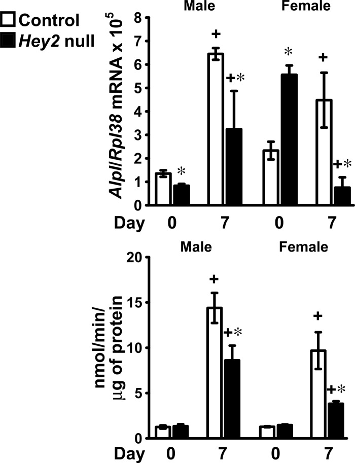
To explore the cellular mechanisms that lead to suppressed bone formation in Oc-Cre+/−;Hey2Δ/Δ mice, primary osteoblastic cells were harvested from Oc-Cre+/−;Hey2Δ/Δ male or female mice and littermate Hey2loxP/loxP controls of the same sex. Alkaline phosphatase transcripts and activity were decreased in the context of Hey2 inactivation in primary calvarial osteoblasts from mice of both sexes (Fig. 2). These findings are in agreement with the decrease in mineral apposition rate exhibited by Hey2 null mice and indicate that HEY2 is required for full osteoblastic function.
FIGURE 2.
Hey2 inactivation inhibits osteoblast function in vitro. Osteoblast-enriched cells were harvested from calvariae of male or female Oc-Cre+/−;Hey2Δ/Δ mice (Hey2 null, black bars), or littermate Hey2loxP/loxP controls of the same sex (Control, white bars) and cultured under conditions favoring osteoblastogenesis. Total RNA was extracted, and mRNA was reverse-transcribed and amplified by qRT-PCR in the presence of specific primers. Data are expressed as Alpl copy number, corrected for Rpl38 copy number. Values are means ± S.E., n = 3–4. Alkaline phosphatase activity was determined in cells extracted with Triton X-100, and data are expressed as nanomoles of p-nitrophenol/min/μg of total protein. Values are means ± S.E., n = 6. +, significantly different between day 7 and day 0, p < 0.05. *, significantly different between HEY2 and control, p < 0.05.
HEY2 Overexpression in Osteoblasts Uncouples Bone Formation from Bone Resorption
To test further the function of HEY2 in the skeleton, the effects of the preferential HEY2 overexpression in osteoblasts were investigated in Col3.6-Hey2 transgenic mice at 1, 3, and 6 months of age. Three transgenic founders were obtained, and only one male founder mouse transmitted the transgene to the offspring, allowing the establishment of a Hey2 transgenic line. Col3.6-Hey2 transgenics were born at the expected Mendelian ratio and appeared normal and healthy. qRT-PCR analysis demonstrated that Hey2 mRNA levels, corrected for Rpl38 expression, in calvariae from 1-month-old Col3.6-Hey2 transgenic males and females mice were increased 32.9 ± 3.4- and 32.2 ± 10.6-fold (p < 0.05), respectively, in comparison with control littermates of the same sex, confirming expression of the Col3.6-Hey2 transgene.
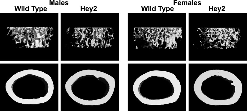
μCT revealed that Col3.6-Hey2 male transgenics did not exhibit a phenotype at 1 or 3 months of age, but were osteopenic at 6 months of age. The SMI value closer to 3 in the Col3.6-Hey2 transgenic male mice indicated a preponderance of rod-like over plate-like trabeculae, suggesting that HEY2 affects bone microarchitecture (Table 3, Fig. 3) (49). Histomorphometric analysis revealed that Col3.6-Hey2 transgenics had increased osteoclast number and eroded surface and decreased bone formation rate at 3 months of age (Table 3). However, HEY2 overexpression did not affect serum levels of CTX-I, a marker of bone resorption, in 1-month-old male mice (data not shown) (34). The changes in the cellular parameters observed at 3 months of age may explain the reduced bone volume observed in 6-month-old male mice, suggesting that overexpression of HEY2 in osteoblasts causes osteopenia due to an uncoupling of osteoblast and osteoclast activities.
TABLE 3.
Microcomputed tomography (μCT) and histomorphometry of the femur of 1, 3 and 6 month old Col3.6-Hey2 transgenic male mice (HEY2), or littermate wild type controls of the same sex
Values are means ± S.E.; n = 5–10. * Significantly between HEY2 and wild type, p < 0.05.
| 1 month |
3 month |
6 month |
||||
|---|---|---|---|---|---|---|
| Wild type | HEY2 | Wild type | HEY2 | Wild type | HEY2 | |
| μCT (males) | ||||||
| Bone volume fraction (%) | 5.5 ± 0.8 | 7.5 ± 0.6 | 4.8 ± 0.5 | 4.7 ± 0.4 | 6.0 ± 0.8 | 3.2 ± 0.6* |
| Trabecular separation (μm) | 212 ± 11 | 204 ± 11 | 270 ± 10 | 259 ± 5 | 366 ± 3 | 356 ± 16 |
| Trabecular number (mm−1) | 4.81 ± 0.24 | 5.03 ± 0.29 | 3.77 ± 0.13 | 3.91 ± 0.07 | 2.78 ± 0.01 | 2.84 ± 0.12 |
| Trabecular thickness (μm) | 24.7 ± 0.7 | 26.5 ± 0.3* | 29.8 ± 1.2 | 29.7 ± 0.9 | 38.2 ± 1.5 | 34.7 ± 2.1 |
| Connectivity density (mm−3) | 213 ± 52 | 338 ± 44 | 138 ± 15 | 140 ± 14 | 103 ± 11 | 48 ± 10* |
| Structure model index (SMI) | 2.85 ± 0.11 | 2.52 ± 0.06* | 2.66 ± 0.13 | 2.75 ± 0.06 | 2.03 ± 0.15 | 2.74 ± 0.14* |
| Cortical thickness (μm) | 119 ± 2 | 117 ± 3 | 182 ± 4 | 177 ± 1 | 192 ± 3 | 189 ± 3 |
| Histomorphometry (males) | ||||||
| Bone volume/tissue volume (%) | 8.8 ± 0.4 | 9.9 ± 1.8 | 6.5 ± 0.6 | 7.6 ± 0.3 | 4.8 ± 0.9 | 3.5 ± 0.4 |
| Osteoblast surface/bone surface (%) | 17.0 ± 1.8 | 15.4 ± 2.2 | 12.7 ± 1.5 | 10.9 ± 1.1 | 11.1 ± 1.0 | 14.5 ± 1.4 |
| Osteoblasts/bone perimeter (mm−1) | 17.2 ± 1.8 | 15.3 ± 2.4 | 15.5 ± 1.8 | 12.8 ± 1.4 | 11.4 ± 1.1 | 15.4 ± 2.4 |
| Osteoid surface/bone surface (%) | 1.0 ± 0.2 | 1.4 ± 0.3 | 0.9 ± 0.1 | 0.7 ± 0.2 | 0.8 ± 0.4 | 0.5 ± 0.2 |
| Osteoclast surface/bone surface (%) | 11.3 ± 0.7 | 10.4 ± 0.6 | 9.3 ± 0.5 | 10.9 ± 0.6 | 8.1 ± 0.4 | 8.3 ± 0.7 |
| Osteoclasts/bone perimeter (mm−1) | 7.3 ± 0.4 | 7.1 ± 0.3 | 6.0 ± 0.3 | 7.2 ± 0.4* | 5.7 ± 0.3 | 5.8 ± 0.6 |
| Eroded surface/bone surface (%) | 23.1 ± 1.1 | 22.2 ± 1.5 | 13.8 ± 0.7 | 17.9 ± 1.0* | 13.8 ± 0.7 | 13.6 ± 1.1 |
| Mineral apposition rate (μm day−1) | 2.53 ± 0.20 | 2.33 ± 0.17 | 0.60 ± 0.04 | 0.53 ± 0.03 | 0.49 ± 0.03 | 0.46 ± 0.04 |
| Mineralizing surface/bone surface (%) | 3.0 ± 0.3 | 3.3 ± 0.5 | 10.9 ± 1.0 | 6.5 ± 1.0* | 5.5 ± 1.2 | 8.2 ± 2.0 |
| Bone formation rate (μm2 μm−3day−1) | 0.08 ± 0.01 | 0.08 ± 0.02 | 0.07 ± 0.01 | 0.04 ± 0.01* | 0.03 ± 0.01 | 0.04 ± 0.01 |
FIGURE 3.
Hey2 overexpression in osteoblasts causes osteopenia in male mice. Microcomputed tomography images of proximal trabecular bone and cortical bone at the midshaft of femurs from representative 6-month-old male or female Col3.6-Hey2 transgenic mice (Hey2) or littermate wild type controls of the same sex (Wild Type) are shown.
μCT and histomorphometric analysis indicated that Col3.6-Hey2 transgenic female mice did not exhibit an obvious skeletal phenotype at 1, 3, and 6 months of age, except for a decrease in bone formation rate at 6 months of age (Table 4, Fig. 3). No changes in osteoclast number, eroded surface, or serum levels of CTX-I were observed, indicating that HEY2 overexpression in osteoblasts regulates osteoclast differentiation and function only in male mice.
TABLE 4.
Microcomputed tomography (μCT) and histomorphometry of the femur of 1, 3 and 6 month old Col3.6-Hey2 transgenic female mice (HEY2) or littermate wild type controls of the same sex
Values are means ± S.E.; n = 4–8. * Significantly between HEY2 and wild type, p < 0.05. + p < 0.51.
| 1 month |
3 month |
6 month |
||||
|---|---|---|---|---|---|---|
| Wild type | HEY2 | Wild type | HEY2 | Wild type | HEY2 | |
| μCT (females) | ||||||
| Bone volume fraction (%) | 7.0 ± 0.6 | 7.2 ± 0.8 | 4.6 ± 0.2 | 4.8 ± 0.4 | 4.7 ± 1.4 | 5.1 ± 0.6 |
| Trabecular separation (μm) | 192 ± 9 | 193 ± 11 | 281 ± 4 | 291 ± 10 | 337 ± 25 | 324 ± 11 |
| Trabecular number (mm−1) | 5.28 ± 0.25 | 5.25 ± 0.31 | 3.63 ± 0.45 | 3.52 ± 0.11 | 3.11 ± 0.26 | 3.17 ± 0.11 |
| Trabecular thickness (μm) | 26.7 ± 0.6 | 26.4 ± 0.8 | 32.8 ± 0.4 | 34.6 ± 0.7 | 35.8 ± 0.9 | 37.0 ± 0.9 |
| Connectivity density (mm−3) | 264 ± 34 | 293 ± 46 | 109 ± 7 | 113 ± 15 | 98 ± 43 | 101 ± 15 |
| Structure model index (SMI) | 2.66 ± 0.06 | 2.66 ± 0.09 | 2.88 ± 0.02 | 2.69 ± 0.09 | 2.72 ± 0.23 | 2.36 ± 0.06 |
| Cortical thickness (μm) | 124 ± 2 | 122 ± 3 | 177 ± 2 | 182 ± 2 | 201 ± 3 | 208 ± 2* |
| Histomorphometry (females) | ||||||
| Bone volume/tissue volume (%) | 10.2 ± 0.8 | 11.5 ± 0.7 | 6.1 ± 0.7 | 5.5 ± 0.5 | 4.4 ± 0.7 | 5.7 ± 0.9 |
| Osteoblast surface/bone surface (%) | 20.7 ± 2.6 | 17.8 ± 1.3 | 18.0 ± 1.3 | 15.0 ± 0.7+ | 16.6 ± 1.0 | 20.0 ± 2.2 |
| Osteoblasts/bone perimeter (mm−1) | 20.7 ± 2.3 | 17.3 ± 1.3 | 21.5 ± 1.4 | 17.7 ± 0.6* | 17.5 ± 0.8 | 19.1 ± 1.9 |
| Osteoid surface/bone surface (%) | 2.8 ± 0.5 | 1.3 ± 0.2* | 2.8 ± 0.6 | 1.7 ± 0.3 | 1.5 ± 0.3 | 1.5 ± 0.6 |
| Osteoclast surface/bone surface (%) | 10.2 ± 0.3 | 10.4 ± 0.8 | 12.7 ± 0.7 | 13.0 ± 0.4 | 11.5 ± 1.0 | 11.3 ± 0.9 |
| Osteoclasts/bone perimeter (mm−1) | 7.1 ± 0.2 | 7.5 ± 0.6 | 8.8 ± 0.5 | 9.1 ± 0.3 | 8.0 ± 0.6 | 7.9 ± 0.6 |
| Eroded surface/bone surface (%) | 21.8 ± 1.4 | 23.4 ± 1.5 | 22.1 ± 1.2 | 22.7 ± 1.0 | 20.3 ± 1.4 | 19.6 ± 1.1 |
| Mineral apposition rate (μm day−1) | 2.80 ± 0.22 | 2.73 ± 0.14 | 0.90 ± 0.03 | 0.76 ± 0.03* | 0.75 ± 0.05 | 0.58 ± 0.04* |
| Mineralizing surface/bone surface (%) | 4.5 ± 0.5 | 5.5 ± 0.7 | 12.2 ± 2.5 | 10.5 ± 0.9 | 16.2 ± 1.3 | 12.8 ± 2.0 |
| Bone formation rate (μm2 μm−3day−1) | 0.12 ± 0.02 | 0.16 ± 0.03 | 0.11 ± 0.02 | 0.08 ± 0.01 | 0.12 ± 0.01 | 0.08 ± 0.01* |
HEY2 Overexpression Impairs Osteoblast Function in Vitro
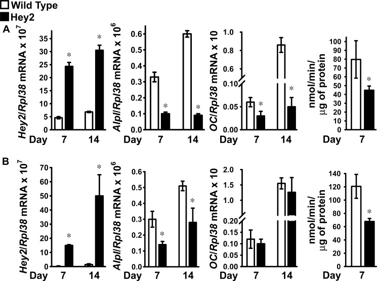
To understand the cellular effects caused by HEY2 overexpression, osteoblast-enriched cells were harvested from calvariae of Hey2 transgenic male or female mice and littermate controls of the same sex. Increased HEY2 transcripts were confirmed by qRT-PCR in osteoblasts from Col3.6-Hey2 transgenics over a 14-day culture period (Fig. 4). Alkaline phosphatase transcripts and activity were suppressed by HEY2 in cells from both sexes (Fig. 4). Osteocalcin expression was detected only at 7 and 14 days of culture and was suppressed in osteoblasts from Col3.6-Hey2 male but not female transgenic mice (Fig. 4). These findings confirm that HEY2 overexpression impairs osteoblast differentiation and function in both sexes and could explain the decreased bone formation and osteoblast number observed in vivo.
FIGURE 4.
HEY2 overexpression suppresses osteoblast function in vitro. A and B, osteoblast-enriched cells were harvested from the calvariae of male (A) or female (B) Col3.6-Hey2 transgenic mice (Hey2, black bars) or littermate wild type controls of the same sex (Wild Type, white bars) and cultured under conditions favoring osteoblastogenesis. Total RNA was extracted, and mRNA was reverse-transcribed and amplified by qRT-PCR in the presence of specific primers. Data are expressed as Hey2, Alpl, and osteocalcin (OC) copy number, corrected for Rpl38 copy number. Values are means ± S.E., n = 4. Alkaline phosphatase activity was determined in cells extracted with Triton X-100, and data are expressed as nanomoles of p-nitrophenol/min/μg of total protein. Values are means ± S.E., n = 6. *, significantly different between HEY2 and wild type, p < 0.05.
HEY2 Overexpression Enhances Osteoclast Function in Vitro
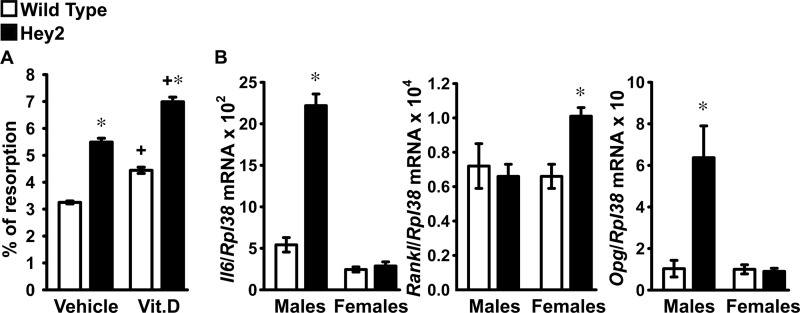
To determine whether the overexpression of HEY2 regulated osteoclast differentiation or function, calvariae from male Col3.6-Hey2 transgenics and littermate wild type controls were co-cultured with wild type FVB splenocytes, a source of mononuclear osteoclast precursors. Treatment with 1,25-dihydroxyvitamin D3 induced resorptive activity of wild type splenocytes, and in agreement with the increased eroded surface and osteoclast number observed in the Col3.6-Hey2 transgenic males, osteoblasts from Col3.6-Hey2 transgenics enhanced resorption (Fig. 5A) (44). To investigate possible mechanisms for the stimulatory effect of HEY2 on bone resorption, interleukin-6 (IL6) Rankl and Opg mRNA levels were determined (50). An increase in IL6 transcripts was observed only in male Col3.6-Hey2 transgenic osteoblasts (Fig. 5B), suggesting that HEY2 increases bone resorption by inducing the expression of IL6. HEY2 caused a modest and unexplained increase in Rankl transcripts in osteoblasts from female mice. In accordance with the effects of Notch on OPG expression, HEY2 induced Opg mRNA levels in cells from male mice (Fig. 5B) (26, 48). This may represent an indirect compensatory mechanism to temper the effects of HEY2 on osteoclastogenesis.
FIGURE 5.
HEY2 overexpression in osteoblasts from male mice induces resorption by co-cultured splenocytes and IL6 expression in vitro. Osteoblast-enriched cells were harvested from calvariae of male or female Col3.6-Hey2 transgenics (Hey2, black bars), or littermate wild type controls of the same sex (Wild Type, white bars) and cultured under conditions favoring osteoblastogenesis. In A, osteoblast-enriched cells from male mice were co-cultured with splenocytes from wild type mice of the same sex in the presence of 10 nm 1,25-dihydroxyvitamin D3 (Vit.D) or vehicle. After 7 days, cells were removed with bleach, the culture substrate was stained by Von Kossa, and digital pictures were acquired for the estimation of the resorbed area. Data are expressed as the percentage of the resorbed area. Values are means ± S.E., n = 4–6. *, significantly different between HEY2 and wild type, p < 0.05. +, significantly different between 1,25-dihydroxyvitamin D3 and vehicle, p < 0.05. In B, total RNA was extracted from osteoblast-enriched cell cultures at confluence, and mRNA was reverse-transcribed and amplified by qRT-PCR in the presence of specific primers. Data are expressed as Il6, Rankl, and Opg copy number, corrected for Gapdh copy number. Values are means ± S.E., n = 4. *, significantly different between HEY2 and wild type, p < 0.05.
DISCUSSION
In this study, we investigated the skeletal function of Hey2, a Notch target gene. Hey2 inactivation in cells of the osteoblastic lineage caused an increase in trabecular bone volume, an effect that is consistent with the osteopenic phenotype caused by the activation of Notch signaling in these cells (16, 48). The skeletal phenotype was mild, transient, and more pronounced in male than in female mice, indicating that Hey2 plays a modest role in skeletal homeostasis. Inactivation of Hey2 at early stages of osteoblastic differentiation did not affect the number or function of skeletal cells, and a developmental nature for the increased bone mass is excluded because the activity of the Osx promoter was suppressed by doxycycline during embryonic development (30). Global HeyL null mice carrying a heterozygous null mutation of Hey1 exhibit increased bone mass, indicating that Hey1, HeyL, and Hey2 have similar functions in the skeleton (24). It is plausible that Hey1 and HeyL compensate for the loss of Hey2 function, preventing detection of subtle changes in osteoblast and osteoclast number and activity, which lead to a modest increase in bone mass. Compensation by Hey1 might explain the sexually dimorphic skeletal phenotype of Hey2 inactivation in immature osteoblastic cells because Hey1 transcript levels significantly increased 1.4-fold in parietal bones when Hey2 was inactivated in female mice, but not in male mice (data not shown). Inactivation of Hey2 in osteoblasts did not affect serum markers of bone remodeling and transiently inhibited bone formation, but the effect was modest, confirming that Hey2 has a limited impact on skeletal function.
Although three Hey2 transgenic founders were obtained, only one transgenic line was established, indicating that excessive levels of HEY2 in cells expressing the 3.6-kb fragment of the Col1a1 promoter are detrimental for embryonic development. The activity of this promoter fragment is not restricted to osteoblastic cells, and expression of HEY2 in nonskeletal tissues may have prevented transmission of the transgene (51, 52). Despite these limitations, the data indicate that HEY2 overexpression affects bone remodeling and uncouples bone formation from bone resorption by increasing osteoclastogenesis in male mice and by suppressing osteoblast number and function in both genders. In accordance with the effects of the Hey2 inactivation in osteoblastic cells, Col3.6-Hey2 transgenic male mice displayed osteopenia and impaired trabecular microarchitecture at 6 months of age. However, the inhibition of osteoblast number and activity in female mice did not result in changes in trabecular volume or structure. There is no immediate explanation for the discrepancy in the skeletal phenotype of male and female transgenics, and it is conceivable that mechanisms necessary for the protection of bone mass are at play in female mice in the context of HEY2 overexpression. Alternatively, a variable penetrance of the Col3.6-Hey2 transgene may be responsible for the absence of changes in trabecular bone of female transgenics.
The differences between the skeletal phenotypes observed in the two sexes also could be explained by a sexually dimorphic mechanism of HEY2 action in osteoblasts. HEY2 is a paralogue of HES1, and HES1 overexpression under the control of the 3.6-kb fragment of the Col1a1 promoter induces osteoclastogenesis in male mice and suppresses osteoblastogenesis in female mice, confirming that the skeletal effects of the HES and HEY families of proteins are sexually dimorphic (22). An explanation for the sexual dimorphism could be provided by the protective effects of estrogens on skeletal mass (53).
In vitro studies exploring the differentiation and function of osteoblastic cells from mice misexpressing Hey2 indicate that HEY2 is dispensable for osteoblastogenesis and that perturbation of its expression in mature cells suppresses osteoblast function in both male and female mice. Although the mechanism was not established, it is possible that HEY2, like HEY1, interacts with transcription factors necessary for osteoblastogenesis, such as Runx2 (15). The decreased osteoblastic function by Hey2 inactivation is not consistent with the effects of Notch on mature cells of the osteoblastic lineage. The suppressive effects of HEY2 on transcription might explain this discrepancy because the absence of HEY2 might lead to increased expression of inhibitors of osteoblastic function (1, 3). Results from cells overexpressing HEY2 are in agreement with the inhibitory effects of Notch overexpression on osteoblast function (4). Overexpression of HEY2 in osteoblasts causes a less severe skeletal phenotype than the one observed following the induction of Notch under the control of the 3.6-kb fragment of the Col1a1 promoter. This may suggest that the skeletal effects of Notch require expression of additional targets of Notch signaling, such as HES1, or are primarily mediated by direct regulation of osteoblast-specific genes by Notch (16).
The increased bone resorption reported in Hey2 transgenic mice does not phenocopy the inhibitory effect of Notch on osteoclastogenesis, which is mediated by an induction of OPG and suppression of RANKL and M-CSF expression by osteoblastic cells (15, 25, 26, 48). HES1 is an inducer of osteoclastogenesis, suggesting that HEY and HES transcription factors carry out functions that are independent from their role as targets of Notch signaling (22). We report an induction of Il6 expression by HEY2 in osteoblasts from male mice, suggesting a possible mechanism for the enhanced bone resorption observed in male Col3.6-Hey2 transgenics. However, HEY2, like Notch, induced OPG expression, and this may be a protective mechanism to prevent excessive osteoclastogenesis. IL6 serum levels were not increased in Col3.6-Hey2 transgenic mice (data not shown), indicating that IL6 acts locally to regulate bone resorption. Although IL6 regulates osteoclastogenesis by inducing RANKL in osteoblastic cells, its direct effects on cells of the osteoclast lineage are less clear, and both a stimulatory function and an inhibitory function of IL6 in the differentiation of osteoclast precursors have been reported (54, 55). Our observations indicate that in the context of HEY2 overexpression, IL6 promotes the resorptive activity of osteoclast precursors.
In conclusion, Hey2 plays a modest role in the regulation of skeletal cell function, whereas HEY2 overexpression in male mice uncouples bone formation from bone resorption, causes osteopenia, and compromises bone microarchitecture.
Acknowledgments
We thank Drs. E. Olson for Hey2loxP/loxP conditional mice, T. Clemens for osteocalcin-CRE transgenics, J. B. Lian for osteocalcin, and R. Wu for GAPDH cDNA. We thank A. Kent, K. Parker, and L. Kranz for technical support and M. Yurczak for secretarial assistance.
This work was supported, in whole or in part, by National Institutes of Health Grant DK045227 from the NIDDK (to E. C.) and Research Fellowship Award 5371 from the Arthritis Foundation (to S. Z.).
- HES
- Hairy Enhancer of Split
- HEY
- HES-related with YRPW motif
- HEYL
- HEY-like
- CTX-I
- collagen type I C-terminal telopeptide
- FVB
- tropism to Friend leukemia virus strain-B
- μCT
- microcomputed tomography
- OPG
- osteoprotegerin
- qRT-PCR
- quantitative reverse transcription-PCR
- RANKL
- receptor activator of NF-κ-B ligand
- Rpl38
- ribosomal protein L38
- SMI
- structure model index.
REFERENCES
- 1. Iso T., Kedes L., Hamamori Y. (2003) HES and HERP families: multiple effectors of the Notch signaling pathway. J. Cell Physiol. 194, 237–255 [DOI] [PubMed] [Google Scholar]
- 2. Kageyama R., Ohtsuka T., Kobayashi T. (2007) The Hes gene family: repressors and oscillators that orchestrate embryogenesis. Development 134, 1243–1251 [DOI] [PubMed] [Google Scholar]
- 3. Fischer A., Gessler M. (2007) Delta–Notch—and then? Protein interactions and proposed modes of repression by Hes and Hey bHLH factors. Nucleic Acids Res. 35, 4583–4596 [DOI] [PMC free article] [PubMed] [Google Scholar]
- 4. Zanotti S., Canalis E. (2010) Notch and the skeleton. Mol. Cell Biol. 30, 886–896 [DOI] [PMC free article] [PubMed] [Google Scholar]
- 5. Kokubo H., Miyagawa-Tomita S., Tomimatsu H., Nakashima Y., Nakazawa M., Saga Y., Johnson R. L. (2004) Targeted disruption of hesr2 results in atrioventricular valve anomalies that lead to heart dysfunction. Circ. Res. 95, 540–547 [DOI] [PubMed] [Google Scholar]
- 6. Xin M., Small E. M., van Rooij E., Qi X., Richardson J. A., Srivastava D., Nakagawa O., Olson E. N. (2007) Essential roles of the bHLH transcription factor Hrt2 in repression of atrial gene expression and maintenance of postnatal cardiac function. Proc. Natl. Acad. Sci. U.S.A. 104, 7975–7980 [DOI] [PMC free article] [PubMed] [Google Scholar]
- 7. Fischer A., Steidl C., Wagner T. U., Lang E., Jakob P. M., Friedl P., Knobeloch K. P., Gessler M. (2007) Combined loss of Hey1 and HeyL causes congenital heart defects because of impaired epithelial to mesenchymal transition. Circ. Res. 100, 856–863 [DOI] [PubMed] [Google Scholar]
- 8. Fischer A., Schumacher N., Maier M., Sendtner M., Gessler M. (2004) The Notch target genes Hey1 and Hey2 are required for embryonic vascular development. Genes Dev. 18, 901–911 [DOI] [PMC free article] [PubMed] [Google Scholar]
- 9. Kokubo H., Miyagawa-Tomita S., Nakazawa M., Saga Y., Johnson R. L. (2005) Mouse hesr1 and hesr2 genes are redundantly required to mediate Notch signaling in the developing cardiovascular system. Dev. Biol. 278, 301–309 [DOI] [PubMed] [Google Scholar]
- 10. Bianco P., Gehron Robey P. (2000) Marrow stromal stem cells. J. Clin. Invest. 105, 1663–1668 [DOI] [PMC free article] [PubMed] [Google Scholar]
- 11. Canalis E., Giustina A., Bilezikian J. P. (2007) Mechanisms of anabolic therapies for osteoporosis. N. Engl. J. Med. 357, 905–916 [DOI] [PubMed] [Google Scholar]
- 12. Canalis E., Economides A. N., Gazzerro E. (2003) Bone morphogenetic proteins, their antagonists, and the skeleton. Endocr. Rev. 24, 218–235 [DOI] [PubMed] [Google Scholar]
- 13. Westendorf J. J., Kahler R. A., Schroeder T. M. (2004) Wnt signaling in osteoblasts and bone diseases. Gene 341, 19–39 [DOI] [PubMed] [Google Scholar]
- 14. Engin F., Yao Z., Yang T., Zhou G., Bertin T., Jiang M. M., Chen Y., Wang L., Zheng H., Sutton R. E., Boyce B. F., Lee B. (2008) Dimorphic effects of Notch signaling in bone homeostasis. Nat. Med. 14, 299–305 [DOI] [PMC free article] [PubMed] [Google Scholar]
- 15. Hilton M. J., Tu X., Wu X., Bai S., Zhao H., Kobayashi T., Kronenberg H. M., Teitelbaum S. L., Ross F. P., Kopan R., Long F. (2008) Notch signaling maintains bone marrow mesenchymal progenitors by suppressing osteoblast differentiation. Nat. Med. 14, 306–314 [DOI] [PMC free article] [PubMed] [Google Scholar]
- 16. Zanotti S., Smerdel-Ramoya A., Stadmeyer L., Durant D., Radtke F., Canalis E. (2008) Notch inhibits osteoblast differentiation and causes osteopenia. Endocrinology 149, 3890–3899 [DOI] [PMC free article] [PubMed] [Google Scholar]
- 17. Lacey D. L., Timms E., Tan H. L., Kelley M. J., Dunstan C. R., Burgess T., Elliott R., Colombero A., Elliott G., Scully S., Hsu H., Sullivan J., Hawkins N., Davy E., Capparelli C., Eli A., Qian Y. X., Kaufman S., Sarosi I., Shalhoub V., Senaldi G., Guo J., Delaney J., Boyle W. J. (1998) Osteoprotegerin ligand is a cytokine that regulates osteoclast differentiation and activation. Cell 93, 165–176 [DOI] [PubMed] [Google Scholar]
- 18. Teitelbaum S. L. (2007) Osteoclasts: what do they do and how do they do it? Am. J. Pathol. 170, 427–435 [DOI] [PMC free article] [PubMed] [Google Scholar]
- 19. Dong Y., Jesse A. M., Kohn A., Gunnell L. M., Honjo T., Zuscik M. J., O'Keefe R. J., Hilton M. J. (2010) RBPjκ-dependent Notch signaling regulates mesenchymal progenitor cell proliferation and differentiation during skeletal development. Development 137, 1461–1471 [DOI] [PMC free article] [PubMed] [Google Scholar]
- 20. Tao J., Chen S., Yang T., Dawson B., Munivez E., Bertin T., Lee B. (2010) Osteosclerosis owing to Notch gain of function is solely Rbpj-dependent. J. Bone Miner Res. 25, 2175–2183 [DOI] [PMC free article] [PubMed] [Google Scholar]
- 21. Zanotti S., Smerdel-Ramoya A., Canalis E. (2011) Reciprocal regulation of Notch and nuclear factor of activated T-cells (NFAT) c1 transactivation in osteoblasts. J. Biol. Chem. 286, 4576–4588 [DOI] [PMC free article] [PubMed] [Google Scholar]
- 22. Zanotti S., Smerdel-Ramoya A., Canalis E. (2011) HES1 (hairy and enhancer of split 1) is a determinant of bone mass. J. Biol. Chem. 286, 2648–2657 [DOI] [PMC free article] [PubMed] [Google Scholar]
- 23. Salie R., Kneissel M., Vukevic M., Zamurovic N., Kramer I., Evans G., Gerwin N., Mueller M., Kinzel B., Susa M. (2010) Ubiquitous overexpression of Hey1 transcription factor leads to osteopenia and chondrocyte hypertrophy in bone. Bone 46, 680–694 [DOI] [PubMed] [Google Scholar]
- 24. Tu X., Chen J., Lim J., Karner C. M., Lee S. Y., Heisig J., Wiese C., Surendran K., Kopan R., Gessler M., Long F. (2012) Physiological notch signaling maintains bone homeostasis via RBPjk and Hey upstream of NFATc1. PLoS. Genet. 8, e1002577. [DOI] [PMC free article] [PubMed] [Google Scholar]
- 25. Yamada T., Yamazaki H., Yamane T., Yoshino M., Okuyama H., Tsuneto M., Kurino T., Hayashi S., Sakano S. (2003) Regulation of osteoclast development by Notch signaling directed to osteoclast precursors and through stromal cells. Blood 101, 2227–2234 [DOI] [PubMed] [Google Scholar]
- 26. Bai S., Kopan R., Zou W., Hilton M. J., Ong C. T., Long F., Ross F. P., Teitelbaum S. L. (2008) NOTCH1 regulates osteoclastogenesis directly in osteoclast precursors and indirectly via osteoblast lineage cells. J. Biol. Chem. 283, 6509–6518 [DOI] [PubMed] [Google Scholar]
- 27. Fukushima H., Nakao A., Okamoto F., Shin M., Kajiya H., Sakano S., Bigas A., Jimi E., Okabe K. (2008) The association of Notch2 and NF-κB accelerates RANKL-induced osteoclastogenesis. Mol. Cell Biol. 28, 6402–6412 [DOI] [PMC free article] [PubMed] [Google Scholar]
- 28. Sethi N., Dai X., Winter C. G., Kang Y. (2011) Tumor-derived JAGGED1 promotes osteolytic bone metastasis of breast cancer by engaging notch signaling in bone cells. Cancer Cell 19, 192–205 [DOI] [PMC free article] [PubMed] [Google Scholar]
- 29. Wei W., Zeve D., Wang X., Du Y., Tang W., Dechow P. C., Graff J. M., Wan Y. (2011) Osteoclast progenitors reside in the peroxisome proliferator-activated receptor γ-expressing bone marrow cell population. Mol. Cell Biol. 31, 4692–4705 [DOI] [PMC free article] [PubMed] [Google Scholar]
- 30. Rodda S. J., McMahon A. P. (2006) Distinct roles for Hedgehog and canonical Wnt signaling in specification, differentiation and maintenance of osteoblast progenitors. Development 133, 3231–3244 [DOI] [PubMed] [Google Scholar]
- 31. Furth P. A., St Onge L., Böger H., Gruss P., Gossen M., Kistner A., Bujard H., Hennighausen L. (1994) Temporal control of gene expression in transgenic mice by a tetracycline-responsive promoter. Proc. Natl. Acad. Sci. U.S.A. 91, 9302–9306 [DOI] [PMC free article] [PubMed] [Google Scholar]
- 32. Kalajzic I., Kalajzic Z., Kaliterna M., Gronowicz G., Clark S. H., Lichtler A. C., Rowe D. (2002) Use of type I collagen green fluorescent protein transgenes to identify subpopulations of cells at different stages of the osteoblast lineage. J. Bone Miner. Res. 17, 15–25 [DOI] [PubMed] [Google Scholar]
- 33. Irwin N. (1989) in Molecular Cloning: A Laboratory Manual (Sambrook J., Fritsch E. F., Maniatis T., eds) Cold Spring Harbor Laboratory Press, Cold Spring Harbor, NY [Google Scholar]
- 34. Garnero P., Borel O., Delmas P. D. (2001) Evaluation of a fully automated serum assay for C-terminal cross-linking telopeptide of type I collagen in osteoporosis. Clin. Chem. 47, 694–702 [PubMed] [Google Scholar]
- 35. Bouxsein M. L., Boyd S. K., Christiansen B. A., Guldberg R. E., Jepsen K. J., Müller R. (2010) Guidelines for assessment of bone microstructure in rodents using micro-computed tomography. J. Bone Miner Res. 25, 1468–1486 [DOI] [PubMed] [Google Scholar]
- 36. Gazzerro E., Pereira R. C., Jorgetti V., Olson S., Economides A. N., Canalis E. (2005) Skeletal overexpression of gremlin impairs bone formation and causes osteopenia. Endocrinology 146, 655–665 [DOI] [PubMed] [Google Scholar]
- 37. Parfitt A. M., Drezner M. K., Glorieux F. H., Kanis J. A., Malluche H., Meunier P. J., Ott S. M., Recker R. R. (1987) Bone histomorphometry: standardization of nomenclature, symbols, and units. Report of the ASBMR Histomorphometry Nomenclature Committee. J. Bone Miner. Res. 2, 595–610 [DOI] [PubMed] [Google Scholar]
- 38. McCarthy T. L., Centrella M., Canalis E. (1990) Cyclic AMP induces insulin-like growth factor I synthesis in osteoblast-enriched cultures. J. Biol. Chem. 265, 15353–15356 [PubMed] [Google Scholar]
- 39. Akazawa C., Sasai Y., Nakanishi S., Kageyama R. (1992) Molecular characterization of a rat negative regulator with a basic helix-loop-helix structure predominantly expressed in the developing nervous system. J. Biol. Chem. 267, 21879–21885 [PubMed] [Google Scholar]
- 40. Lian J., Stewart C., Puchacz E., Mackowiak S., Shalhoub V., Collart D., Zambetti G., Stein G. (1989) Structure of the rat osteocalcin gene and regulation of vitamin D-dependent expression. Proc. Natl. Acad. Sci. U.S.A. 86, 1143–1147 [DOI] [PMC free article] [PubMed] [Google Scholar]
- 41. Iso T., Sartorelli V., Poizat C., Iezzi S., Wu H. Y., Chung G., Kedes L., Hamamori Y. (2001) HERP, a novel heterodimer partner of HES/E(spl) in Notch signaling. Mol. Cell Biol. 21, 6080–6089 [DOI] [PMC free article] [PubMed] [Google Scholar]
- 42. Kouadjo K. E., Nishida Y., Cadrin-Girard J. F., Yoshioka M., St-Amand J. (2007) Housekeeping and tissue-specific genes in mouse tissues. BMC. Genomics 8, 127. [DOI] [PMC free article] [PubMed] [Google Scholar]
- 43. Tso J. Y., Sun X. H., Kao T. H., Reece K. S., Wu R. (1985) Isolation and characterization of rat and human glyceraldehyde-3-phosphate dehydrogenase cDNAs: genomic complexity and molecular evolution of the gene. Nucleic Acids Res. 13, 2485–2502 [DOI] [PMC free article] [PubMed] [Google Scholar]
- 44. Takahashi N., Akatsu T., Udagawa N., Sasaki T., Yamaguchi A., Moseley J. M., Martin T. J., Suda T. (1988) Osteoblastic cells are involved in osteoclast formation. Endocrinology 123, 2600–2602 [DOI] [PubMed] [Google Scholar]
- 45. Lee S. K., Kalinowski J., Jastrzebski S., Lorenzo J. A. (2002) 1,25(OH)2 vitamin D3-stimulated osteoclast formation in spleen-osteoblast cocultures is mediated in part by enhanced IL-1α and receptor activator of NF-κB ligand production in osteoblasts. J. Immunol. 169, 2374–2380 [DOI] [PubMed] [Google Scholar]
- 46. Wyzga N., Varghese S., Wikel S., Canalis E., Sylvester F. A. (2004) Effects of activated T cells on osteoclastogenesis depend on how they are activated. Bone 35, 614–620 [DOI] [PubMed] [Google Scholar]
- 47. Sokal R. R., Rohlf F. J. (1981) Biometry, 2nd Edition, W. H. Freeman, San Francisco, CA [Google Scholar]
- 48. Canalis E., Parker K., Feng J. Q., Zanotti S. (2013) Osteoblast lineage-specific effects of notch activation in the skeleton. Endocrinology 154, 623–634 [DOI] [PMC free article] [PubMed] [Google Scholar]
- 49. Hildebrand T., Rüegsegger P. (1997) Quantification of bone microarchitecture with the Structure Model Index. Comput. Methods Biomech. Biomed. Engin. 1, 15–23 [DOI] [PubMed] [Google Scholar]
- 50. Mundy G. R. (2007) Osteoporosis and inflammation. Nutr. Rev. 65, S147–S151 [DOI] [PubMed] [Google Scholar]
- 51. Boban I., Jacquin C., Prior K., Barisic-Dujmovic T., Maye P., Clark S. H., Aguila H. L. (2006) The 3.6 kb DNA fragment from the rat Col1a1 gene promoter drives the expression of genes in both osteoblast and osteoclast lineage cells. Bone 39, 1302–1312 [DOI] [PubMed] [Google Scholar]
- 52. Scheller E. L., Leinninger G. M., Hankenson K. D., Myers M. G., Jr., Krebsbach P. H. (2011) Ectopic expression of Col2.3 and Col3.6 promoters in the brain and association with leptin signaling. Cells Tissues Organs 194, 268–273 [DOI] [PMC free article] [PubMed] [Google Scholar]
- 53. Manolagas S. C., Kousteni S., Jilka R. L. (2002) Sex steroids and bone. Recent Prog. Horm. Res 57, 385–409 [DOI] [PubMed] [Google Scholar]
- 54. O'Brien C. A., Lin S. C., Bellido T., Manolagas S. C. (2000) Expression levels of gp130 in bone marrow stromal cells determine the magnitude of osteoclastogenic signals generated by IL-6-type cytokines. J. Cell Biochem. 79, 532–541 [DOI] [PubMed] [Google Scholar]
- 55. Yoshitake F., Itoh S., Narita H., Ishihara K., Ebisu S. (2008) Interleukin-6 directly inhibits osteoclast differentiation by suppressing receptor activator of NF-κB signaling pathways. J. Biol. Chem. 283, 11535–11540 [DOI] [PubMed] [Google Scholar]


