Abstract
15-deoxy-Δ12,14-prostaglandin J2 (15d-PGJ2) is an anti-inflammatory downstream product of the cyclooxygenase enzymes. It has been implicated to play a protective role in a variety of inflammatory mediated diseases, including rheumatoid arthritis, neural damage, and myocardial infarctions. Here we show that 15d-PGJ2 also plays a role in Salmonella infection. Salmonella enterica Typhimurium is a Gram-negative facultative intracellular pathogen that is able to survive and replicate inside phagocytic immune cells, allowing for bacterial dissemination to systemic sites. Salmonella species cause a wide range of morbidity and mortality due to gastroenteritis and typhoid fever. Previously we have shown that in mouse models of typhoid fever, Salmonella infection causes a major perturbation in the prostaglandin pathway. Specifically, we saw that 15d-PGJ2 production was significantly increased in both liver and feces. In this work we show that 15d-PGJ2 production is also significantly increased in macrophages infected with Salmonella. Furthermore, we show that the addition of 15d-PGJ2 to Salmonella infected RAW264.7, J774, and bone marrow derived macrophages is sufficient to significantly reduce bacterial colonization. We also show evidence that 15d-PGJ2 is reducing bacterial uptake by macrophages. 15d-PGJ2 reduces the inflammatory response of these infected macrophages, as evidenced by a reduction in the production of cytokines and reactive nitrogen species. The inflammatory response of the macrophage is important for full Salmonella virulence, as it can give the bacteria cues for virulence. The reduction in bacterial colonization is independent of the expression of Salmonella virulence genes SPI1 and SPI2, and is independent of the 15d-PGJ2 ligand PPAR-γ. 15d-PGJ2 also causes an increase in ERK1/2 phosphorylation in infected macrophages. In conclusion, we show here that 15d-PGJ2 mediates the outcome of bacterial infection, a previously unidentified role for this prostaglandin.
Introduction
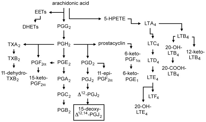
Prostaglandins (PG) are a class of lipid hormones responsible for a wide range of functions within the body. PGs are synthesized from arachidonic acid that is released from the cell membrane by phospholipase A2 and then modified by the cyclooxygenase enzymes (COX1 and COX2) to enter the PG pathway (Figure 1) [1], [2]. COX1 is constitutively active, whereas COX2 is induced under inflammatory conditions [2]. COX2-derived PGs are involved in a variety of pro- and anti-inflammatory processes [2], [3]. The involvement of COX1 and COX2 in regulating inflammation is evidenced by the increased cardiovascular risk associated with the inhibition of COX2 [4], and the increased susceptibility to colitis in mice lacking these two enzymes [5]. Two waves of COX2 activity have been identified: the first (early) activity is associated with the pro-inflammatory response, whereas the second wave mediates the resolution of inflammation [6], and is associated with high levels of PGD2 and 15-deoxy-Δ12,14-PGJ2 (hereafter referred to as 15d-PGJ2) [1], [6].
Figure 1. Arachidonic acid metabolism and formation of prostaglandins and leukotrienes.
15d-PGJ2 is non-enzymatically produced from PGD2.
15d-PGJ2 has recently been identified as an anti-inflammatory PG. By forming adducts with various molecules within the cell, 15d-PGJ2 is able to modulate a variety of cellular signaling pathways [7]. 15d-PGJ2 is an endogenous ligand that activates the nuclear receptor peroxisome proliferator-activated receptor gamma (PPAR-γ) transcription factor, thus inhibiting the NF-κB, STAT, and AP1 signaling pathways, and reducing the production of inflammatory mediators such as iNOS, TNFα, and IL-6 [7]–[9]. 15d-PGJ2 has also been found to modify the production of reactive nitrogen species (RNS), the NF-κB pathway, heat shock proteins, JNK signaling, ERK signaling, and cytokine production [6], [9]–[20]. Both RAW264.7 macrophages and HeLa epithelial cells do not produce quantifiable amounts of PPAR-γ [8], [15], [18], which is not necessary for the anti-inflammatory effects of 15d-PGJ2 in these cells [18]. In addition, for 15d-PGJ2 to activate PPAR-γ, it must be present at relatively high concentrations [21]. Several PPAR-γ independent functions of 15d-PGJ2 have recently been described [11], [12], [15]–[20], [22]–[25].
15d-PGJ2 inhibits the synthesis of iNOS in activated and peritoneal macrophages, which is at least partially dependent on NF-κB [8], [11], [16]. In RAW 264.7 and J774A.1 macrophages, 15d-PGJ2 increases ROS formation, which may inhibit phagocytosis and induce apoptosis at later time points [20], [22]. Furthering its role as an anti-inflammatory mediator, 15d-PGJ2 reduces the production of cytokines [10], and reduces the recruitment of bone marrow monocytes during liver inflammation [25]. It was also found that 15d-PGJ2 reduces the phagocytic activities of bone marrow macrophages (BMMO) in vitro [25]. Recently, the use of nanocapsules loaded with 15d-PGJ2 has proved an effective strategy to reduce neutrophil migration, IL-1β, TNF-α, and IL-12p70 production during inflammation [26]. In fact, 15d-PGJ2 is so vital to the resolution phase of the inflammatory process, that when it is added back to animals treated with COX2 inhibitors, it is sufficient to restore the normal resolution that occurs after inflammation, which is prevented by COX2 inhibitors [6].
Since 15d-PGJ2 has been found to reduce inflammation in such a variety of models, it has been explored as a potential therapeutic in a number of inflammatory diseases. Liu and colleges (2012) concluded that since 15d-PGJ2 reduces the general activity of both RAW264.7 and J774A.1 macrophages, it has the potential to be an effective therapeutic for inflammatory diseases [20]. More specifically, the role of 15d-PGJ2 and its potential applications in therapy have been explored in rheumatoid arthritis, atherosclerosis, myocardial infarctions, cerebral injury, and gastrointestinal inflammation [8], [10], [27]. 15d-PGJ2 has also been found to protect enteric glial cells from oxidative stress, to reduce hepatic inflammation and fibrosis, and to reduce symptoms of COPD in rats [24], [25], [27]. 15d-PGJ2 may also be useful in the treatment of cancers, as it has been found to inhibit cell growth and tumorigenicity [28]. In a model of periodontitis, 15d-PGJ2 nanocapsules were found to reduce inflammation caused by infection with Actinobacillus actinomycetemcomitans, but no effect on bacterial colonization was seen [23]. Despite this wealth of knowledge and exploration into the roles of 15d-PGJ2 in inflammatory diseases, little is known about 15d-PGJ2 in bacterial infections.
15d-PGJ2 has been studied in models of sepsis and septic shock. In models of polymicrobial sepsis, 15d-PGJ2 treatment leads to increases in blood pressure, reductions in vascular injury, neutrophil infiltration, cytokine production, renal and liver dysfunction and injury, resulting in increased survival [29], [30]. In rat macrophages treated with heat killed S. aureus and E. coli, 15d-PGJ2 treatment leads to reductions in NO production, TBXB2 production, and ERK1/2 and NF-κB activity [31]. In bacterial sepsis, PMN migration is reduced, and this was found to be mediated by PPARγ, and 15d-PGJ2 treatment reduced PMN adherence to fibrinogen, another aspect of PMN migration [32]. The role of 15d-PGJ2 in microglial inflammatory response to S. aureus was examined, and 15d-PGJ2 was found to inhibit a variety of cytokines including IL-1β, TNFα, IL-12p40, and MCP1, while in this model the levels of PPARγ were unaffected by either 15d-PGJ2 or S. aureus treatment [33]. The role of 15d-PGJ2 in H. pylori infected epithelial cells was also studied, and it was found that 15d-PGJ2 treatment reduced JAK/STAT signaling, RANTES production, and NADPH oxidase activity [34]. In this study, the involvement of PPARγ was not determined [34]. Interestingly, 15d-PGJ2 treatment of mice one day after infection with the influenza virus was found to significantly reduce morbidity and mortality, in a PPARγ dependent fashion [35]. In this study, 15d-PGJ2 reduced the production of chemokines and cytokines, as well as reducing viral titers [35]. They also found that 15d-PGJ2 decreased inflammatory infiltrate in the lungs and reduced the production of IL-6, TNFα, CCL2, CCL3, CCL4, and CXCL10, but had no effect on IFNγ production [35]. GW9662, a PPARγ specific inhibitor, was used, and this inhibitor abolished the protection afforded by 15d-PGJ2 treatment [35]. These studies show the potential use of 15d-PGJ2 in a variety of microbial associated disease conditions, however, it seems that there have not been any studies looking at the role of 15d-PGJ2 in Salmonella infection.
Salmonella is a Gram-negative enteric pathogen that is transmitted by contaminated food or water [36]. Once ingested, the bacteria replicate in the small intestine, and in cases of systemic disease, such as typhoid fever, the bacteria cross the intestinal barrier and are taken up by phagocytes [36], [37]. By means of the Salmonella Pathogenicity Island 2 (SPI2) type III-secretion system, Salmonella is able to replicate inside macrophages in a special vacuole termed the Salmonella containing vacuole [37]–[39]. From inside these macrophages, Salmonella is able to disseminate to systemic sites such as the spleen and liver, causing severe disease and bacteremia [36].
We have recently performed a high-throughput metabolomics study to determine the effect of Salmonella enterica serovar Typhimurium infection of mice on the chemical composition of multiple body fluids and organs [40]. We found that the PG pathway was greatly perturbed by Salmonella infection and that 15d-PGJ2 production was greatly increased in infected mice [40]. Therefore, we sought to study the impact of this hormone on the pathogenesis of Salmonella. In this study we show that 15d-PGJ2 production is increased during Salmonella infection of cultured macrophages. Additionally, we examined the roles of individual PGs on bacterial colonization of macrophages, and show that 15d-PGJ2 causes a marked decrease in Salmonella colonization, despite its well-known role in reducing macrophage activity. We also show that, like many activities of 15d-PGJ2, this effect is PPAR-γ independent. Furthermore, we present evidence showing that this reduction in colonization is not due to inhibition of SPI2. Altogether, our data shows a novel role for 15d-PGJ2 in infectious disease, and provides further evidence for the importance of inflammation to Salmonella pathogenesis.
Materials and Methods
Chemical reagents
Streptomycin and dimethyl sulfoxide (DMSO) were purchased from Sigma-Aldrich (St. Louis, USA). 15d-PGJ2 was obtained from Cayman Chemical (Ann Arbor, USA).
Tissue culture
RAW264.7 and J774 macrophages, as well as HeLa epithelial cells, were obtained from the American Type Culture Collection (Manassas, USA). Cells were grown in Dulbecco's Modified Eagle Medium (DMEM; HyClone, Waltham, USA) supplemented with 10% fetal bovine serum (FBS; HyClone), 1% non-essential amino acids (Gibco, Carlsbad, USA) and 1% GlutaMAX (Gibco). Cells were seeded approximately 20 hours before experiments in 24-well plates at a density of 105 cells per well. 15d-PGJ2 was dissolved in DMSO and concentrations of 2 µM were used, unless otherwise indicated. Controls without 15d-PGJ2 contained the same amounts of DMSO. For infection assays, bacterial cells grown in LB, in mid-logarithmic growth were spun down and resuspended in phosphate-buffered saline (PBS) and diluted in tissue culture medium. Cells were infected at a multiplicity of infection of 10 for 30 minutes at 37°C, 5% CO2. Subsequently, cells were washed with PBS and incubated at 37°C, 5% CO2 in growth medium containing 100 µg/mL gentamycin (Sigma-Aldrich) for 1 hour. Medium was replaced to decrease the gentamycin concentration to 10 µg/mL for later time points. All media contained (or did not contain for controls) the indicated concentration of 15d-PGJ2. At the appropriate times, supernatants were collected and cells were lysed in 250 µL of 1% Triton X-100 (BDH, Yorkshire, UK), 0.1% sodium dodecyl sulfate (Sigma-Aldrich). Serial dilutions were plated on LB plates containing 100 µg/mL of streptomycin (Sigma-Aldrich) for bacterial enumeration. For fold replication assays, CFUs were determined at 2 and 24 hours post-infection and fold replication was calculated by dividing the number of CFUs at 24 hours by the average of the corresponding 2-hour CFU counts.
Bone marrow macrophage (BMMO) collection and infection
Age-matched C57BL/6 female mice were euthanized by CO2 asphyxiation and femurs were removed. Femurs were cleaned, and marrow was removed in Hank's balanced salt solution (Gibco) with 2% FBS. Animal experiments were approved by the Animal Care Committee of the University of British Columbia and performed in accordance with institutional guidelines. Cells were spun down and resuspended in BMMO media [DMEM (HyClone), 20% FBS, 2 mM Glutamax, 1 mM Sodium Pyruvate (Gibco), (5%) penicillin/streptomycin (Gibco), 20% L-conditioned media]. Cells were grown for 7–10 days before use. For infection, BMMO's were seeded in 24-well plates at 1×10⁶ cells/well in BMMO media without penicillin/streptomycin and L-conditioned media. BMMOs were infected with Salmonella at a multiplicity of infection of 10, and the gentamycin protection assay was completed as above. CFU was determined at 2, 6, and 10 hours post-infection.
Cytokine analysis
Cytometric bead assay (CBA) for mouse inflammation (BD Biosciences) was performed following the recommended assay procedure. Supernatants from macrophage infections were used for CBAs.
Enzyme-linked immunosorbent assays (ELISAs)
ELISAs were performed on culture supernatants from uninfected and infected cells using commercially available ELISA kits to determine concentrations of 15d-PGJ2 (Assay Designs, Ann Arbor, USA). ELISAs (BD Biosciences) were also used to examine the concentrations of cytokines (TNF-α, MCP1, IL-10, IL-6) in the supernatants of infected, 15d-PGJ2-treated and untreated macrophages. Manufacturer's recommendations and procedures were followed for all ELISAs.
Quantitative Real-time PCR (qRT-PCR)
RNA was purified using the RNeasy Mini Kit (Qiagen, Hilden, Germany), with the on-column DNA digestion (Qiagen). cDNA was synthesized using the QuantiTect Reverse Transcription Kit (Qiagen). For qRT-PCRs, we used the QuantiTect SYBR Green PCR Kit (Qiagen) and the Applied Biosystems (Foster City, USA) 7500 system. Reactions contained forward and reverse primers at 0.4 µM each. All results were normalized using the mRNA levels of the acidic ribosomal phosphoprotein PO as baseline. Averages of the data obtained with untreated samples were normalized to 1 and the data from each sample (untreated or treated) was normalized accordingly. Primer sequences are available upon request.
Immunofluorescence microscopy
Macrophages were seeded as mentioned previously, but on glass coverslips. Infections were carried out as above. Cells were fixed using 4% paraformaldehyde (Canemco Supplies, Quebec, Canada) overnight. Cells were then stained using a rabbit, polyclonal, anti-Salmonella LPS antibody (BD Biosciences). Prolong Gold containing DAPI (Invitrogen) was used to attach coverslips to the slides. The Zeiss Axioplan Fluorescence Microscope was then used to enumerate the bacteria in each infected macrophage for a total of 50 infected macrophages per sample.
Trypan blue exclusion
At the appropriate time points after infection, macrophages were released from the bottom of plates using cell scrapers, and stained with Trypan Blue (Gibco). The number of cells were then counted using the Countess automated cell counter (Invitrogen).
LDH release assay
CytoTox96 Non-Radioactive Cytotoxicity Assay (Promega) was performed on supernatants from infected or uninfected, 15d-PGJ2 treated or untreated macrophages. The manufacturer's protocol was followed.
Salmonella growth in 15d-PGJ2
Salmonella was grown in LB overnight with aeration at 37°C in the presence or absence of 15d-PGJ2. Salmonella was also grown in DMEM with or without 15d-PGJ2, without aeration, in 5% CO2 at 37°C for the indicated time points. Bacterial growth was monitored through measurements of absorbance at 600 nm.
hilA, phoP, ssrA reporter assays
Salmonella strains containing fusions between the promoters of hilA, ssrA or phoP and gfp, as previously described [41] were sub-cultured in liquid LB culture for 4 hours in the absence or presence of 2 µM 15d-PGJ2, and GFP production was analyzed through flow cytometry of bacterial cultures using a FACSCalibur (BD Biosciences, Franklin Lakes, NJ), as indicated. All cultures contained carbenicillin (100 µg/ml) and were incubated at 37°C with shaking (225 rpm). In each experiment, 50,000 events were collected per sample. Also, the ssrA reporter plasmid was introduced into Salmonella strain MCS004, which constitutively expresses the mKO red/orange protein. This strain was then used to infect RAW264.7 macrophages, as indicated above. Macrophages were lysed and bacteria were washed with PBS containing 2% FBS. GFP and RFP production was analyzed through flow cytometry, performed using an LSR II (BD Biosciences), and data were analyzed with FlowJo 8.7 software (TreeStar, Ashland, OR). In each experiment, 100,000 events were collected per sample.
Reactive nitrogen and oxygen species production
To determine reactive nitrogen species, the Griess reaction was performed on supernatants taken from macrophages infected as indicated above.
PPAR-γ inhibitor
RAW264.7 macrophages were seeded as above and GW9662 was used at 4 µM, where indicated.
Protein extraction and ERK1/2 western blot
RAW264.7 macrophages were seeded as above, overnight without 15d-PGJ2. 2 hours before infection cells were treated with indicated concentrations of 15d-PGJ2, 10 ng/mL of EGF (Sigma), or 10 mM PD98059 (CalBiochem Billerica USA). Uninfected samples were treated either with DMSO, 15d-PGJ2, or EGF. EGF treated samples were used as a positive control for ERK1/2 phosphorylation. PD98059, a MEK inhibitor, was used as a negative control for prevention of ERK1/2 phosphorylation, in the presence of Salmonella. Macrophages were infected for 1 hour, then washed with PBS, and lysed in 50 µL of lysis buffer (PBS, 1% Triton X-100 (BDH), 0.1% sodium dodecyl sulfate (Sigma-Aldrich), with protease inhibitor (Roche), and sodium orthovanandate (Sigma). Lysates were collected and spun at 4°C for 20 minutes, supernatants were collected, Bradford assays were performed, and SDS-PAGE loading buffer containing DTT (Sigma) was added. Samples were boiled for 5 minutes, then proteins were separated using denaturing SDS-PAGE. Proteins were then transferred to methanol-activated polyvinylidene difluoride membranes (Bio-Rad, Hercules USA) using wet transfer. Membranes were blocked with rocking for 1 hour using 5% nonfat milk in TBST (Tris-Buffered saline with 0.1% Tween 20). Primary antibodies were added to blocking buffer at 1∶1,000 (Phospho and total p44/42 Map Kinase (ERK1/2) (Cell signaling Techologies, Danvers USA) and anti-Calnexin (Enzo Life Sciences, Farmingdale USA)), and membranes were incubated at 4°C over night with rocking. Membranes were washed 3 times with TBST, then incubated with 1∶5,000 dilution of goat anti-rabbit horseradish peroxidase-conjugated antibodies for 1 hour with rocking in blocking buffer. Membranes were washed 3 times with TBST, then Immun-Star Western C kit (Bio-Rad) was used. Imaging was performed on Bio-Rad ChemiDoc MP Imaging System, and Image Lab (Bio-Rad) software was used.
Statistical analysis
Data were analyzed by unpaired t tests with 95% confidence intervals using GraphPad Prism version 4.0 (GraphPad Software Inc., San Diego, USA).
Results
Salmonella infection induces 15d-PGJ2 production
We have previously shown that the prostaglandin pathway is perturbed in mice infected with Salmonella [40]. Specifically, 15d-PGJ2 levels were increased during infection in both liver and feces [40]. To further characterize the interactions between the anti-inflammatory molecule 15d-PGJ2 and Salmonella, so we first established a simplified cell culture system. Because Salmonella actively replicates in macrophages, we examined RAW264.7 macrophage cells infected with Salmonella to determine if 15d-PGJ2 production was induced in these cells, as observed in mice. Similar to mice, we observed a significant increase in the amount of 15d-PGJ2 produced by cultured macrophages in response to Salmonella (Figure 2).
Figure 2. 15d-PGJ2 is increased during Salmonella infection of RAW264.7 macrophages at 20 hours post-infection.
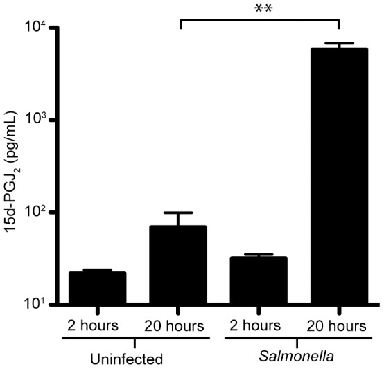
Supernatants were collected at 2 and 20 hours post-infection and levels of 15d-PGJ2 were determined through ELISA. Results shown are averages of four measurements, with standard errors of means. (**p<0.001).
Addition of exogenous 15d-PGJ2 reduces Salmonella colonization of macrophages
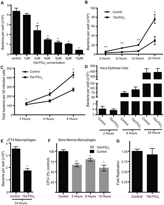
Given the role of 15d-PGJ2 in reducing the inflammatory response, we wanted to determine if 15d-PGJ2 had any effect on Salmonella interactions with host cells. To test this, we added increasing concentrations of 15d-PGJ2 to RAW264.7 macrophages prior to and during Salmonella infection and monitored colonization through bacterial enumeration by selective plating. We saw a dose-dependent decrease in Salmonella colonization of macrophages 24 hours post infection (Figure 3a). At the high concentration of 15d-PGJ2 cell lifting was slightly increased (data not shown). To determine when during the infection process 15d-PGJ2 exerts its effect on Salmonella colonization and to understand the kinetics of this phenomenon, we examined bacterial loads in macrophages at 2, 6, 10, and 24 hours post infection in the absence or presence of 2 µM 15d-PGJ2. These time points were chosen to give a general overview of Salmonella colonization. The time course showed that 15d-PGJ2 reduces Salmonella colonization as early as 2 hours post infection, and continues to exert its effect until 24 hours post infection (Figure 3b). To confirm that 15d-PGJ2 was not killing the macrophages, we used Trypan blue and LDH release assays to measure cell viability. When we used the Trypan blue exclusion assay to count the number of cells in infected, 15d-PGJ2 treated and untreated, macrophage cultures, no differences were seen (Figure S1A). An LDH-release assay was also used to ensure that 15d-PGJ2 was not causing cell death at 24 hours post-infection. No significant difference was seen in the amount of LDH released by 15d-PGJ2 treated cells as compared to untreated control cells (Figure S1B). We also used immunofluorescence microscopy to enumerate the Salmonella inside individual macrophages. By counting the bacteria inside 50 macrophages untreated or treated with 2 µM 15d-PGJ2 at 2, 4, and 8 hours post infection we saw significantly fewer Salmonella in the 15d-PGJ2 treated RAW264.7 macrophages (Figure 3c), confirming our CFU observations. Later time points were not used because bacteria became to numerous to accurately count. Therefore, the reduction in Salmonella colonization is due to 15d-PGJ2 and not to increased macrophage cell death.
Figure 3. Salmonella colonization of macrophages is significantly reduced by the addition of 15d-PGJ2.
(A) Salmonella colonization of RAW264.7 macrophages with the addition of increasing concentrations of 15d-PGJ2 at 24 hours post infection. (B) The effect of 2 µM 15d-PGJ2 on Salmonella colonization of RAW264.7 macrophages over time as determined by CFU analysis. (C) Immunofluorescence microscopy was used to enumerate bacterial colonization in individual macrophages at 2, 4 and 8 hours post-infection. (D) Salmonella colonization of HeLa epithelial cells treated with 15d-PGJ2 at 2, 6, and 24 hours post-infection. (E) The effect of 15d-PGJ2 on Salmonella colonization of J774 macrophages cells, as determined by CFU analysis at 24 hours. (F) Salmonella colonization of bone marrow macrophages at 2, 6, and 10 hours post-infection, with 15d-PGJ2 treatment. (G) Fold replication of Salmonella in RAW264.7 macrophages treated with 15d-PGJ2. Averages of at least 8 measurements are shown with standard errors of means. (*p<0.05, **p<0.001).
15d-PGJ2 does not inhibit Salmonella growth directly
The above results indicate that 15d-PGJ2 inhibits Salmonella colonization of macrophages. This could occur through a number of distinct mechanisms, the simplest of which would be direct inhibition of bacterial viability and growth. To determine if this was the case, we tested the effect of 15d-PGJ2 on Salmonella growth in culture media in the absence of macrophages. 15d-PGJ2 did not affect the growth of Salmonella alone in either LB or DMEM (Figure S2), suggesting that the effect of this hormone on Salmonella colonization of macrophages is not due to a direct inhibition of Salmonella viability and growth.
The effect of 15d-PGJ2 on Salmonella colonization of host cells is dependent on cell type
To determine whether the effect of 15d-PGJ2 was dependent on cell type, we infected both HeLa epithelial cells and J774 macrophages with and without 15d-PGJ2. We found that 15d-PGJ2 had no effect on Salmonella colonization of HeLa epithelial cells (Figure 3d), but, like that observed with RAW macrophages, 15d-PGJ2 reduced colonization in J774 macrophages (Figure 3e). We also tested the effect of 15d-PGJ2 on activated, IFN-γ pre-treated, RAW264.7 macrophages, and found that Salmonella colonization was also significantly reduced by 15d-PGJ2 treatment (Figure S3). We also wanted to use a model that would more closely represent the murine infection model previously used in our original metabolomics study [40]. To do this, we used bone marrow derived macrophages from C57BL/6 mice, and infected them with Salmonella with or without 15d-PGJ2 treatment. At 2, 6, and 10 hours post infection there was a significant reduction in Salmonella in the 15d-PGJ2 treated samples (Figure 3f). Because bone marrow macrophages are highly bactericidal, later time points were not used. Therefore, while 15d-PGJ2 significantly reduces Salmonella colonization of macrophages, it has no effect on Salmonella replication in epithelial cells.
15d-PGJ2 reduces Salmonella entry into macrophages
To determine if 15d-PGJ2 was affecting bacterial replication or entry in RAW264.7 macrophages, we determined the fold replication of Salmonella (Figure 3G). Fold replication was calculated by comparing CFUs at 2 and 24 hours post-infection. Interestingly, we found that the 15d-PGJ2 treated samples had a fold replication similar to control treated samples. This implies that 15d-PGJ2 is reducing the entry of Salmonella into macrophages.
15d-PGJ2 affects the immune response of macrophages infected with Salmonella
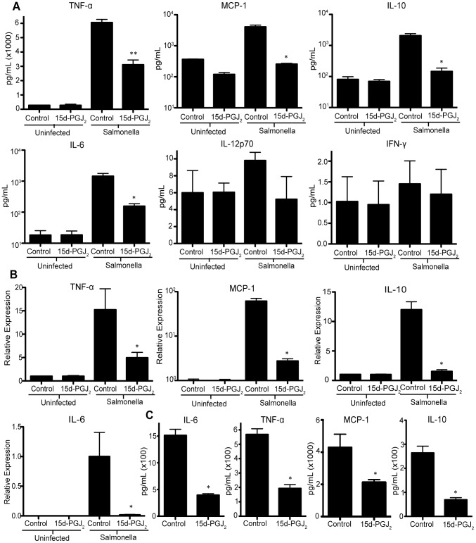
As the effect of 15d-PGJ2 seemed to be restricted to macrophages, we examined the effects of 15d-PGJ2 on the macrophage inflammatory response. By performing a cytometric bead assay (CBA) on supernatants from Salmonella infected RAW264.7 macrophages, we found that the 15d-PGJ2 treated macrophages produced significantly lower levels of TNF-α, MCP-1, IL-10, and IL-6, whereas levels of IFN-γ and IL-12 were unaffected (Figure 4a). This was confirmed using qRT-PCR (Figure 4b), and ELISA (Figure 4c). IL-12 was also examined using ELISA, and levels were too low to detect (data not shown), corroborating the CBA data. Together, this indicates that 15d-PGJ2 is in fact reducing specific cytokines produced in response to Salmonella infection. In addition to reducing the cytokines produced during infection, we also tested whether 15d-PGJ2 would reduce other macrophage mechanisms aimed at responding to pathogens. To this end, we show that RNS production in response to Salmonella infection was significantly reduced by the addition of 15d-PGJ2 (Figure 5).
Figure 4. The effect of 2 µM 15d-PGJ2 treatment during Salmonella infection on cytokine production.
RAW264.7 macrophages were examined at 24 hours post infection, cytokine production was determined by; (A) CBA assay, (B) quantitative real-time PCR, and (C) ELISA performed on supernatants from infected cells. Averages of 8 measurements are shown with standard errors of means. (*p<0.05, **p<0.001).
Figure 5. 15d-PGJ2 reduces the production of reactive nitrogen species.
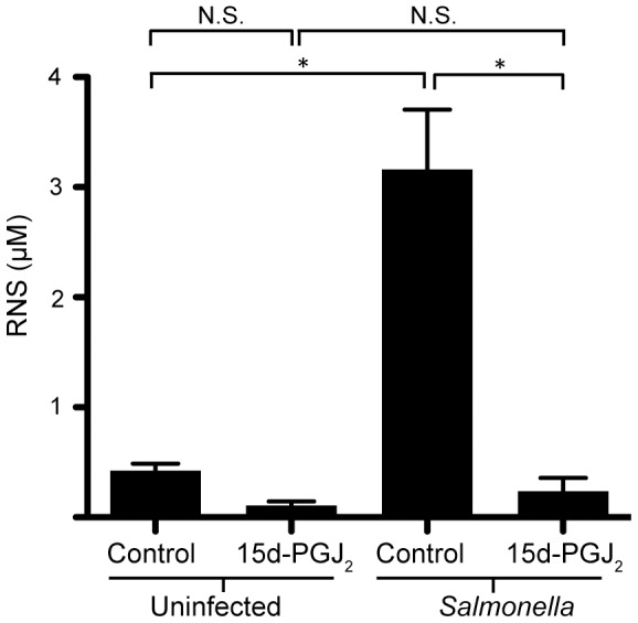
The Griess reaction was used to determine the amount of reactive nitrogen species produced by RAW264.7 macrophages treated with 2 µM 15d-PGJ2 and infected with Salmonella. Averages of 8 measurements are shown with standard errors of means. (*p<0.05).
15d-PGJ2 does not affect Salmonella virulence gene expression
We also considered the possibility that in addition to dampening the immune response, 15d-PGJ2 may also have an effect on virulence gene expression thus affecting the ability of Salmonella to invade and replicate in macrophages. Therefore, we used Salmonella reporter strains to determine if the regulation of virulence genes was directly affected by 15d-PGJ2 treatment. For these experiments, we chose the SPI1 regulatory gene hilA, the SPI2 regulatory gene ssrA, and the two-component regulatory gene phoP to examine the expression of virulence genes in the presence of 15d-PGJ2, as these genes play major roles in the regulation of the SPI1 and SPI2 virulence regulons during the infection process. To study their expression, reporter fusions between the promoters of these genes and gfp were used as previously described [41]. We found that their expression was not affected by the addition of 15d-PGJ2 (Figure 6a). Additionally, because SPI2 is highly induced inside the Salmonella containing vacuole, where it is known to play a major role in systemic virulence and the formation of a hospitable intracellular niche in phagocytes [36], [39], we determined the activity of SPI2 in 15d-PGJ2 treated macrophages. First we infected 15d-PGJ2 treated or untreated macrophages with either the wild-type Salmonella strain, or the ΔssaR strain, which does not secrete any SPI2 effectors into the macrophage. Since we thought that 15d-PGJ2 may be affecting Salmonella colonization by inhibiting SPI2, we anticipated that infecting host cells with a strain already missing a SPI2 component would abolish the colonization defect seen with 15d-PGJ2 treatment. Interestingly, this was not the case; in fact, macrophage colonization by the ΔssaR strain was inhibited to the same extent as the wild-type infections when compared to the samples that did not receive 15d-PGJ2 (Figure 6b). We also wanted to test if the pathway by which Salmonella was taken up by the macrophages was being affected by 15d-PGJ2 treatment. To this end, we infected macrophages with a ΔinvA strain, which does not secrete SPI1 effectors, and therefore bacterial uptake occurs through phagocytosis alone. Our data show that the ΔinvA strain's colonization was inhibited by 15d-PGJ2 to the same extent as wild-type Salmonella. In Figure 6b the data are expressed as a percentage of the respective control samples, to illustrate that the extent of the inhibition caused by 15d-PGJ2 is equivalent, even though the ΔssaR and ΔinvA strain colonized at a lower levels than the wild-type Salmonella. To further ensure that SPI2 expression was not affected in 15d-PGJ2 treated macrophages we used the ssrA reporter fusion in constitutive mKO expressing bacteria (red/orange), and looked at ssrA expression after macrophage infection. Our data did not show any differences in ssrA expression in untreated or 15d-PGJ2 treated macrophages (Figure 6c). Therefore our data indicates that despite 15d-PGJ2 generally reducing the inflammatory response, the expression of virulence genes is not directly affected.
Figure 6. Expression of Salmonella virulence genes are unaffected by 15d-PGJ2 treatment.
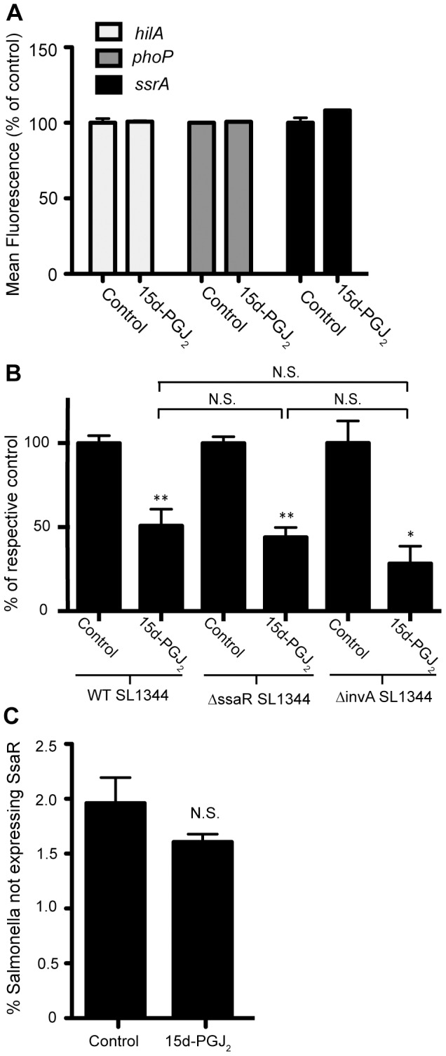
(A) Wild-type Salmonella carrying hilA-, phoP-, and ssrA-gfp reporter transcriptional fusions were used to analyze the effect of 15d-PGJ2 on virulence gene expression. Cultures were grown in LB for 4 hours. No changes in expression were seen. (B) 15d-PGJ2 reduced bacterial colonization of macrophages by the wild-type Salmonella strain, the ΔssaR strain, and the ΔinvA strain. Data are expressed as a percentage of the respective control samples, to illustrate that the extent of the inhibition caused by 15d-PGJ2 is equivalent in both strains. (C) Flow cytometry analysis of ssrA gene expression in Salmonella after infection of RAW264.7 macrophages with or without 15d-PGJ2 treatment. Averages of 8 measurements are shown with standard errors of means. (*p<0.05, **p<0.001).
15d-PGJ2 affects Salmonella colonization via a PPAR-γ independent mechanism
15d-PGJ2 is known to bind to and alter PPAR-γ activity, however, it is also not considered to be important in RAW264.7 macrophages. Ricote et. al. have shown that PPAR-γ is not expressed to a significant extent in these cells [8]. We wanted to ensure that PPAR-γ was not involved in our system. To do so, we added the PPAR-γ inhibitor GW9662 to RAW264.7 macrophages infected with Salmonella and treated with 15d-PGJ2 and monitored bacterial colonization, as above. The inhibitor was unable to restore the colonization defect seen with 15d-PGJ2 (Figure 7), indicating that the effect of 15d-PGJ2 on macrophage colonization by Salmonella may be through a PPAR-γ independent mechanism.
Figure 7. PPAR-γ inhibitor has no effect on Salmonella colonization.
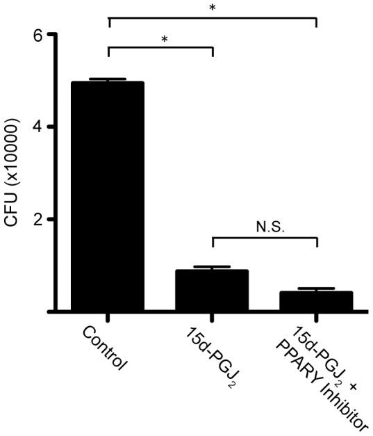
The effect of the addition of a PPAR-γ inhibitor to 15d-PGJ2 treated macrophages infected with Salmonella on bacterial colonization. Averages of 8 measurements are shown with standard errors of means. (*p<0.05, **p<0.001).
15d-PGJ2 induces ERK1/2 phosphorylation in macrophages infected with Salmonella
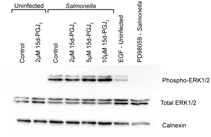
15d-PGJ2 has been shown to alter the activity of ERK1/2. Therefore we examined the phosphorylation of ERK1/2 in RAW264.7 macrophages treated with 15d-PGJ2 and infected with Salmonella (Figure 8). ERK1/2 phosphorylation was not seen in uninfected samples. ERK1/2 phosphorylation increased with increasing concentrations of 15d-PGJ2. EGF was used as a positive control of ERK1/2 phosphorylation, and PD98059, an ERK1/2 inhibitor, was used as a negative control. This indicates that 15d-PGJ2 is inducing the activity of ERK1/2. Because increased concentrations of 15d-PGJ2 can lead to increased cell death, cells were treated for 2 hours prior to infection, instead of overnight, and no increase was seen in cell lifting (data not shown).
Figure 8. Phosphorylated ERK1/2 levels increase with 15d-PGJ2 treatment.
Phosphorylated ERK1/2 levels are increased 1 hour after Salmonella infection of RAW264.7 macrophages treated with increasing concentrations of 15d-PGJ2. Figure is representative of 3 independent experiments.
Discussion
The potential role of 15d-PGJ2 during bacterial infection was initially considered because of the results of a metabolomics screen recently performed by our lab [40]. Because we identified the PG pathway to be highly responsive to Salmonella infection in our metabolomics analysis, we went on to examine if this pathway played a role in the establishment of infection by Salmonella. Here, we show that at 20 hours post-infection macrophages produce high levels of 15d-PGJ2 in response to Salmonella infection, which coincides with our previous data showing that 15d-PGJ2 is highly induced by Salmonella infection in mice. We then hypothesized that the high level of 15d-PGJ2 production observed would likely have a significant effect during the course of infection. To test this, we added 15d-PGJ2 exogenously and monitored its effects on Salmonella colonization of macrophages. Our data demonstrated a significant impact of 15d-PGJ2 on Salmonella burden and also showed a dose-dependent decrease in Salmonella colonization, clearly indicating that 15d-PGJ2 is sufficient to prevent bacterial colonization of macrophages. Some reports have claimed that 15d-PGJ2 treatment causes apoptosis in macrophages [17], and in fact at high concentrations of 15d-PGJ2 we did begin to see increased macrophage cell death (data not shown). Therefore, we used 2 µM 15d-PGJ2 in our experiments because this was the lowest concentration at which we still saw a decrease in colonization without an increase in cell death (Figure S1).
The effect of 15d-PGJ2 on Salmonella colonization was not limited to RAW264.7 macrophages. In fact, 15d-PGJ2 reduced bacterial colonization in both J774 macrophages and BMMOs. BMMOs are considered more ‘physiologically relevant’, and are able to clear Salmonella more rapidly and effectively than RAW264.7 macrophages (data not shown). It is also interesting that 15d-PGJ2 appears to be impacting bacterial entry, as indicated by Figure 3g. Furthermore, 15d-PGJ2 did not affect Salmonella replication in HeLa epithelial cells. This could indicate that the 15d-PGJ2–induced resistance to Salmonella is cell type specific. It is possible that 15d-PGJ2 alters a macrophage specific response to bacteria, thus inhibiting bacterial infection. It is interesting to note that Straus and colleagues [12] found that 15d-PGJ2 had a dramatically different effect on NF-κB inhibition in RAW264.7 and HeLa cells. The Salmonella life cycle inside of these two cell types is also very different [36] and this may be the reason for the significantly different responses.
Previously published results indicate that 15d-PGJ2 is able to reduce the production of cytokines in response to LPS [20], [27]. Here, we show the same effect with live, replicating bacteria. Specifically, we saw a reduction in IL-10 production in 15d-PGJ2 treated cells. IL-10 is increased via a SPI2 dependent mechanism during Salmonella infection, and may inhibit ROS and RNS in macrophages [42], [43]. We also saw a reduction in the amount of IL-6 and MCP-1 produced by macrophages treated with 15d-PGJ2 and infected with Salmonella. Interestingly, we did not see a change in IL-12, which can stimulate IFN-γ production [43]. IFN-γ is very important for the defense against Salmonella, and is produced predominantly by NK cells and T cells [43]. Since IFN-γ plays such an important role in anti-Salmonella defenses we were surprised to see that 15d-PGJ2 did not significantly alter either IL-12 or IFN-γ production. We also show that TNF-α, which is known to be important for anti-Salmonella defenses and is involved in triggering NO production [43], [44], was decreased with 15d-PGJ2 treatment. Generally these data indicate a reduction in pro-inflammatory molecules.
Similar to the results presented here, Cloutier et al. found that 15d-PGJ2 treatment reduced the production of IL-6, and TNFα in mice infected with the influenza virus, but also showed no effect on IFN-γ production [35]. In addition, Kielian et al., showed that 15d-PGJ2 selectively inhibited the inflammatory response of microglia in response to S. aureus [33]. This group showed 15d-PGJ2 dependent reduction in the production of IL-12p40, MCP1, and TNFα [33].
There is increasing evidence that Salmonella induced inflammation can actually benefit the pathogen in both intestinal colonization and systemic disease. Stecher et al. showed that intestinal inflammation is both necessary and sufficient in allowing Salmonella to outcompete the microbiota [45]. More specifically, Winter and colleagues (2010) showed that Salmonella induced gut inflammation resulted in the production of tetrathionate, which Salmonella is able to use as an electron acceptor, thus showing a mechanism by which inflammation benefits this pathogen [46]. Salmonella also gains a growth advantage by the production of ethanolamine and nitrate, which Salmonella is able to respire [47], [48]. It was also recently shown that Salmonella induces the recruitment of neutrophils to the intestinal lumen [49]. These neutrophils produce neutrophil elastase, which shifts the microbiota to favour Salmonella colonization [49]. At the systemic level, Arpaia and colleagues (2011) showed that TLR induced innate immunity in response to Salmonella induces virulence in the pathogen, allowing bacterial growth leading to systemic disease [50]. These studies, like the one we present here, show that inflammation is an important aspect of Salmonella infection.
Another immune mechanism generally considered to be critical to the host's defense against Salmonella are RNS. RNS are normally produced during Salmonella infection and are integral to bacterial killing as they modify components of the bacterial electron transport chain, metabolic enzymes, transcription factors, DNA, and DNA associated proteins [51]–[53]. Furthermore, IFN-γ pretreated macrophages have a stronger RNS response to Salmonella than untreated macrophages [53]. Intriguingly, we see both a reduction in RNS as well as a reduction in Salmonella burden in 15d-PGJ2 treated macrophages. In addition, we see this effect in both untreated and IFN-γ treated macrophages, which is interesting since IFN-γ treated macrophages are thought to have a much stronger RNS response to Salmonella. The reduction in RNS is also in line with the reduction in TNF-α that is caused by 15d-PGJ2 addition.
Our data also indicates that the 15d-PGJ2 mediated changes in bacterial colonization are SPI2 independent. We initially explored the possibility of SPI2 involvement due to the apparent reduction in the inflammatory response of the macrophages, and were surprised to see that SPI2 does not appear to be involved. We have also examined the potential role of PPAR-γ in the 15d-PGJ2 mediated reduction in Salmonella colonization. Not surprisingly, we found that the effects of 15d-PGJ2 on bacterial colonization were PPAR-γ independent. Furthermore, RAW264.7 macrophages do not appear to produce physiologically relevant amounts of PPAR-γ [8]. However, in the future siRNA knock-down and over-expression strains could be used to ensure that PPAR-γ is not involved in the 15d-PGJ2 mediated reduction in Salmonella growth.
We have also shown that the levels of phosphorylated ERK1/2 increase with increasing concentrations of 15d-PGJ2 treatment of Salmonella infected macrophages. Salmonella infection is known to lead to the activation of the ERK MAPK pathway [54]. Furthermore, the MEK/ERK pathway regulates changes in the actin cytoskeleton [55]. It is possible that 15d-PGJ2 is resulting in dis-regulation of ERK1/2 activity, which reduces Salmonella entry into macrophages; however this remains to be elucidated.
Research into the use of 15d-PGJ2 for the treatment of inflammatory diseases is already underway, and recently the use of nanocapsules as a mechanism of delivery has shown promise [23], [26]. In the future, the use of these or other delivery mechanisms may provide a way to effectively administer 15d-PGJ2 during Salmonella infection. Such research may provide insights into a novel mechanism of treating salmonellosis and possibly other bacterial infections. Our work sheds light onto a new role of 15d-PGJ2, namely the control of Salmonella pathogenesis and replication within phagocytic immune cells. The role of 15d-PGJ2 in bacterial infections is uncharacterized, and our work lays the foundation for further research into this area.
Supporting Information
Enumeration of live RAW264.7 macrophages (A) using Trypan Blue exclusion after treatment with 2 µM 15d-PGJ2 and infection with Salmonella . (B) LDH released from macrophages infected with Salmonella in the absence or presence of 15d-PGJ2.
(TIF)
Salmonella growth curves in (A) LB and (B) DMEM, with and without 2 µM 15d-PGJ2 treatment.
(TIF)
The effect of 15d-PGJ2 on Salmonella colonization of 2 ng/mL IFN-γ activated RAW264.7 macrophages 24 hours post infection. Averages of 8 measurements are shown with standard errors of means. (*p<0.05).
(TIF)
Acknowledgments
The authors would like to thank the anonymous reviewers for their helpful and constructive recommendations. We would also like to thank Hongbing Yu, Yanet Valdez, Wanyin Deng, Rosana Ferreira, and Jose Puente for suggestions and critical reading of this manuscript.
Funding Statement
This work was funded by the Canadian Institutes of Health Research (CIHR). MMCB is supported by a graduate scholarship from the National Sciences and Engineering Research Council of Canada (NSERC). LCMA is supported by a postdoctoral fellowship from the CIHR. NG is supported by fellowships from CIHR and Michael Smith Foundation for Health Research. SLR is supported by a graduate scholarship from the CIHR. SRS is supported by a graduate scholarship from the NSERC. The funders had no role in study design, data collection and analysis, decision to publish, or preparation of the manuscript.
References
- 1. Yoshikai Y (2001) Roles of prostaglandins and leukotrienes in acute inflammation caused by bacterial infection. Curr Opin Infect Dis 14: 257–263. [DOI] [PubMed] [Google Scholar]
- 2. Funk CD (2001) Prostaglandins and leukotrienes: advances in eicosanoid biology. Science 294: 1871–1875. [DOI] [PubMed] [Google Scholar]
- 3. Matsuoka T, Narumiya S (2008) The roles of prostanoids in infection and sickness behaviors. J Infect Chemother 14: 270–278. [DOI] [PubMed] [Google Scholar]
- 4. Cannon CP, Cannon PJ (2012) Physiology. COX-2 inhibitors and cardiovascular risk. Science 336: 1386–1387. [DOI] [PubMed] [Google Scholar]
- 5. Morteau O, Morham SG, Sellon R, Dieleman LA, Langenbach R, et al. (2000) Impaired mucosal defense to acute colonic injury in mice lacking cyclooxygenase-1 or cyclooxygenase-2. J Clin Invest 105: 469–478. [DOI] [PMC free article] [PubMed] [Google Scholar]
- 6. Gilroy DW, Colville-Nash PR, Willis D, Chivers J, Paul-Clark MJ, et al. (1999) Inducible cyclooxygenase may have anti-inflammatory properties. Nat Med 5: 698–701. [DOI] [PubMed] [Google Scholar]
- 7. Kansanen E, Kivela AM, Levonen AL (2009) Regulation of Nrf2-dependent gene expression by 15-deoxy-Delta12,14-prostaglandin J2. Free Radic Biol Med 47: 1310–1317. [DOI] [PubMed] [Google Scholar]
- 8. Ricote M, Li AC, Willson TM, Kelly CJ, Glass CK (1998) The peroxisome proliferator-activated receptor-gamma is a negative regulator of macrophage activation. Nature 391: 79–82. [DOI] [PubMed] [Google Scholar]
- 9. Waku T, Shiraki T, Oyama T, Morikawa K (2009) Atomic structure of mutant PPARgamma LBD complexed with 15d-PGJ2: novel modulation mechanism of PPARgamma/RXRalpha function by covalently bound ligands. FEBS Lett 583: 320–324. [DOI] [PubMed] [Google Scholar]
- 10. Jiang C, Ting AT, Seed B (1998) PPAR-gamma agonists inhibit production of monocyte inflammatory cytokines. Nature 391: 82–86. [DOI] [PubMed] [Google Scholar]
- 11. Petrova TV, Akama KT, Van Eldik LJ (1999) Cyclopentenone prostaglandins suppress activation of microglia: down-regulation of inducible nitric-oxide synthase by 15-deoxy-Delta12,14-prostaglandin J2. Proc Natl Acad Sci U S A 96: 4668–4673. [DOI] [PMC free article] [PubMed] [Google Scholar]
- 12. Straus DS, Pascual G, Li M, Welch JS, Ricote M, et al. (2000) 15-deoxy-delta 12,14-prostaglandin J2 inhibits multiple steps in the NF-kappa B signaling pathway. Proc Natl Acad Sci U S A 97: 4844–4849. [DOI] [PMC free article] [PubMed] [Google Scholar]
- 13. Negishi M, Koizumi T, Ichikawa A (1995) Biological actions of delta 12-prostaglandin J2. J Lipid Mediat Cell Signal 12: 443–448. [DOI] [PubMed] [Google Scholar]
- 14. Rossi A, Elia G, Santoro MG (1996) 2-Cyclopenten-1-one, a new inducer of heat shock protein 70 with antiviral activity. J Biol Chem 271: 32192–32196. [DOI] [PubMed] [Google Scholar]
- 15. Rossi A, Kapahi P, Natoli G, Takahashi T, Chen Y, et al. (2000) Anti-inflammatory cyclopentenone prostaglandins are direct inhibitors of IkappaB kinase. Nature 403: 103–108. [DOI] [PubMed] [Google Scholar]
- 16. Castrillo A, Diaz-Guerra MJ, Hortelano S, Martin-Sanz P, Bosca L (2000) Inhibition of IkappaB kinase and IkappaB phosphorylation by 15-deoxy-Delta(12,14)-prostaglandin J(2) in activated murine macrophages. Mol Cell Biol 20: 1692–1698. [DOI] [PMC free article] [PubMed] [Google Scholar]
- 17. Hortelano S, Castrillo A, Alvarez AM, Bosca L (2000) Contribution of cyclopentenone prostaglandins to the resolution of inflammation through the potentiation of apoptosis in activated macrophages. J Immunol 165: 6525–6531. [DOI] [PubMed] [Google Scholar]
- 18. Crosby MB, Svenson JL, Zhang J, Nicol CJ, Gonzalez FJ, et al. (2005) Peroxisome proliferation-activated receptor (PPAR)gamma is not necessary for synthetic PPARgamma agonist inhibition of inducible nitric-oxide synthase and nitric oxide. J Pharmacol Exp Ther 312: 69–76. [DOI] [PubMed] [Google Scholar]
- 19. Ruiz PA, Kim SC, Sartor RB, Haller D (2004) 15-deoxy-delta12,14-prostaglandin J2-mediated ERK signaling inhibits gram-negative bacteria-induced RelA phosphorylation and interleukin-6 gene expression in intestinal epithelial cells through modulation of protein phosphatase 2A activity. J Biol Chem 279: 36103–36111. [DOI] [PubMed] [Google Scholar]
- 20. Liu X, Yu H, Yang L, Li C, Li L (2012) 15-Deoxy-Delta(12,14)-prostaglandin J(2) attenuates the biological activities of monocyte/macrophage cell lines. Eur J Cell Biol 91: 654–661. [DOI] [PubMed] [Google Scholar]
- 21. Bell-Parikh LC, Ide T, Lawson JA, McNamara P, Reilly M, et al. (2003) Biosynthesis of 15-deoxy-delta12,14-PGJ2 and the ligation of PPARgamma. J Clin Invest 112: 945–955. [DOI] [PMC free article] [PubMed] [Google Scholar]
- 22. Castrillo A, Mojena M, Hortelano S, Bosca L (2001) Peroxisome proliferator-activated receptor-gamma-independent inhibition of macrophage activation by the non-thiazolidinedione agonist L-796,449. Comparison with the effects of 15-deoxy-delta(12,14)-prostaglandin J(2). J Biol Chem 276: 34082–34088. [DOI] [PubMed] [Google Scholar]
- 23. Napimoga MH, da Silva CA, Carregaro V, Farnesi-de-Assuncao TS, Duarte PM, et al. (2012) Exogenous administration of 15d-PGJ2-loaded nanocapsules inhibits bone resorption in a mouse periodontitis model. J Immunol 189: 1043–1052. [DOI] [PubMed] [Google Scholar]
- 24. Abdo H, Mahe MM, Derkinderen P, Bach-Ngohou K, Neunlist M, et al. (2012) The omega-6 fatty acid derivative 15-deoxy-Delta(1)(2),(1)(4)-prostaglandin J2 is involved in neuroprotection by enteric glial cells against oxidative stress. J Physiol 590: 2739–2750. [DOI] [PMC free article] [PubMed] [Google Scholar]
- 25. Han Z, Zhu T, Liu X, Li C, Yue S, et al. (2012) 15-deoxy-Delta12,14 -prostaglandin J2 reduces recruitment of bone marrow-derived monocyte/macrophages in chronic liver injury in mice. Hepatology 56: 350–360. [DOI] [PubMed] [Google Scholar]
- 26. Alves C, de Melo N, Fraceto L, de Araujo D, Napimoga M (2011) Effects of 15d-PGJ(2)-loaded poly(D,L-lactide-co-glycolide) nanocapsules on inflammation. Br J Pharmacol 162: 623–632. [DOI] [PMC free article] [PubMed] [Google Scholar]
- 27. Surh YJ, Na HK, Park JM, Lee HN, Kim W, et al. (2011) 15-Deoxy-Delta(1)(2),(1)(4)-prostaglandin J(2), an electrophilic lipid mediator of anti-inflammatory and pro-resolving signaling. Biochem Pharmacol 82: 1335–1351. [DOI] [PubMed] [Google Scholar]
- 28. Bui T, Straus DS (1998) Effects of cyclopentenone prostaglandins and related compounds on insulin-like growth factor-I and Waf1 gene expression. Biochim Biophys Acta 1397: 31–42. [DOI] [PubMed] [Google Scholar]
- 29. Zingarelli B, Sheehan M, Hake PW, O'Connor M, Denenberg A, et al. (2003) Peroxisome proliferator activator receptor-gamma ligands, 15-deoxy-Delta(12,14)-prostaglandin J2 and ciglitazone, reduce systemic inflammation in polymicrobial sepsis by modulation of signal transduction pathways. J Immunol 171: 6827–6837. [DOI] [PubMed] [Google Scholar]
- 30. Dugo L, Collin M, Cuzzocrea S, Thiemermann C (2004) 15d-prostaglandin J2 reduces multiple organ failure caused by wall-fragment of Gram-positive and Gram-negative bacteria. Eur J Pharmacol 498: 295–301. [DOI] [PubMed] [Google Scholar]
- 31. Guyton K, Zingarelli B, Ashton S, Teti G, Tempel G, et al. (2003) Peroxisome proliferator-activated receptor-gamma agonists modulate macrophage activation by gram-negative and gram-positive bacterial stimuli. Shock 20: 56–62. [DOI] [PubMed] [Google Scholar]
- 32. Reddy RC, Narala VR, Keshamouni VG, Milam JE, Newstead MW, et al. (2008) Sepsis-induced inhibition of neutrophil chemotaxis is mediated by activation of peroxisome proliferator-activated receptor-{gamma}. Blood 112: 4250–4258. [DOI] [PMC free article] [PubMed] [Google Scholar]
- 33. Kielian T, McMahon M, Bearden ED, Baldwin AC, Drew PD, et al. (2004) S. aureus-dependent microglial activation is selectively attenuated by the cyclopentenone prostaglandin 15-deoxy-Delta12,14- prostaglandin J2 (15d-PGJ2). J Neurochem 90: 1163–1172. [DOI] [PMC free article] [PubMed] [Google Scholar]
- 34. Cha B, Lim JW, Kim KH, Kim H (2011) 15-deoxy-D12,14-prostaglandin J2 suppresses RANTES expression by inhibiting NADPH oxidase activation in Helicobacter pylori-infected gastric epithelial cells. J Physiol Pharmacol 62: 167–174. [PubMed] [Google Scholar]
- 35. Cloutier A, Marois I, Cloutier D, Verreault C, Cantin AM, et al. (2012) The prostanoid 15-deoxy-Delta12,14-prostaglandin-j2 reduces lung inflammation and protects mice against lethal influenza infection. J Infect Dis 205: 621–630. [DOI] [PubMed] [Google Scholar]
- 36. Haraga A, Ohlson MB, Miller SI (2008) Salmonellae interplay with host cells. Nat Rev Microbiol 6: 53–66. [DOI] [PubMed] [Google Scholar]
- 37. McGhie EJ, Brawn LC, Hume PJ, Humphreys D, Koronakis V (2009) Salmonella takes control: effector-driven manipulation of the host. Curr Opin Microbiol 12: 117–124. [DOI] [PMC free article] [PubMed] [Google Scholar]
- 38. Buckner MM, Croxen MA, Arena ET, Finlay BB (2011) A comprehensive study of the contribution of Salmonella enterica serovar Typhimurium SPI2 effectors to bacterial colonization, survival, and replication in typhoid fever, macrophage, and epithelial cell infection models. Virulence 2: 208–216. [DOI] [PMC free article] [PubMed] [Google Scholar]
- 39. van der Heijden J, Finlay BB (2012) Type III effector-mediated processes in Salmonella infection. Future Microbiol 7: 685–703. [DOI] [PubMed] [Google Scholar]
- 40.Antunes LC, Arena ET, Menendez A, Han J, Ferreira RB, et al.. (2011) The impact of Salmonella infection on host hormone metabolism revealed by metabolomics. Infect Immun. [DOI] [PMC free article] [PubMed]
- 41. Antunes LC, Buckner MM, Auweter SD, Ferreira RB, Lolic P, et al. (2010) Inhibition of Salmonella host cell invasion by dimethyl sulfide. Appl Environ Microbiol 76: 5300–5304. [DOI] [PMC free article] [PubMed] [Google Scholar]
- 42. Uchiya K, Groisman EA, Nikai T (2004) Involvement of Salmonella pathogenicity island 2 in the up-regulation of interleukin-10 expression in macrophages: role of protein kinase A signal pathway. Infect Immun 72: 1964–1973. [DOI] [PMC free article] [PubMed] [Google Scholar]
- 43. Eckmann L, Kagnoff MF (2001) Cytokines in host defense against Salmonella. Microbes Infect 3: 1191–1200. [DOI] [PubMed] [Google Scholar]
- 44. Coburn B, Grassl GA, Finlay BB (2007) Salmonella, the host and disease: a brief review. Immunol Cell Biol 85: 112–118. [DOI] [PubMed] [Google Scholar]
- 45. Stecher B, Robbiani R, Walker AW, Westendorf AM, Barthel M, et al. (2007) Salmonella enterica serovar typhimurium exploits inflammation to compete with the intestinal microbiota. PLoS Biol 5: 2177–2189. [DOI] [PMC free article] [PubMed] [Google Scholar]
- 46. Winter SE, Thiennimitr P, Winter MG, Butler BP, Huseby DL, et al. (2010) Gut inflammation provides a respiratory electron acceptor for Salmonella. Nature 467: 426–429. [DOI] [PMC free article] [PubMed] [Google Scholar]
- 47. Thiennimitr P, Winter SE, Winter MG, Xavier MN, Tolstikov V, et al. (2011) Intestinal inflammation allows Salmonella to use ethanolamine to compete with the microbiota. Proc Natl Acad Sci U S A 108: 17480–17485. [DOI] [PMC free article] [PubMed] [Google Scholar]
- 48.Lopez CA, Winter SE, Rivera-Chavez F, Xavier MN, Poon V, et al. (2012) Phage-mediated acquisition of a type III secreted effector protein boosts growth of salmonella by nitrate respiration. MBio 3.. [DOI] [PMC free article] [PubMed] [Google Scholar]
- 49. Gill N, Ferreira RB, Antunes LC, Willing BP, Sekirov I, et al. (2012) Neutrophil elastase alters the murine gut microbiota resulting in enhanced Salmonella colonization. PLoS One 7: e49646. [DOI] [PMC free article] [PubMed] [Google Scholar]
- 50. Arpaia N, Godec J, Lau L, Sivick KE, McLaughlin LM, et al. (2011) TLR signaling is required for Salmonella typhimurium virulence. Cell 144: 675–688. [DOI] [PMC free article] [PubMed] [Google Scholar]
- 51. Shi L, Chowdhury SM, Smallwood HS, Yoon H, Mottaz-Brewer HM, et al. (2009) Proteomic investigation of the time course responses of RAW 264.7 macrophages to infection with Salmonella enterica. Infect Immun 77: 3227–3233. [DOI] [PMC free article] [PubMed] [Google Scholar]
- 52. Bourret TJ, Song M, Vazquez-Torres A (2009) Codependent and independent effects of nitric oxide-mediated suppression of PhoPQ and Salmonella pathogenicity island 2 on intracellular Salmonella enterica serovar typhimurium survival. Infect Immun 77: 5107–5115. [DOI] [PMC free article] [PubMed] [Google Scholar]
- 53. Henard CA, Vazquez-Torres A (2011) Nitric oxide and salmonella pathogenesis. Front Microbiol 2: 84. [DOI] [PMC free article] [PubMed] [Google Scholar]
- 54. Mynott TL, Crossett B, Prathalingam SR (2002) Proteolytic inhibition of Salmonella enterica serovar typhimurium-induced activation of the mitogen-activated protein kinases ERK and JNK in cultured human intestinal cells. Infect Immun 70: 86–95. [DOI] [PMC free article] [PubMed] [Google Scholar]
- 55. Kim H, White CD, Li Z, Sacks DB (2011) Salmonella enterica serotype Typhimurium usurps the scaffold protein IQGAP1 to manipulate Rac1 and MAPK signalling. Biochem J 440: 309–318. [DOI] [PMC free article] [PubMed] [Google Scholar]
Associated Data
This section collects any data citations, data availability statements, or supplementary materials included in this article.
Supplementary Materials
Enumeration of live RAW264.7 macrophages (A) using Trypan Blue exclusion after treatment with 2 µM 15d-PGJ2 and infection with Salmonella . (B) LDH released from macrophages infected with Salmonella in the absence or presence of 15d-PGJ2.
(TIF)
Salmonella growth curves in (A) LB and (B) DMEM, with and without 2 µM 15d-PGJ2 treatment.
(TIF)
The effect of 15d-PGJ2 on Salmonella colonization of 2 ng/mL IFN-γ activated RAW264.7 macrophages 24 hours post infection. Averages of 8 measurements are shown with standard errors of means. (*p<0.05).
(TIF)


